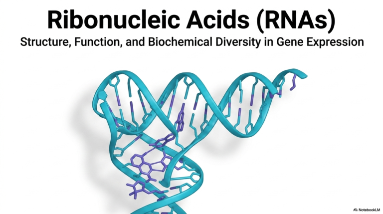
29. Enzymatic Analysis: Principles, Assays, and Applications
Imagine trying to orchestrate a complex symphony without knowing your musicians’ tempos. In the vibrant world of biochemistry, understanding the exact speed of cellular catalysts is essential for everything from diagnosing diseases to developing new biological drugs. The core purpose of this slide deck is to demystify the fundamental mechanisms of enzymatic analysis by illustrating how proteins transform specific substrates into measurable products. This first section lays the essential groundwork for assay kinetics.
Slide 1 – Enzymatic Analysis: Unveiling the Foundations of Assay Kinetics
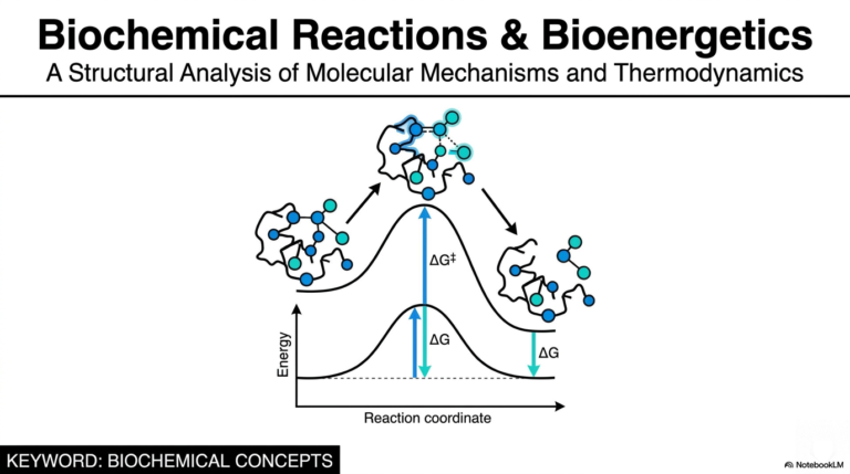
The bedrock of any enzymatic analysis is the classical kinetic equation. It begins when a free enzyme (E) naturally encounters its specific substrate (S). These molecules must perfectly align and physically bind to form a transient, highly specific intermediate structure known as the enzyme-substrate complex (ES). This temporary, locked state represents the critical transition phase in all biochemical reactions. The structural mechanics that govern this binding dictate the overall pace of the reaction, providing a clear target for scientific measurement.
Following formation of the enzyme-substrate complex, the catalytic event occurs. The enzyme drastically lowers the activation energy of the reaction, swiftly transforming the substrate into a final product (P). Crucially, the enzyme itself emerges from this chemical reaction completely unchanged and ready to immediately initiate another cycle. The rapid, cyclical turnover of this process is exactly what makes enzymatic analysis incredibly sensitive and universally useful in modern medical and research settings.
Because a single enzyme molecule can continuously generate thousands of product molecules, the resulting chemical signal amplifies rapidly over time. Whether researchers are using advanced spectrophotometry or basic clinical tests, mastering this foundational kinetic cycle is mandatory. Through rigorous enzymatic analysis, medical professionals can track these microscopic interactions in real time, turning invisible molecular events into quantifiable data that drive patient care and pharmacological innovation.
Slide 2 – Enzymatic Analysis: Isolating Targets in Complex Biological Mixtures
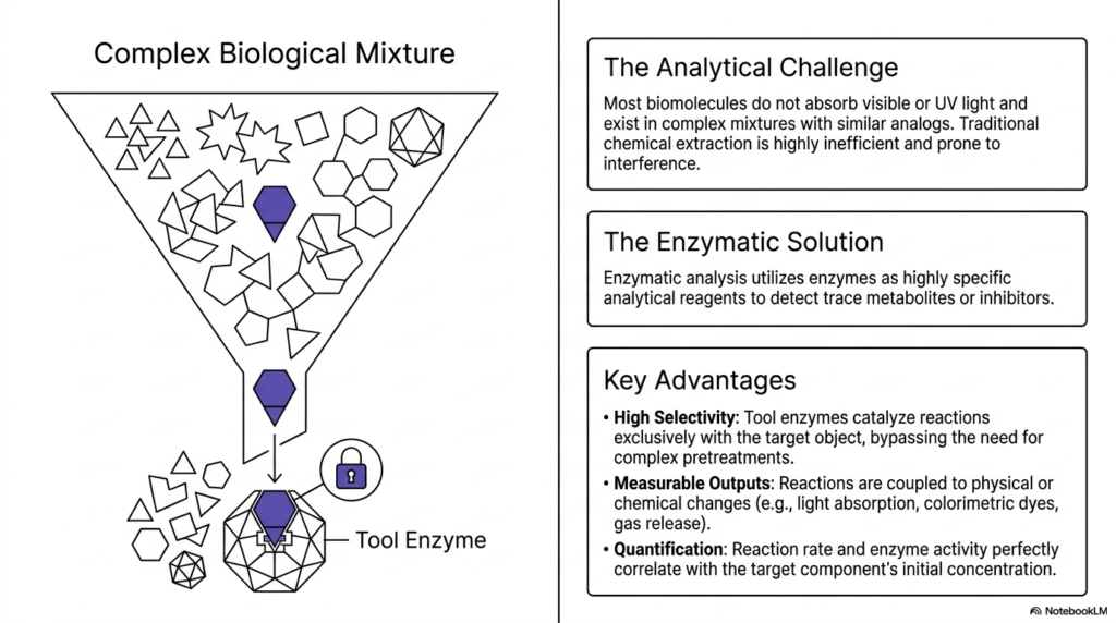
Finding a single microscopic needle in a massive molecular haystack is a daunting task, yet living cells perform this feat millions of times a second. Biological samples are incredibly chaotic, filled with thousands of similar molecules that obscure standard chemical detection. The purpose of this slide is to explore how enzymatic analysis addresses this analytical challenge by using highly specific enzymes to selectively identify and measure trace metabolites hidden within complex physiological mixtures.
Traditional chemical extraction methods are notoriously inefficient and highly prone to massive interference when dealing with complex biological mixtures. Most biomolecules do not naturally absorb visible or ultraviolet light, rendering standard optical detection completely useless for direct measurement. To overcome this hurdle, modern science turns to enzymatic analysis. By introducing a carefully selected tool enzyme into the chaotic cellular mixture, biochemists can perfectly isolate a single target object without needing to perform complex, time-consuming sample pretreatments.
The true diagnostic genius of enzymatic analysis lies in its unparalleled high selectivity. A specific enzyme acts like a microscopic lock-and-key, catalyzing chemical reactions exclusively with its perfectly matched target substrate while ignoring all other background noise. Once the targeted reaction occurs, it is intelligently coupled to a measurable physical or chemical output. This could involve an observable shift in light absorption, the generation of vibrant colorimetric dyes, or even the localized release of gas, providing a very clear optical signal.
Ultimately, this highly specific interaction allows for precise molecular quantification in clinical settings. Because the enzyme-substrate pair reacts only with the target, the overall reaction rate and measured enzyme activity correlate perfectly with the target component’s initial concentration. Mastering this aspect of enzymatic analysis empowers future doctors and scientists to accurately analyze blood, tissue, and cellular extracts, ensuring highly reliable diagnostic readouts.
Slide 3 – Enzymatic Analysis: Standardizing Catalytic Power and Specific Activity
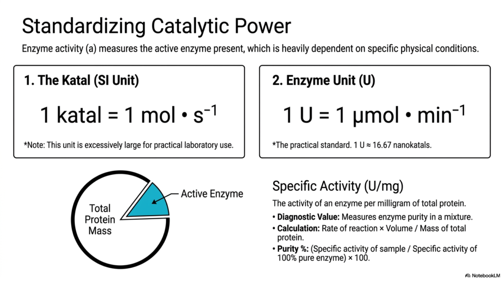
How do scientists agree on a standard measurement when every laboratory globally operates under slightly different conditions? Simply measuring the raw mass of an enzyme tells us absolutely nothing about its actual biological capability or health. The core purpose of this slide is to define the standardized metric units used in enzymatic analysis, ensuring that catalytic power is universally understood. By mastering these metrics, researchers can accurately communicate enzyme efficiency across different studies.
To bring absolute order to the chaotic world of biochemistry, scientists rely on highly specific units to quantify active enzymes. The official International System (SI) unit for catalytic activity is the katal, strictly defined as the amount of enzyme that converts one mole of substrate per second. However, one katal represents a massive, almost absurdly large amount of biological activity for standard laboratory work. Therefore, practical enzymatic analysis typically utilizes the significantly more manageable Enzyme Unit (U).
One standard Enzyme Unit (U) is mathematically defined as the amount of enzyme required to catalyze the conversion of one micromole of substrate per minute under ideal conditions. For reference, one standard unit is mathematically equivalent to approximately 16.67 nanokatals. This standardized metric is the absolute backbone of quantitative enzymatic analysis, allowing medical researchers to evaluate enzyme kinetics consistently. Yet, simply knowing the total units is not always enough, especially when evaluating crude biological mixtures containing numerous background proteins.
This brings us to the crucial biological concept of specific activity, measured in units per milligram of total protein (U/mg). Specific activity acts as a direct, reliable measure of overall enzyme purity. By dividing the measured reaction rate by the total protein mass, biochemists can determine the exact concentration of their target enzyme. In diagnostic enzymatic analysis, comparing the specific activity of a freshly drawn sample with that of a 100% pure enzyme yields the exact purity percentage, a vital quality-control step.
Slide 4 – Enzymatic Analysis: Bridging Molecules and Light with Spectrophotometry
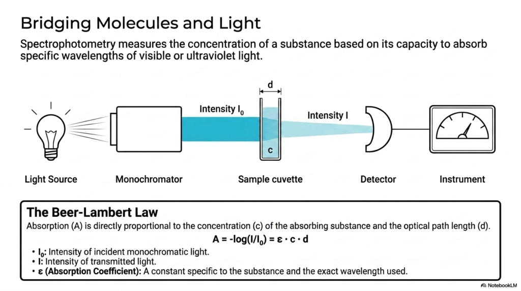
Have you ever wondered how scientists actually ‘see’ invisible chemical reactions taking place inside a sterile test tube? Because we cannot observe individual molecules with the naked eye, we must rely entirely on the interaction between molecular matter and electromagnetic radiation. The core purpose of this slide is to introduce spectrophotometry, a foundational optical technique in enzymatic analysis that quantitatively relates a substance’s concentration to its ability to absorb specific wavelengths of light.
Spectrophotometry serves as the primary analytical bridge between microscopic molecular events and macroscopic, highly quantifiable diagnostic data. An instrument known as a spectrophotometer uses a controlled light source and a precise monochromator to beam a specific, single wavelength of visible or ultraviolet light directly through a sample cuvette. The fundamental operating principle of this enzymatic analysis method relies on continuously comparing the incident light’s original intensity entering the sample with the transmitted light’s weakened intensity exiting it.
The mathematical heart of this optical enzymatic analysis is the Beer-Lambert Law. This crucial scientific law dictates that light absorption is directly proportional to both the concentration of the absorbing chemical substance and the total optical path length of the sample cuvette. The governing equation incorporates an absorption coefficient, a unique mathematical constant specific to the given substance and the exact wavelength of light used in the laboratory procedure.
By successfully utilizing the Beer-Lambert Law, biochemists can rapidly translate seemingly arbitrary changes in light intensity into highly accurate chemical concentration values. If a biological reaction produces a light-absorbing product, the sensitive optical detector will record a steady, mathematical increase in absorbance over time. This provides a continuous, real-time window into the reaction’s kinetic rate. Mastering spectrophotometry is an absolute necessity for anyone conducting robust enzymatic analysis in modern clinical environments.
Slide 5 – Enzymatic Analysis: Decoding the Continuous Direct Assay Using LDH
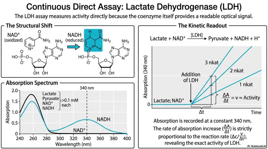
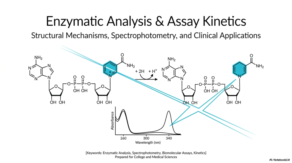
Imagine watching a crowded race where the fastest runners naturally glow in the dark—you wouldn’t need a complex camera system to track their progress. In the realm of biochemistry, some natural reactions produce their own readable signals, making it incredibly straightforward and fast to track them. The purpose of this slide is to dissect a continuous direct assay within enzymatic analysis, using Lactate Dehydrogenase (LDH) as the perfect model for how natural coenzyme shifts provide real-time diagnostic optical data.
The LDH assay is a premier clinical example of continuous enzymatic analysis because it measures activity directly, without requiring secondary marker dyes or complex engineered chemical steps. The diagnostic secret lies in the structural shift of its vital biological coenzyme. During the reaction, Lactate Dehydrogenase rapidly oxidizes lactate into pyruvate, simultaneously reducing the coenzyme NAD+ into NADH. While NAD+ remains optically silent at specific ultraviolet wavelengths, the newly formed NADH molecule undergoes a subtle structural change that allows it to absorb ultraviolet light more strongly.
By carefully examining the chemical absorption spectrum, we see that both NAD+ and NADH absorb light strongly around 260 nanometers. However, a massive optical divergence occurs at exactly 340 nanometers. At this specific ultraviolet wavelength, NADH shows a distinct, massive absorption peak, while NAD+, lactate, and pyruvate show absolutely zero absorption. Therefore, medical researchers conducting this specific enzymatic analysis can confidently set their spectrophotometer to a fixed wavelength of 340 nm to monitor the generation of NADH exclusively.
The resulting kinetic readout is beautifully simple, elegantly clean, and mathematically precise. Following the initial addition of the LDH enzyme to the mixture, ultraviolet light absorption strictly increases over time. The mathematical rate of this absorption increase is directly and strictly proportional to the overall biochemical reaction rate. Through this direct enzymatic analysis, medical scientists can continuously monitor systemic LDH activity, a critical diagnostic biomarker for severe tissue damage and cellular necrosis in hospitals.
Slide 6 – Enzymatic Analysis: Engineering Visibility via Continuous Coupled Assays
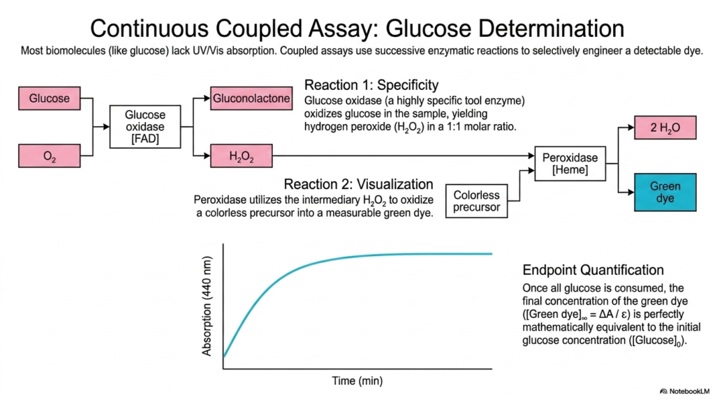
What happens when the vital biological target you desperately need to measure is entirely invisible to standard light detectors? Unfortunately, most essential biomolecules, like human blood sugar, do not naturally absorb ultraviolet or visible light. The core purpose of this slide is to demonstrate how enzymatic analysis boldly circumvents this issue by using a continuous coupled assay that strategically links multiple biochemical reactions to engineer a highly visible, trackable colorimetric dye for diagnostics.
Clinical glucose determination is a textbook, life-saving example of a coupled assay utilized in daily enzymatic analysis. Because glucose lacks any natural UV/Vis absorption, the diagnostic process requires two distinct, flawlessly linked chemical steps. Reaction 1 provides the necessary specificity. A highly specific enzyme, glucose oxidase, is introduced into the biological sample. This enzyme selectively oxidizes the invisible glucose, producing gluconolactone and, crucially, hydrogen peroxide in a perfect one-to-one molar ratio.
While the generated hydrogen peroxide is also entirely invisible, it acts as the essential reactive trigger for Reaction 2, the true visualization phase. A second tool enzyme, peroxidase, is added alongside a completely colorless chemical precursor. The peroxidase uses the intermediate hydrogen peroxide to oxidize the colorless precursor, transforming it into a highly measurable, dark green dye. This ingenious linked strategy allows enzymatic analysis to transform a completely invisible metabolic event into a vibrant, trackable optical signal.
The analytical endpoint of this specific enzymatic analysis is incredibly elegant and mathematically sound. As the coupled reaction progresses, the spectrophotometer continuously monitors the increasing absorbance of the newly formed green dye. Once all the initial glucose is fully consumed by the first enzyme, the absorption curve rigidly flattens out at the endpoint. At this exact moment, the final concentration of the generated green dye is mathematically equivalent to the initial glucose concentration, providing life-saving diagnostic information for diabetic patients globally.
Slide 7 – Enzymatic Analysis: Tracking Cell Viability with Colorimetric Assays
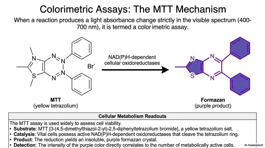
How can biomedical researchers definitively tell if a microscopic population of cells is thriving, actively dying, or positively responding to a newly developed experimental drug? Instead of attempting to count individual cells one by one, biochemists use clever metabolic tricks to make living cells reveal themselves visually. The core purpose of this slide is to explore colorimetric assays within enzymatic analysis, focusing specifically on the robust MTT mechanism as a global standard for visually quantifying cellular viability.
A colorimetric assay is a highly specific branch of enzymatic analysis in which a biochemical reaction deliberately produces a measurable change in light absorbance within the visible spectrum, typically between 400 and 700 nanometers. The MTT assay is widely used globally to assess cellular health. The primary chemical substrate is MTT, a water-soluble, highly visible yellow tetrazolium salt. When introduced to a biological cellular environment, this vibrant yellow molecule serves as a direct target for active metabolic machinery.
The diagnostic magic of this particular enzymatic analysis relies entirely on NAD(P)H-dependent cellular oxidoreductases. These vital, hard-working cellular enzymes are only biologically active and functioning in healthy, living cells. When the yellow MTT substrate enters a viable cell, these robust oxidoreductases rapidly cleave the delicate tetrazolium ring via a rapid reduction reaction. This highly specific catalytic event completely transforms the soluble yellow salt into an entirely new biological product: an insoluble, deeply purple crystal known scientifically as formazan.
Because dead or dying cells rapidly lose their internal oxidoreductase activity, they absolutely cannot process the MTT substrate, leaving the entire solution a clear yellow. However, vibrant, rapidly dividing cellular populations will generate massive, visible amounts of the purple formazan product. In this highly visual enzymatic analysis, the final optical intensity of the purple color, once solubilized, directly and mathematically correlates to the exact number of metabolically active cells. This provides pharmaceutical researchers with a rapid, scalable method for screening large-scale drug toxicity trials.
Slide 8 – Enzymatic Analysis: Navigating the Typology of Assays
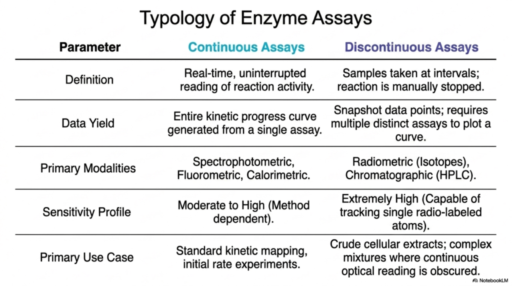
Not all biochemical experiments can be run using the exact same playbook; different molecular environments absolutely demand very different analytical approaches. A major challenge for any budding biochemist or medical student is successfully choosing the right diagnostic tool for the specific job at hand. The core purpose of this slide is to break down the overarching typology of enzymatic analysis and compare modern continuous and traditional discontinuous assay formats to help students select the optimal methodology for specific experimental parameters.
In the fast-paced realm of modern enzymatic analysis, continuous assays are rigidly defined by their real-time, uninterrupted reading of chemical reaction activity. These popular methods generate an entire, highly detailed kinetic progress curve from a single, smoothly running biological assay. Major diagnostic modalities such as spectrophotometry, fluorometry, and calorimetry fall squarely into this continuous category. They offer sensitivity ranging from moderate to high and are the absolute gold standard for mapping standard kinetics and conducting initial rate experiments in clear, highly controlled laboratory solutions.
Conversely, discontinuous assays represent an older, yet highly critical branch of enzymatic analysis. Here, busy researchers must physically extract tiny sample volumes at highly specific time intervals and then manually, abruptly stop the ongoing biochemical reaction. Because this labor-intensive method only yields frozen snapshot data points, generating a full kinetic curve requires performing multiple, entirely distinct assays and plotting them manually together. While highly complex, discontinuous assays employ powerful modalities such as radiometry and chromatography to achieve extreme scientific precision.
Why would a scientist purposefully choose a difficult discontinuous method over a simple continuous one? The clear answer lies entirely within the experimental environment. Discontinuous enzymatic analysis is functionally necessary when working directly with crude cellular extracts or complex, highly opaque biological mixtures in which continuous optical reading is physically impossible. Furthermore, discontinuous radiometric assays offer exceptionally high sensitivity, capable of tracing single radio-labeled atoms, making them indispensable for highly specialized, cutting-edge metabolic pathway mapping.
Slide 9 – Enzymatic Analysis: Exploring Advanced Continuous Modalities
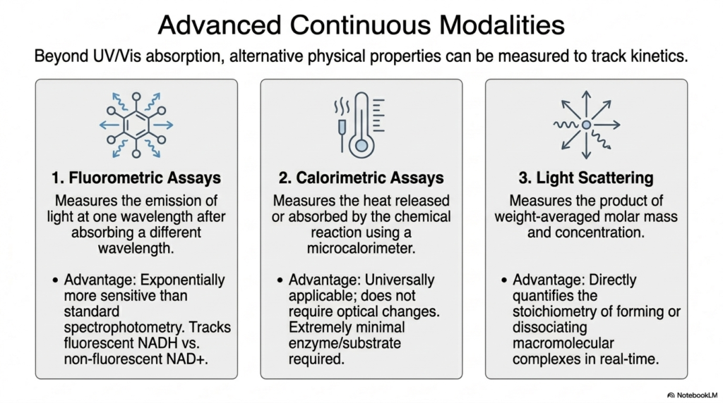
Standard UV/Vis spectrophotometry is a fantastic foundational tool, but what happens when a biological signal is simply too faint to see or doesn’t involve light absorption at all? To aggressively push the boundaries of modern biochemical research, scientists must continually rely on alternative physical properties to track hidden molecular motion. The core purpose of this slide is to delve deeply into advanced continuous modalities in enzymatic analysis, highlighting how cutting-edge fluorometry, calorimetry, and light scattering readily overcome the rigid limitations of traditional optical absorption.
Advanced fluorometric assays represent an absolutely massive technological leap in raw sensitivity for modern enzymatic analysis. Instead of passively measuring absorbed light, active fluorometry precisely tracks the rapid emission of bright light at a single, highly specific wavelength immediately after the target molecule absorbs light at a completely different starting wavelength. This advanced methodology is exponentially more sensitive than standard baseline spectrophotometry. It is particularly adept at tracking highly fluorescent target molecules such as NADH while completely ignoring non-fluorescent background molecules, enabling precise measurement of ultra-trace molecular concentrations.
Modern calorimetric assays deliberately eliminate the need for optical changes, making them universally applicable across all branches of clinical enzymatic analysis. Using a highly sensitive digital microcalorimeter, researchers continuously measure the microscopic amounts of heat released or absorbed by a running chemical reaction. Because every single chemical bond formation or sudden breakage involves a strict thermodynamic shift, this advanced modality requires extremely minimal enzyme and substrate concentrations, naturally thriving in opaque mixtures where standard light simply cannot penetrate.
Finally, the technique of light scattering adds a profound physical structural dimension to standard enzymatic analysis. This robust technique directly measures the exact product of a biomolecule’s weight-averaged molar mass and its overall, real-time concentration. By intentionally scattering powerful lasers off a biological sample, scientists can directly and mathematically quantify the physical stoichiometry of rapidly forming or dissociating macromolecular complexes in absolute real-time. Together, these three highly advanced diagnostic modalities ensure that modern enzymology possesses the exact tools to track any reaction.
Slide 10 – Enzymatic Analysis: Thermodynamic Kinetics via Microscale Thermophoresis
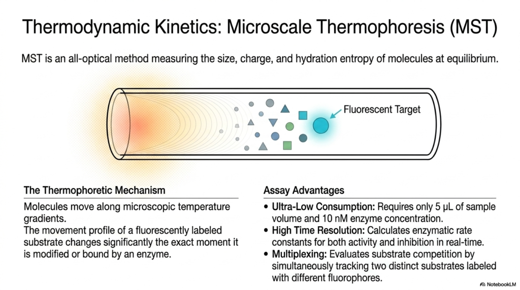
How do you accurately measure delicate molecular binding in an experimental environment so incredibly tiny that even a single drop of water seems like a massive ocean? Modern drug discovery constantly demands novel technologies that use incredibly small sample sizes while simultaneously yielding massive amounts of highly accurate kinetic data. The core purpose of this slide is to introduce Microscale Thermophoresis (MST), a cutting-edge technique in enzymatic analysis that leverages microscopic temperature gradients to cleanly evaluate molecular dynamics.
Modern Microscale Thermophoresis is a truly brilliant, highly sophisticated, all-optical method utilized strictly in advanced enzymatic analysis. The underlying physical thermophoretic mechanism relies entirely on the universal scientific principle that molecules will naturally and predictably move along microscopic, artificially generated temperature gradients created by a highly focused infrared laser. Crucially, the specific spatial movement profile of a fluorescently labeled target substrate changes significantly the exact moment it is chemically modified or physically bound by an active enzyme, providing a crystal-clear thermodynamic readout.
MST offers truly profound, undeniable advantages for high-level clinical enzymatic analysis. First, it routinely boasts ultra-low biological consumption, requiring only 5 microliters of total sample volume and an incredibly low starting enzyme concentration of 10 nanomolar. This is a massive financial game-changer when working with exceedingly rare or incredibly expensive human proteins. Furthermore, the technology seamlessly provides high time resolution, rapidly calculating highly accurate enzymatic rate constants for both catalytic activity and competitive drug inhibition in real time.
Another massive scientific benefit of this specific form of enzymatic analysis is its inherent capacity for rapid, advanced multiplexing. Researchers can easily evaluate complex, multi-level substrate competition by simultaneously tracking two completely distinct target substrates, each carefully labeled with a totally different colored fluorophore. By closely observing how these distinct molecules behave within the thermophoretic gradient, biochemists continually gain unprecedented, deep structural insights into the size, charge, and hydration entropy of complex biomolecules, thereby vastly accelerating modern drug screening.
Slide 11 – Enzymatic Analysis: Merging Immunology with EIA and ELISA
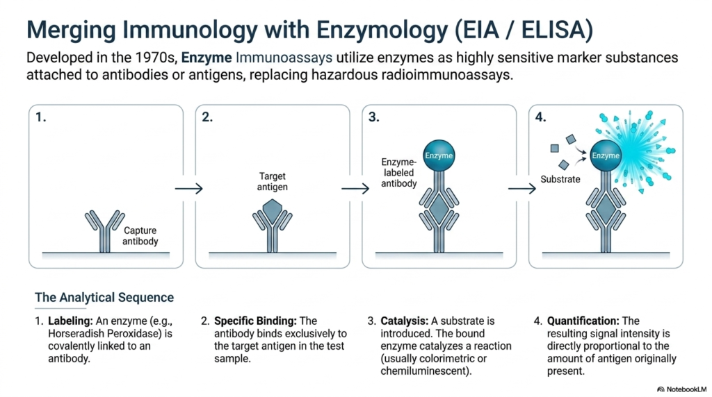
Imagine flawlessly combining the extreme, rapid sensitivity of an enzyme with the utterly perfect molecular targeting system of the human immune system. In the innovative 1970s, brilliant scientists did exactly that, permanently revolutionizing global medical diagnostics by entirely replacing highly dangerous radioactive materials with remarkably safe, highly active biological catalysts. The core purpose of this slide is to clearly explain the underlying mechanics of Enzyme Immunoassays (EIA/ELISA), a remarkably powerful hybrid form of enzymatic analysis that utilizes antibodies to detect incredibly minute concentrations of viral antigens.
The absolute foundation of this unique, highly effective enzymatic analysis lies directly in a highly specific, four-step analytical sequence. The critically important first step involves strategic chemical labeling, where a robust, highly active enzyme, typically Horseradish Peroxidase, is permanently and covalently linked to a synthetic laboratory antibody. The vital second step relies entirely on specific immunological binding. When safely introduced to a patient’s fluid sample, this specialized antibody acts exactly like a guided biological missile, hunting down and binding exclusively to the specific target antigen being tested for.
Once the specialized antibody binds tightly to its biological target, the true, magnificent power of enzymatic analysis is finally unleashed. A completely colorless chemical substrate, specifically designed for the permanently linked enzyme, is carefully introduced into the testing well. The bound enzyme rapidly catalyzes a fierce chemical reaction, completely transforming the invisible substrate into a highly visible, vibrant colorimetric or intensely chemiluminescent product. This massive catalytic amplification ensures that even a microscopic trace of the target viral antigen produces a readily detectable optical signal.
In the final, absolutely crucial quantification step of this enzymatic analysis, a highly calibrated spectrophotometer precisely measures the intensity of the resulting color or light. Because the linked enzyme strictly only remains in the testing well if the antibody successfully binds a real antigen, the resulting signal intensity is directly and flawlessly mathematically proportional to the precise amount of antigen originally present. Today, robust ELISA methodologies remain the absolute unquestioned gold standard in modern hospitals worldwide for rapidly detecting dangerous viruses and tracking hormones.
Slide 12 – Enzymatic Analysis: Navigating Kinetic Variables and Constraints
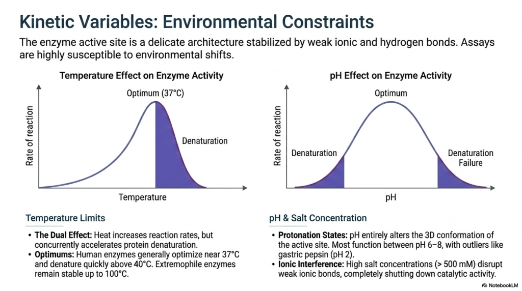
Even the absolutely most perfectly designed biological machine will critically fail if you abruptly throw it into a freezing volcano or drop it entirely in burning acid. Enzymes are incredibly delicate, highly nuanced protein architectures, heavily reliant on a fragile, microscopic web of weak chemical bonds to maintain their perfect 3D shape. The core purpose of this slide is to closely explore the severe, highly destructive environmental constraints heavily impacting enzymatic analysis, detailing exactly how minor environmental shifts in temperature and pH can entirely dismantle a biochemical assay.
External temperature constantly exerts a powerful, highly dangerous dual effect during any enzymatic analysis procedure. As environmental heat increases, molecular collisions naturally become more frequent, thereby accelerating overall reaction rates. Human enzymes generally optimize their kinetic rate near 37 degrees Celsius. However, this same exact heat concurrently accelerates incredibly destructive, irreversible protein denaturation. Pushing the ambient temperature rapidly above 40 degrees Celsius violently destroys the weak ionic and hydrogen bonds that stabilize the fragile active site, melting the protein structure and bringing the catalytic reaction to an immediate halt.
Extreme, sudden pH shifts are equally highly destructive in any robust enzymatic analysis. The specific pH of an experimental environment can entirely alter the delicate protonation states of the vital amino acids directly lining the enzyme’s active site. If the 3D biological conformation shifts even slightly due to incorrect protonation, the rigid substrate can no longer bind physically. While most human cellular enzymes function optimally strictly between a neutral pH of 6 and 8, specific, unique outliers like gastric pepsin are structurally evolved to remarkably thrive in highly acidic environments, aggressively near pH 2.
Additionally, clinical researchers constantly conducting enzymatic analysis must very carefully monitor chaotic ionic interference. Extremely high external salt concentrations can immediately and aggressively disrupt the vital weak ionic bonds that hold the enzyme’s tertiary structure together, completely shutting down all real catalytic activity. Recognizing and strictly controlling these critical, highly dangerous environmental variables—rigidly locking down the exact physical temperature, absolute salt concentration, and delicate pH buffer—is essential to ensuring that laboratory diagnostic data is reproducible, highly accurate, and scientifically valid.
Slide 13 – Enzymatic Analysis: Saturation Kinetics and Macromolecular Crowding
Have you ever been waiting in a massive grocery store where every single checkout lane is completely full with customers? It absolutely doesn’t matter exactly how many more new shoppers enter the busy store; the checkout line physically won’t move any faster. Enzymes constantly operate under these exact same rigid biological traffic laws. The core purpose of this slide is to sharply analyze highly critical kinetic variables in enzymatic analysis, specifically exploring the rigid mathematical limits of pure enzyme saturation and the chaotic reality of cellular crowding.
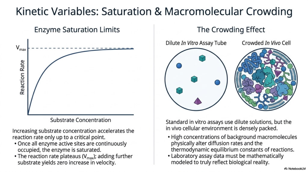
A truly fundamental, incredibly important principle of modern enzymatic analysis is a deep understanding of rigid enzyme saturation limits. As you steadily increase the raw concentration of a target substrate in a clear solution, the overall biological reaction rate aggressively accelerates proportionally. However, this rapid acceleration strictly only continues up to a highly critical, predefined biological point. Eventually, every single open enzyme active site is continuously and physically occupied by a rapidly arriving substrate molecule. At this exact moment of total, undeniable saturation, the enzyme is physically working at its absolute maximum kinetic capacity.
Once absolute maximum saturation is successfully reached, the recorded reaction rate rigidly and permanently plateaus at a standard metric heavily known as Vmax. At total Vmax, adding any additional substrate yields absolutely no measurable increase in the reaction velocity. In standard laboratory enzymatic analysis, accurately mapping this exact mathematical curve helps medical scientists rigorously calculate precise drug efficacy. However, a truly massive, undeniable physical discrepancy constantly exists between a pristine laboratory test tube and a highly active, living human cell. Standard in vitro assays strictly use highly dilute solutions, whereas the actual in vivo cellular environment is incredibly densely packed.
This dense, chaotic biological packing instantly creates the crowding effect, a remarkably major complicating physical factor in robust enzymatic analysis. High, massive concentrations of background cellular macromolecules physically alter the standard diffusion rates and the basic thermodynamic equilibrium constants of highly vital cellular reactions. Because the crowded, dense environment severely restricts molecular movement, standard laboratory assay data must be rigorously, continuously, and mathematically modeled to accurately reflect biological reality. Recognizing this density disparity directly ensures that pharmaceutical researchers don’t wildly overestimate an enzyme’s true kinetic rate.
Slide 14 – Enzymatic Analysis: Synthesizing and Designing a Custom Assay
Building an incredibly reliable, highly accurate scientific experiment is heavily analogous to strictly following a highly precise master recipe; violently skipping a single tiny step, and the entire chemical dish is horribly ruined. Flawlessly translating raw biochemical scientific theory into a functional, highly robust real-world laboratory procedure requires absolutely meticulous planning and highly rigorous logical sequencing. The core purpose of this slide is to proudly provide a highly comprehensive, strict four-step synthesis strictly for designing a flawless enzymatic analysis, deeply empowering new students to safely construct custom assays for novel biological research.
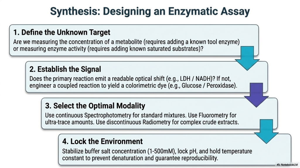
The absolutely critical, very first step in designing a highly effective enzymatic analysis is to explicitly and precisely define the exact biological target. Diligent researchers must rigorously decide if they are strictly measuring the hidden chemical concentration of a highly specific metabolite, which requires immediately adding a known, highly purified tool enzyme. Alternatively, they might be measuring an entirely unknown underlying enzyme activity continuously within a diseased tissue sample, which strongly demands abruptly adding highly controlled, maximally saturated substrates. Clearly and rigorously identifying the target directly dictates the entire future trajectory of the experimental protocol.
Step two strictly involves aggressively establishing a crystal clear, highly measurable chemical signal. Does the primary biochemical reaction naturally and predictably emit a highly readable physical optical shift, remarkably so, such as the well-known LDH and NADH transition? If the specific target molecules are completely optically silent, the brilliant researcher must ingeniously and meticulously engineer a robust coupled reaction directly into their complex enzymatic analysis, flawlessly linking multiple tool enzymes to successfully yield a highly vibrant, intensely readable colorimetric dye. Step three then strongly requires strictly selecting the absolute optimal experimental modality—wisely choosing continuous standard spectrophotometry for clear standard mixtures or highly discontinuous advanced radiometry for thick, opaque biological tissue extracts.
Finally, step four is arguably the most highly critical for fiercely ensuring absolute scientific validity: rigidly locking down the highly chaotic experimental environment. As discussed in detail previously, all cellular enzymes are incredibly and remarkably chemically fragile. To guarantee absolute reproducibility in an enzymatic analysis, dedicated researchers must rigorously stabilize the exact buffer salt concentration, strictly maintain the precise biological pH, and maintain the laboratory temperature constant. By rigorously and flawlessly controlling the biological environment to completely prevent any trace of rapid denaturation, medical scientists ensure that their resulting vital kinetic data is flawlessly accurate and fully ready for immediate medical publication.
Slide 15 – Enzymatic Analysis: Charting the High-Tech Future of Diagnostics
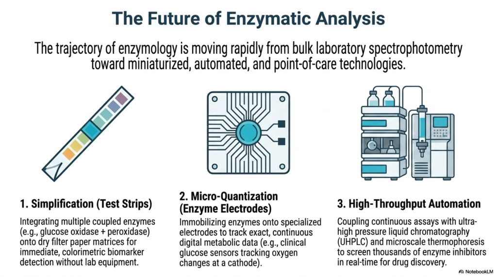
The archaic days of relying heavily on massive, highly expensive, room-sized laboratory equipment and enduring days-long, frustrating waiting periods for vital diagnostic test results are rapidly and finally coming to an end. Modern clinical biochemistry is undergoing a rapid technological renaissance, driven by the intense clinical need for significantly faster, remarkably smaller, and highly intelligent diagnostic screening tools. The core purpose of this final slide is to deeply explore the highly promising future of modern enzymatic analysis, sharply highlighting the dramatic shift toward remarkably miniaturized, highly automated, and ultra-rapid point-of-care medical technologies.
The absolutely first major progressive trajectory in cutting-edge modern enzymatic analysis is extreme, highly rapid physical simplification. We clearly see this remarkable biochemical progress daily with highly modern, cheap clinical test strips. By flawlessly chemically integrating multiple robust coupled enzymes—such as the classic glucose oxidase and peroxidase—directly and permanently onto completely dry filter paper matrices, highly brilliant scientists have successfully created immediate, vibrant colorimetric biomarker diagnostic detection systems. These incredibly simple paper strips bypass the need for highly expensive laboratory and hospital equipment, placing immense, rapid diagnostic power directly into the eager hands of ordinary patients for robust, real-time physiological health monitoring.
Another remarkably revolutionary, massively impactful leap in highly advanced enzymatic analysis is the rapid global rise of extreme micro-quantization enabled by highly advanced digital enzyme electrodes. By flawlessly, permanently immobilizing incredibly fragile chemical-tool enzymes directly onto highly specialized digital electrical electrodes, clinical scientists can precisely track continuous metabolic biological data strictly inside the active human body. Vital clinical digital glucose sensors that constantly, flawlessly track precise microscopic oxygen shifts directly at a tiny microscopic electrical cathode are perfect examples of this massive tech. It perfectly merges highly wet clinical biochemistry with advanced digital silicon circuitry, strongly enabling absolutely life-saving, totally continuous metabolic biological tracking.
Finally, incredibly massive, totally robust high-throughput robotic automation is completely and wildly reshaping the entire global pharmaceutical research industry. By flawlessly coupling highly robust continuous enzymatic analysis assays directly with cutting-edge ultra-high-pressure liquid chromatography (UHPLC) and highly advanced microscale thermophoresis robotic systems, massive medical supercomputers can now rapidly screen hundreds of thousands of potential vital enzyme inhibitors in mere seconds. This is totally incredible, unbelievably rapid real-time biological data generation that wildly accelerates critical global drug discovery, ensuring that the remarkable next generation of highly targeted, biochemically advanced therapeutics will definitively reach critically ill hospital patients incredibly faster and remarkably safer than ever before in history.
Please read our Content Disclaimer Statement.
Check out our social media channels: