61. Xenobiotic Metabolism: Detoxification Pathways & Cytochrome P450
Every time a person takes medication, eats processed food, or breathes city air, their body faces a barrage of foreign chemicals. Without a defense system, these compounds would accumulate and cause fatal damage. This post explores the critical biochemical pathways that locate, alter, and expel these invaders. It breaks down complex cellular mechanisms into understandable concepts, showing exactly how the body protects itself on a molecular level.
Slide 1: Xenobiotic Metabolism: The Biochemical Blueprint for Detoxification
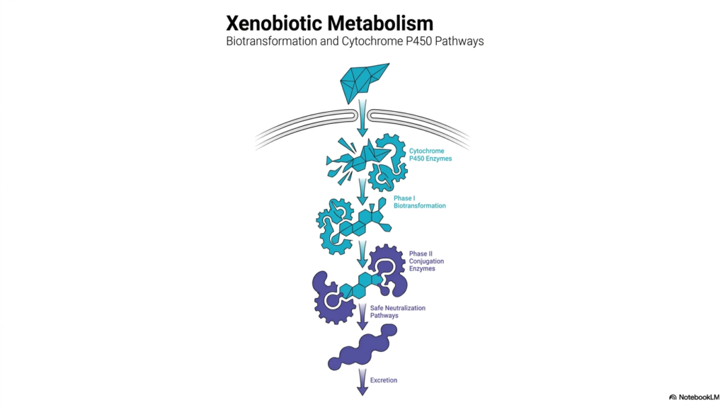
The human body constantly encounters foreign substances that require immediate processing and elimination. This essential function, known as xenobiotic metabolism, is beautifully illustrated in the opening diagram. The visual represents the journey of a foreign compound as it enters the liver, the primary organ responsible for this physiological defense. The liver acts as a sophisticated chemical processing plant. By breaking down the mechanisms of xenobiotic metabolism, the graphic establishes a strong foundation for understanding how biological systems transform harmful invaders into harmless waste. The interlocking gears symbolize the enzymatic machinery at work, highlighting a highly coordinated assembly line.
Central to this defensive process are the Cytochrome P450 enzymes, depicted as the first set of gears in the biotransformation pathway. These specialized enzymes initiate the first phase of structural modification. As the diagram demonstrates, the initial foreign molecule undergoes deliberate physical changes, preparing it for the next critical step. This is the fundamental principle of xenobiotic metabolism: altering the chemical structure of a compound to make it more reactive. The visual perfectly captures the transition from a complex, non-polar molecule into a slightly modified intermediate that is ready for downstream processing.
The biochemical journey concludes with the Phase II conjugation enzymes, which attach large, highly polar groups to the intermediate molecule. This vital step creates safe neutralization pathways, as shown by the final gear assembly in the slide. The ultimate goal of xenobiotic metabolism is excretion, actively transforming a stubborn, fat-soluble toxin into a water-soluble compound that the organism can quickly remove. This slide provides an excellent macroscopic view of a microscopic battle, setting the stage for the specific chemical reactions explored in subsequent sections.
Slide 2: Xenobiotic Metabolism: The Hydrophobic Challenge
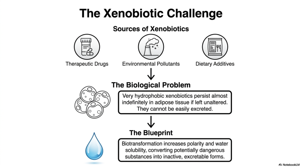
Understanding the source and chemical nature of foreign compounds is crucial in the study of xenobiotic metabolism. This slide categorizes the primary invaders into three common groups: therapeutic drugs, environmental pollutants, and dietary additives. Biological systems face a constant bombardment from these substances. The core issue, identified here as the biological problem, lies in the chemical properties of these invaders. Many of these compounds are highly hydrophobic, meaning they naturally repel water and dissolve readily in fats. Without an intervention system, these dangerous substances would persist almost indefinitely in adipose tissue.
This persistent nature of hydrophobic compounds highlights the absolute necessity of xenobiotic metabolism. If left unaltered, the permanent accumulation of these lipid-soluble toxins would disrupt cellular membranes and lead to severe physiological damage. The organism must therefore deploy a specific strategy to handle this persistent chemical threat. This slide defines that physiological strategy as the blueprint. The primary goal of this biochemical blueprint is biotransformation. Biotransformation involves a deliberate series of enzymatic reactions designed to structurally modify the offending molecule at the cellular level.
The ultimate objective of this specific phase of xenobiotic metabolism is to drastically increase the polarity and water solubility of the foreign compound. The visual of a simple water droplet perfectly encapsulates this biological goal. By converting potentially dangerous, fat-soluble substances into inactive, highly polar forms, the body ensures they can be flushed out of the system. This conversion is the elegant solution to the biological problem, utilizing xenobiotic metabolism to transform a permanent physiological threat into a temporary visitor that is safely excreted.
Slide 3: Xenobiotic Metabolism: The Three-Phase Assembly Line
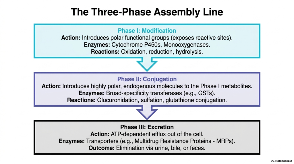
The intricate process of xenobiotic metabolism operates much like a highly organized factory assembly line, neatly divided into three distinct phases. This slide outlines the complete tripartite cellular system. Phase I acts as the initial modification stage. Here, the primary enzymatic action is the introduction of polar functional groups to the foreign molecule. This step essentially exposes reactive chemical sites on the compound. Enzymes such as Cytochrome P450s and monooxygenases drive these early reactions, which primarily include oxidation, reduction, and hydrolysis. This first step does not always neutralize the threat immediately.
Following initial modification, the altered metabolite enters Phase II of xenobiotic metabolism, recognized universally as the conjugation stage. This critical step involves attaching highly polar, endogenous molecules directly to the functional groups exposed during Phase I. Broad-specificity transferases manage these targeted reactions. Common biological processes include glucuronidation, sulfation, and glutathione conjugation. This conjugation step is absolutely vital because it drastically increases the water solubility of the molecule. It effectively neutralizes the reactive intermediate created in the previous phase and prepares it for safe transport.
The final stage of xenobiotic metabolism is Phase III, designated as the excretion phase. This involves the active, ATP-dependent efflux of the transformed molecule out of the local cellular environment. Specialized transporter proteins, specifically Multidrug Resistance Proteins, act as biological pumps to physically push the water-soluble conjugate across the lipid cell membrane. The ultimate outcome of this entire three-phase assembly line is the successful elimination of the altered compound from the body via urine, bile, or feces.
Slide 4: Xenobiotic Metabolism: Diagnosing Phase I and Phase II Reactions
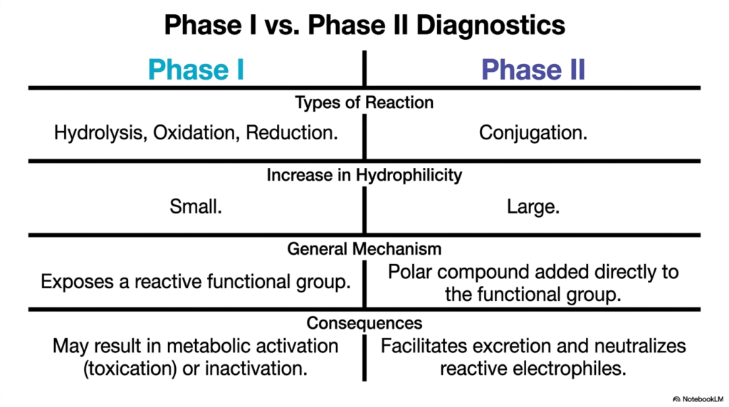
Distinguishing between the primary stages of xenobiotic metabolism is essential for understanding drug clearance and underlying toxicology. This slide provides a clear diagnostic comparison between Phase I and Phase II biochemical reactions. Phase I is characterized primarily by hydrolysis, oxidation, and reduction reactions. These structural modifications result in only a very small increase in overall hydrophilicity. The general mechanism revolves around exposing or adding a reactive functional group to the base molecule. Because it creates a reactive site, Phase I can sometimes result in dangerous metabolic activation.
In stark contrast, the second major stage of xenobiotic metabolism focuses entirely on molecular conjugation. Phase II reactions take the newly prepared molecule and covalently add a substantial polar compound directly to the exposed functional group. This structural addition results in a massive increase in hydrophilicity, making the molecule highly water-soluble. Unlike the potentially dangerous intermediates formed earlier, the consequences of Phase II in xenobiotic metabolism are almost universally protective. This stage successfully neutralizes reactive electrophiles and ensures the compound is safe for systemic excretion.
By placing these two distinct phases side-by-side, the slide highlights the dual nature of xenobiotic metabolism. Phase I acts as a necessary preparatory step that carries an inherent risk of toxication, while Phase II serves as the true detoxification and chemical neutralization stage. Understanding this delicate dynamic is crucial for medical students, as many severe drug-induced liver injuries occur when Phase I activity vastly outpaces Phase II capacity, allowing dangerous intermediate compounds to accumulate locally.
Slide 5: Xenobiotic Metabolism: The Cytochrome P450 Engine
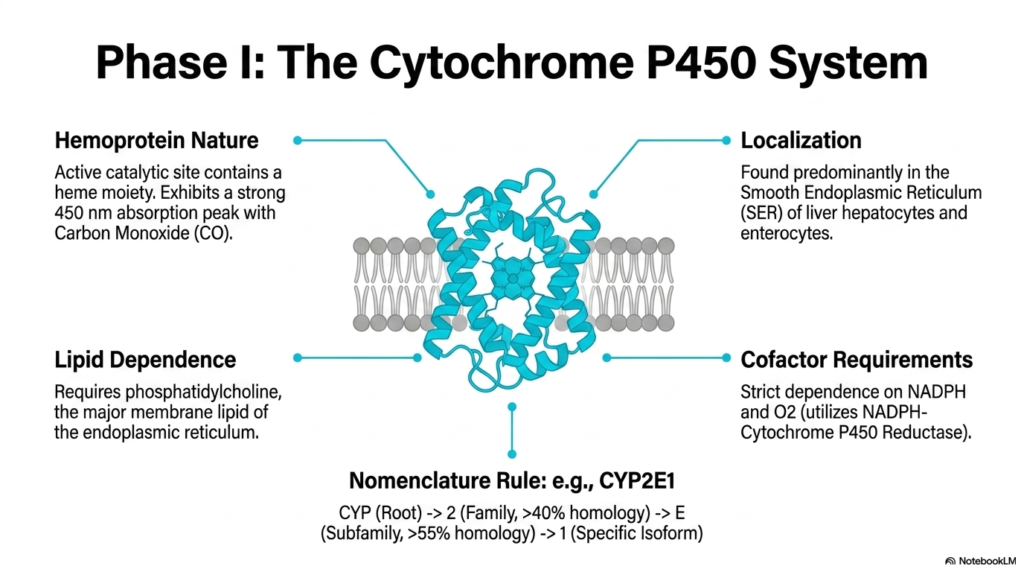
The absolute workhorse of early-stage xenobiotic metabolism is the renowned Cytochrome P450 system. This slide details the fundamental biochemical characteristics of these remarkable enzymes. Cytochrome P450s are categorized as hemoproteins, meaning their active catalytic site contains a critical heme moiety. They are famous for exhibiting a strong absorption peak at exactly 450 nanometers when artificially bound to carbon monoxide. These essential enzymes are predominantly localized in the Smooth Endoplasmic Reticulum of liver hepatocytes and enterocytes, placing them precisely at the forefront of the organism’s chemical defense grid.
A unique feature of this phase of xenobiotic metabolism is its strict structural and chemical requirements for successful activation. The Cytochrome P450 system exhibits a very strong lipid dependence. It strictly requires phosphatidylcholine, the major membrane lipid of the endoplasmic reticulum, to function properly. Furthermore, the catalytic cycle has absolute physiological cofactor requirements. The enzyme depends entirely on the presence of molecular oxygen and NADPH, utilizing an accessory protein to transfer essential electrons for the oxidation reaction.
Finally, the slide decodes the complex nomenclature used globally in xenobiotic metabolism research. Taking CYP2E1 as an academic example, the naming convention follows strict genetic homology rules. The root “CYP” identifies the system. The number “2” indicates the protein family, requiring greater than forty percent sequence homology. The letter “E” denotes the subfamily, demanding greater than fifty-five percent homology. The final number “1” identifies the specific active enzyme isoform, helping scientists track specific metabolic pathways.
Slide 6: Xenobiotic Metabolism: Oxidation and Reduction Pathways
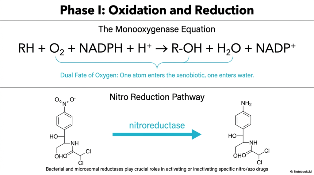
The fundamental chemistry of Phase I xenobiotic metabolism involves precise, deliberate alterations to electron configurations. This slide breaks down the core structural reactions of oxidation and reduction. The top section clearly displays the classic Monooxygenase Equation: the xenobiotic plus oxygen plus NADPH yields a hydroxylated xenobiotic, water, and oxidized NADP. This represents the absolute most common reaction catalyzed by Cytochrome P450. A crucial theoretical concept here is the dual fate of oxygen. One oxygen atom enters the foreign molecule, while the second is safely reduced to form water.
While oxidation dominates early xenobiotic metabolism, specific reduction reactions also play a vital role in processing particular compounds. The bottom section of the slide highlights the Nitro Reduction Pathway. Using a specific pharmacological molecule as an example, it demonstrates how nitroreductase enzymes physically target nitro groups. In this specific alternative pathway, the nitrogen-oxygen double bonds are sequentially reduced, eventually replacing the oxygen atoms with hydrogen atoms to form an entirely new amine group.
Both bacterial and local microsomal reductases are essential for this specific pathway in xenobiotic metabolism. They play critical roles in activating or inactivating specific nitro and azo drugs within the gastrointestinal tract and the local liver environment. Whether inserting oxygen to create a reactive hydroxyl site or stripping oxygen away to form an amine, these vital Phase I mechanisms provide the necessary chemical handles that downstream conjugation enzymes require.
Slide 7: Xenobiotic Metabolism: Inducing and Inhibiting the System
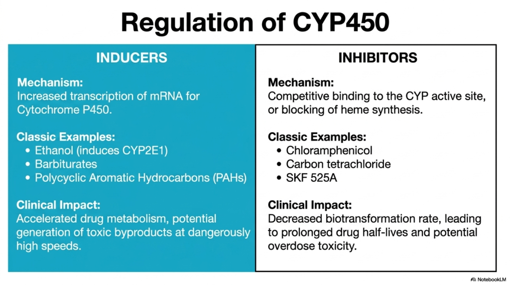
The enzymes responsible for xenobiotic metabolism are not static structural entities; their activity levels change dramatically based on acute environmental exposures. This slide explores the critical pharmacological concepts of enzyme induction and inhibition. Inducers work by actively increasing the cellular transcription of messenger RNA for Cytochrome P450 enzymes. When an organism consumes substances like ethanol, barbiturates, or polycyclic aromatic hydrocarbons, the body responds rapidly by synthesizing more metabolic enzymes. This adaptation attempts to clear the offending chemicals much more rapidly.
However, artificially accelerating xenobiotic metabolism carries significant clinical and physiological risks. The increased presence of CYP450 enzymes leads to highly accelerated drug processing. While this clears the parent compound very quickly, it can easily generate toxic Phase I byproducts at dangerously high speeds. If these reactive molecular intermediates are produced faster than Phase II pathways can handle them, severe cellular damage inevitably occurs. Therefore, metabolic induction increases processing capacity while simultaneously elevating the localized risk of acute toxicity.
Conversely, biological inhibitors actively decrease the standard rate of xenobiotic metabolism. These chemical agents typically work through direct competitive binding at the CYP active site or by physically blocking heme synthesis altogether. Classic identified inhibitors include chloramphenicol and carbon tetrachloride. The direct clinical impact of enzymatic inhibition is a severely decreased biotransformation rate. This failure leads to prolonged drug half-lives in the systemic bloodstream, meaning a standard therapeutic dose can easily accumulate to fatal toxic levels.
Slide 8: Xenobiotic Metabolism: The Power of Glucuronidation and Sulfation
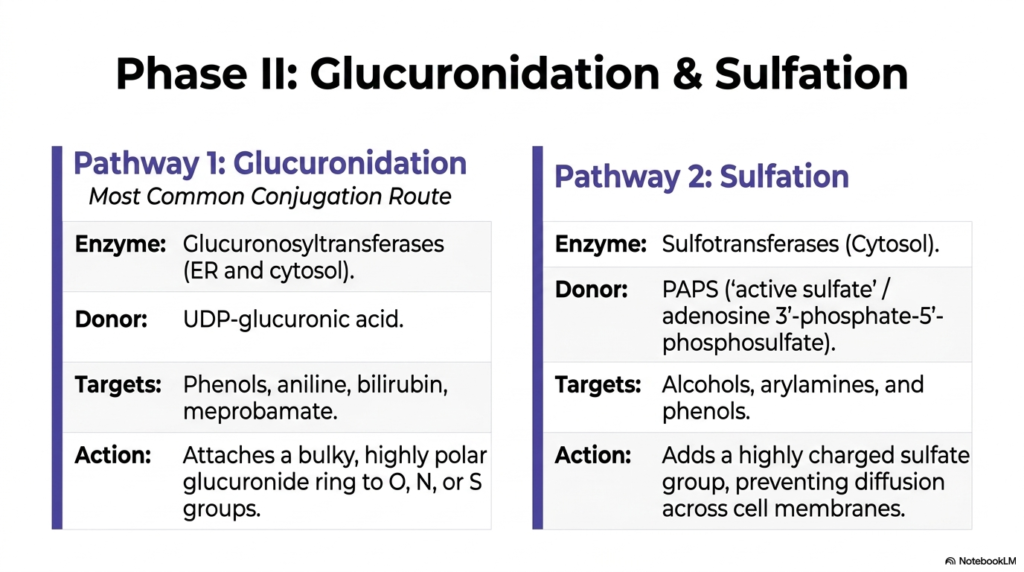
Once a foreign molecule is prepared, it must be neutralized and made highly water-soluble. This slide details the two most prominent Phase II conjugation pathways found in xenobiotic metabolism: Glucuronidation and Sulfation. Pathway 1, Glucuronidation, acts as the absolute most common conjugation route in the human body. Driven by Glucuronosyltransferase enzymes located securely in both the endoplasmic reticulum and cytosol, this specific pathway uses UDP-glucuronic acid as the vital donor molecule. It aggressively targets phenols, aniline, bilirubin, and countless medications.
This massive structural addition is a recognized hallmark of successful Phase II xenobiotic metabolism. The attached glucuronide ring immediately shifts the core physical properties of the foreign compound, transforming a potentially dangerous, lipophilic molecule into a safe, bulky, water-soluble complex. The sheer size and extreme polarity of the newly formed conjugate prevent it from diffusing back across sensitive cellular membranes. It effectively traps the neutralized waste in the aqueous environment of the blood or bile for rapid elimination.
Pathway 2 highlights Sulfation, another absolutely critical defense mechanism utilized in xenobiotic metabolism. Cytosolic sulfotransferase enzymes meticulously manage this reaction, utilizing PAPS, often referred to as active sulfate, as the primary donor molecule. This targeted pathway primarily seeks out alcohols, arylamines, and phenols. By adding a highly charged sulfate group, the molecule becomes extremely polar. Similar to glucuronidation, this structural addition physically prevents the toxic compound from crossing lipid bilayers, fully neutralizing the active threat.
Slide 9: Xenobiotic Metabolism: Glutathione Defense and Methylation
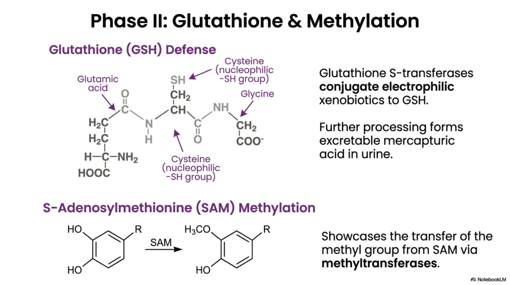
Beyond adding bulky molecular rings or charged sulfates, the cell utilizes specific molecular traps to actively capture dangerous intermediates. This slide highlights two highly specialized Phase II reactions critical to xenobiotic metabolism. The top section showcases the essential Glutathione Defense system. Glutathione is a tripeptide made of glutamic acid, cysteine, and glycine. The central cysteine residue features a highly nucleophilic sulfhydryl group. Glutathione S-transferase enzymes use this reactive sulfur to quickly capture and conjugate dangerous, electrophilic xenobiotics before they destroy proteins.
This immediate capturing mechanism is a crucial cellular failsafe in xenobiotic metabolism. Once the toxic electrophile is firmly bound to the glutathione molecule, the localized danger is completely neutralized. The newly formed conjugate undergoes further enzymatic processing within the kidneys to form a highly water-soluble compound known as mercapturic acid, which is safely expelled in the urine. This exact pathway acts as the primary cellular defense against reactive oxygen species and highly destructive drug metabolites.
The bottom visual illustrates active Methylation, utilizing S-Adenosylmethionine as the primary active methyl donor. Methyltransferases facilitate the direct transfer of this specific methyl group onto target molecules. Unlike other standard Phase II pathways found in xenobiotic metabolism, methylation does not significantly increase systemic water solubility. Instead, its primary biological function is to heavily mask reactive functional groups, thereby immediately terminating the pharmacological or toxicological activity of the molecule.
Slide 10: Xenobiotic Metabolism: The Pathways of Cellular Toxicity
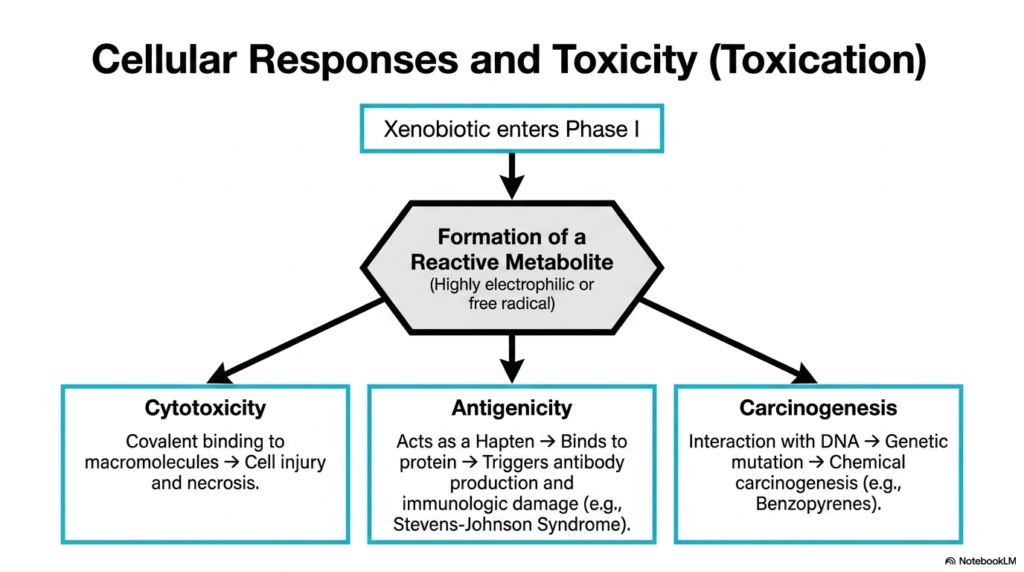
While the metabolic system is designed primarily for cellular protection, it occasionally creates the very poison it seeks to destroy. This slide maps the dark side of xenobiotic metabolism, a catastrophic event known scientifically as toxication. The process begins when a relatively benign foreign compound enters standard Phase I processing. Instead of forming a safe intermediate, the local enzymes occasionally generate a highly electrophilic reactive metabolite or a volatile free radical. This rogue molecule violently seeks out cellular structures to steal electrons.
The biological consequences of this absolute failure in xenobiotic metabolism are severe and manifest in three primary ways. The first is direct Cytotoxicity. The highly reactive metabolite binds covalently to essential cellular macromolecules, destroying their structural function and triggering rapid cell injury and necrotic death. The second severe consequence is Antigenicity. The errant metabolite acts as a hapten, binding to a normal protein. The immune system flags this new structure, triggering destructive immunologic reactions.
The most insidious long-term outcome of misdirected xenobiotic metabolism is true Carcinogenesis. If the highly reactive electrophile successfully bypasses cytoplasmic protein structures and enters the deep nucleus, it can interact directly with the organism’s DNA. This irreversible covalent binding causes deep, permanent genetic mutations. If these specific mutations affect critical cell cycle regulators, chemical carcinogenesis is rapidly initiated, potentially leading to tumor development through compounds like benzopyrenes.
Slide 11: Xenobiotic Metabolism: The Mechanisms of Paracetamol Toxicity
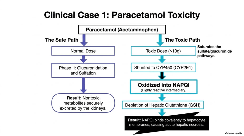
To truly understand the real-world impact of metabolic pathways, medical students must examine clinical case studies. This slide completely breaks down the specific xenobiotic metabolism of paracetamol, commonly known as acetaminophen. The flowchart splits into two distinct, life-altering scenarios. The safe path illustrates standard, normal processing. At regular therapeutic doses, paracetamol safely bypasses the dangerous Phase I enzymes entirely and proceeds directly to Phase II. Through standard glucuronidation and sulfation, the drug transforms into nontoxic metabolites for excretion.
The physiological situation changes catastrophically during an acute overdose. The toxic path reveals the absolute limits of xenobiotic metabolism. When a patient consumes a massive toxic dose, the primary sulfate and glucuronide pathways become entirely saturated and fail. The excess drug is forcibly shunted toward the Cytochrome P450 system, specifically the CYP2E1 enzyme. This alternative Phase I oxidation immediately generates a highly reactive and uniquely dangerous intermediary molecule known specifically as NAPQI.
In a final, desperate attempt to survive, the cell leverages its remaining glutathione reserves to neutralize the chemical threat. However, the sheer massive volume of NAPQI rapidly causes severe depletion of hepatic glutathione. This is the absolute breaking point of xenobiotic metabolism. Without the protective glutathione shield, free NAPQI binds covalently to vital hepatocyte membranes and structural proteins. This massive internal structural damage causes acute hepatic necrosis, literally destroying the liver tissue.
Slide 12: Xenobiotic Metabolism: The Principal Route of Ethanol Clearance
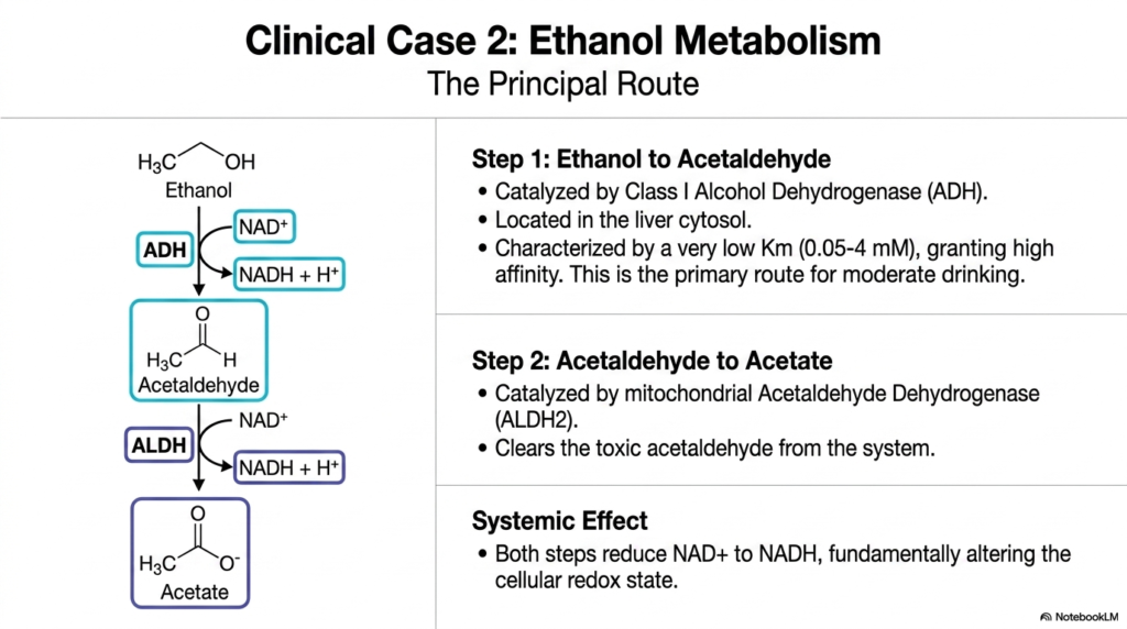
The cellular processing of alcohol represents one of the absolute most prominent examples of daily metabolic function. This slide clearly outlines the principal route of ethanol processing, acting as a true cornerstone of human xenobiotic metabolism. The first major step occurs directly in the liver cytosol, where Class I Alcohol Dehydrogenase actively catalyzes the fast conversion of ethanol into acetaldehyde. This specific enzyme is uniquely characterized by a very low Michaelis constant, meaning it has an extremely high affinity for ethanol molecules.
However, the resulting acetaldehyde is highly toxic and must be actively eliminated rapidly from the cellular environment. The second critical step of this xenobiotic metabolism pathway takes place deep inside the mitochondria. Here, Acetaldehyde Dehydrogenase successfully converts the toxic intermediate into harmless acetate. This specific enzyme essentially clears the dangerous poison from the system entirely. Acetate can then be safely dispersed through the bloodstream and effectively utilized by peripheral tissues as a basic energy source.
While this highly efficient pathway successfully clears the toxin, it simultaneously exerts a massive systemic effect on general cellular biology. Both sequential steps of this specific xenobiotic metabolism fundamentally alter the delicate cellular redox state. The dehydrogenase enzymes constantly strip electrons from the alcohol molecules and transfer them directly to NAD+, rapidly reducing it to NADH. This vast accumulation of NADH dramatically shifts the chemical balance inside the liver cell, deeply disrupting normal metabolic functions.
Slide 13: Xenobiotic Metabolism: The MEOS Pathway and Heavy Drinking
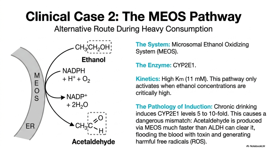
When the primary metabolic defense systems are completely overwhelmed, the human body activates emergency backup protocols. This slide investigates the Microsomal Ethanol Oxidizing System, a major alternative route of xenobiotic metabolism activated exclusively during incredibly heavy alcohol consumption. The primary enzymatic engine for this backup system is the Cytochrome P450 enzyme CYP2E1, located deep in the endoplasmic reticulum. Unlike sensitive cytosolic enzymes, CYP2E1 possesses a profoundly high Michaelis constant, keeping the pathway mostly dormant until concentrations peak.
The physiological reliance on this secondary system rapidly exposes a dangerous, inherent flaw in human xenobiotic metabolism. Chronic heavy drinking actively induces the mass production of CYP2E1, increasing cellular levels massively. This creates a highly severe pathology of induction. As the MEOS system expands physically, it begins actively converting ethanol into highly toxic acetaldehyde at a massive, completely accelerated rate. This hyperactive Phase I cellular response rapidly outpaces the Phase II mitochondrial enzymes.
This specific, deep metabolic mismatch is the absolute biological hallmark of alcoholic liver damage. Because acetaldehyde is produced via MEOS much faster than the subsequent enzymes can possibly clear it, the raw, unbound toxin literally floods the local cellular environment. Furthermore, because this specific form of xenobiotic metabolism actively utilizes molecular oxygen, the hyperactive CYP2E1 enzymes generate massive amounts of harmful reactive oxygen species. The cell is simultaneously poisoned and physically bombarded.
Slide 14: Xenobiotic Metabolism: The Pathology of Alcohol-Induced Liver Disease
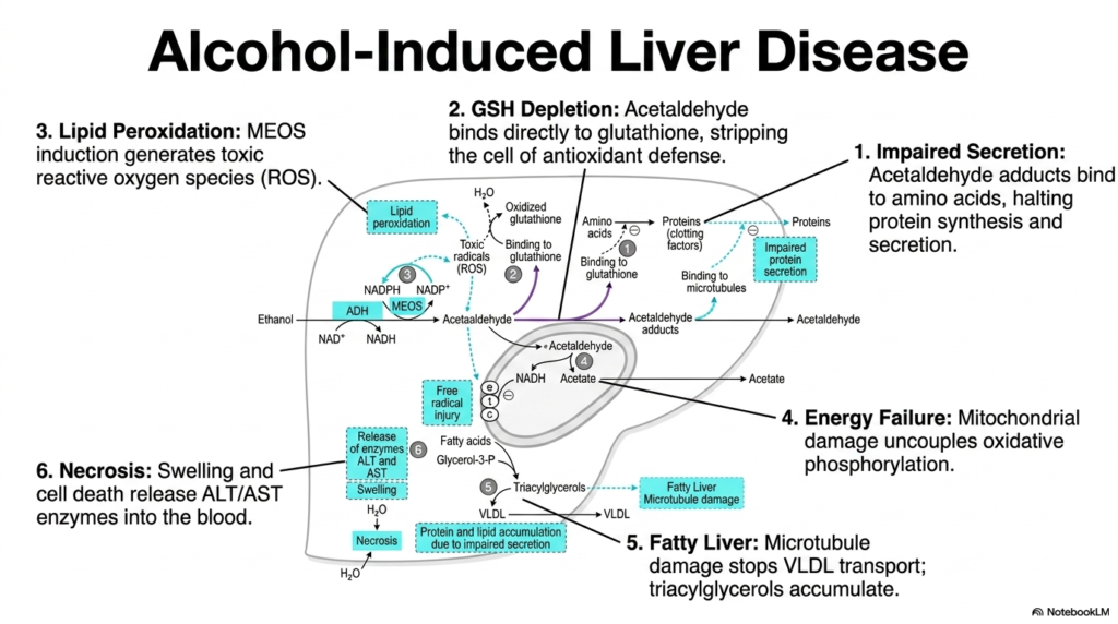
When metabolic defense systems completely fail, acute cellular destruction follows a highly predictable biochemical cascade. This slide maps the extremely severe consequences of overwhelmed xenobiotic metabolism, detailing the specific, destructive mechanisms of alcohol-induced liver disease. The cellular diagram meticulously illustrates six interconnected points of biological failure. The true crisis begins immediately when excess acetaldehyde actively forms chemical adducts, binding directly to essential amino acids and vital microtubules, physically halting essential protein secretion.
Simultaneously, the entirely overwhelmed cellular system exhausts its absolute last defenses. The excess local acetaldehyde directly binds to glutathione, completely stripping the cell of its primary antioxidant shield. Without this absolutely crucial component of xenobiotic metabolism, the toxic reactive oxygen species generated by the overactive MEOS pathway run completely rampant, immediately causing widespread lipid peroxidation. Further compounding the massive disaster, the rapidly accumulating toxins physically damage the mitochondria, which rapidly uncouples oxidative phosphorylation.
The comprehensive visual clearly highlights the highly visible physical hallmarks of a completely failed system of xenobiotic metabolism. Acute microtubule damage permanently stops the active transport of very-low-density lipoproteins, causing massive internal cellular accumulations of triacylglycerols. Ultimately, the immense combined stress of energy failure, toxic protein buildup, and total membrane destruction forces the cell to swell rapidly and violently rupture. This final necrotic event heavily releases liver enzymes into the bloodstream.
Slide 15: Xenobiotic Metabolism: Sensing the Threat and Restoring Homeostasis
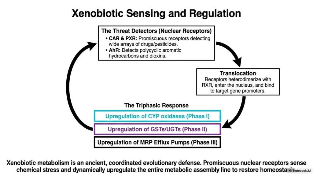
The human body does not simply wait passively for toxins to cause cellular damage; it highly actively detects and forcefully responds to systemic chemical threats. This final slide explains the absolutely vital regulatory master switches of xenobiotic metabolism. The complex process begins actively with Threat Detectors, specifically highly sophisticated nuclear receptors located inside the cell. Specialized receptors like CAR and PXR are highly promiscuous, elegantly designed to detect a massive, diverse array of different modern drugs and toxic pesticides.
Upon actively detecting a chemical threat, these biological sensors immediately initiate a massive, profound physiological shift. The sensitive receptors undergo rapid translocation, heterodimerizing with structural RXR proteins to forcefully enter the cell nucleus. There, they immediately bind directly to target gene promoters, initiating a highly comprehensive triphasic response. This exact genetic process is the master control mechanism of all xenobiotic metabolism. The DNA instantly upregulates the rapid cellular production of Phase I, Phase II, and Phase III proteins.
This elegant genetic sensing network conclusively proves that xenobiotic metabolism is not a simple, passive physical filter, but a highly ancient, deeply coordinated evolutionary defense system. By actively utilizing promiscuous nuclear receptors to accurately sense acute chemical stress, the biological organism can dynamically and rapidly upregulate its entire metabolic machinery on pure demand. This incredible mechanism ensures that the cell can rapidly amplify its molecular defenses to successfully restore internal physiological homeostasis.
Please read our Content Disclaimer Statement.
Check out our social media channels: