62. Prokaryotic vs Eukaryotic Cells: Key Differences and Structure
Students often wonder what exactly makes human bodies exponentially more complex than simple, microscopic bacteria. The answer fundamentally relies on the precise structural blueprints of biological life. The core purpose of this slide deck is to comprehensively explore the distinct biochemical and structural elements that define modern cellular life. By actively analyzing prokaryotic and eukaryotic cells side by side, medical and college students can clearly understand the foundational spatial organization that enables complex multicellular lifeforms to thrive.
Slide 1: Biochemical Structure in Prokaryotic vs Eukaryotic Cells: Why Cellular Architecture Matters

When observing general cellular biology, the primary distinction between Prokaryotic vs. eukaryotic cells becomes a fascinating story of internal biological organization. This first visual serves as a beautifully comprehensive map of a complex eukaryotic animal cell. It perfectly illustrates a highly organized interior space in which specialized biochemical tasks are carefully divided among distinct, isolated internal compartments. The selectively permeable plasma membrane acts as the critical outer physical boundary, while the fluid cytoplasm completely fills the internal biological environment.
Within this dense cellular space, a vast multitude of membrane-bound organelles function harmoniously like tiny internal organs. The nucleus visibly sits prominently in the cellular center, safely guarding the vital genetic material. It remains completely surrounded by a massive network of supporting structures, explicitly including the endoplasmic reticulum, the Golgi apparatus, and energy-producing mitochondria. Additionally, a pervasive cytoskeleton supports the entire microscopic structure, constantly accompanied by specialized lysosomes and peroxisomes that safely carry out discrete metabolic reactions.
Maintaining focus on this highly compartmentalized interior image is incredibly crucial as the detailed analysis progresses. It perfectly captures the profound structural complexity of eukaryotic biology, which stands in stark contrast to the extremely simple, open floor plan routinely found in basic microorganisms. Understanding this stark visual contrast in Prokaryotic vs Eukaryotic Cells is the absolutely crucial first educational step for dedicated biology students aiming to deeply master advanced cellular physiology.
Slide 2: Taxonomic Divergence in Prokaryotic vs Eukaryotic Cells: Categorizing the Tree of Life
Sorting millions of incredibly unique living organisms into just two primary architectural categories seems impossible, yet biologists do exactly this when examining the fundamental building blocks of biology. The core purpose of this slide deck is to meticulously break down these complex taxonomic classifications into easily understandable educational pieces. By closely examining the fundamental architectural groups, researchers clarify the strict biological boundaries safely separating simple microscopic life from intricate multicellular organisms.
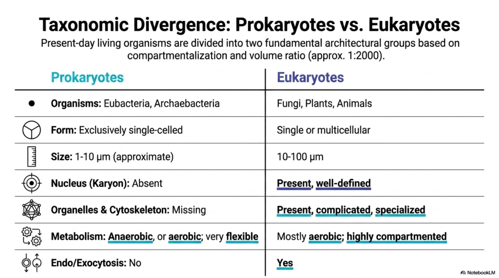
The primary architectural difference between Prokaryotic and Eukaryotic Cells lies in strict internal compartmentalization and a massive internal volume ratio difference of approximately 1:2,000. Eubacteria and archaebacteria perfectly represent the much simpler prokaryotic category. These microscopic organisms are exclusively single-celled and incredibly small, typically measuring between 1 and 10 micrometers in length. In stark contrast, larger fungi, intricate plants, and complex animals consist entirely of eukaryotic cellular units, ranging widely from 10 to 100 micrometers.
This highly significant physical size difference in Prokaryotic vs Eukaryotic Cells is permanently accompanied by an enormous evolutionary leap in internal structural complexity. Eukaryotes uniquely possess a well-defined, heavily guarded nucleus, officially referred to as a karyon, alongside numerous specialized, complicated internal organelles. They also feature a pervasive dynamic cytoskeleton explicitly designed for internal structural support. Furthermore, modern eukaryotic metabolism is highly compartmentalized and relies predominantly on highly efficient aerobic processes.
Conversely, simpler prokaryotes inherently boast a highly flexible basic metabolism that can function either aerobically or anaerobically, depending on the external environment. Finally, highly complex intracellular transport mechanisms, such as endocytosis and exocytosis, remain exclusive to eukaryotes. When rigorously studying Prokaryotic vs Eukaryotic Cells, it is evident that this profound biological divergence ultimately enabled eukaryotes to develop large, structurally complex bodies capable of dominating the planet.
Slide 3: Genomic Architecture in Prokaryotic vs Eukaryotic Cells: Storing Biological Data

DNA functions precisely like an essential, highly detailed instruction manual uniquely designed to build a functioning living organism. Some genetic manuals are surprisingly simple, microscopic pamphlets, while others resemble incredibly massive, multi-volume encyclopedias. The core purpose of this slide deck is to visually illustrate exactly how this critical biological data is stored in completely different ways across evolutionary domains. Understanding this unique genomic storage remains absolutely critical for deeply mastering modern genetics and complex molecular biology.
A profoundly defining feature in the ongoing academic study of Prokaryotic vs Eukaryotic Cells explicitly involves how these diverse organisms physically structure their delicate genetic blueprints. Prokaryotic DNA exists freely floating in the fluid cytoplasm as small, circular, ring-shaped structures, often accompanied by tiny supplementary plasmids. This highly simplistic genomic arrangement is relatively short, containing up to roughly five million base pairs.
Importantly, this simpler prokaryotic DNA entirely lacks non-coding introns, meaning that almost the entire biological sequence encodes strictly usable information. This specific structural simplicity allows for the direct, immediate spatial coupling of rapid RNA synthesis and protein generation. Conversely, modern eukaryotic genomic architecture remains physically massive, highly protected, and incredibly rigidly organized. In the continuous comparison of Prokaryotic vs Eukaryotic Cells, complex eukaryotes strictly store their incredibly large, distinctively linear DNA molecules safely inside a dedicated, membrane-bound nucleus.
This specific eukaryotic DNA tightly wraps around specialized histone proteins to effortlessly form highly complicated chromatin structures. The eukaryotic genome is astronomically massive, spanning up to tens of billions of base pairs. Furthermore, analyzing Prokaryotic vs Eukaryotic Cells clearly highlights that eukaryotic genes contain numerous non-coding regions known as introns. This demanding biological complexity strictly requires highly sophisticated RNA maturation processes safely inside the nucleus long before functional proteins can ever be properly synthesized.
Slide 4: Spatial Compartmentalization in Prokaryotic vs Eukaryotic Cells: The Power of Internal Boundaries

A chaotic chemical manufacturing factory attempting to safely carry out vastly different, highly volatile chemical reactions in a single giant, unorganized open room would instantly prove disastrous. The core purpose of this slide deck is to comprehensively demonstrate how advanced biological life ingeniously solves this exact physical problem through highly sophisticated spatial engineering. This section actively explores how strictly divided internal physical boundaries elegantly allow biological cells to perform multiple complex metabolic reactions safely and simultaneously.
Strict spatial compartmentalization permanently provides a massive, unprecedented evolutionary advantage for highly complex biological organisms. When carefully analyzing the internal architecture of Prokaryotic vs Eukaryotic Cells, this strict internal physical division beautifully stands out as a primary distinguishing evolutionary factor. Advanced eukaryotic cells brilliantly utilize highly defined, rigidly membrane-bound subcellular compartments, professionally known as organelles.
These rigid internal biological boundaries meticulously confine highly specific, potentially dangerous reaction spaces. This safely ensures that delicate macromolecular synthesis and aggressive cellular metabolism are strictly separately regulated without disastrous spatial chemical interference. The selectively permeable exterior plasma membrane serves as the critical outer boundary, expertly mediating crucial biological interactions with the highly unpredictable external environment. Within this heavily protective wall, the large internal space, the fluid cytoplasm, safely holds a highly concentrated liquid solution of vital biomolecules.
Unlike dramatically simpler bacterial organisms, this highly compartmentalized physical structure successfully prevents direct interference between competing interior biochemical pathways. In the detailed academic study of Prokaryotic vs Eukaryotic Cells, biochemistry students quickly learn that this specific physical division of biological labor remains absolutely essential for sustained multicellular survival. Understanding the fundamentally distinct internal floor plans directly separating Prokaryotic vs Eukaryotic Cells greatly helps developing students properly grasp exactly how intricate biological factories operate with such maximum chemical efficiency.
Slide 5: The Nucleus in Prokaryotic vs Eukaryotic Cells: Guarding the Master Blueprint

Every highly secure, incredibly valuable government facility strictly requires a heavily guarded, isolated central command center to perfectly protect its most sensitive proprietary information. In cellular biochemistry, the nucleus flawlessly serves this exact physical protective role. The core purpose of this slide deck is to intimately dissect these absolutely critical biological command centers. This section meticulously unpacks how safely isolating fragile genetic material protects it from accidental metabolic damage while enabling highly sophisticated gene regulation.
The heavily protected nucleus beautifully serves as the literal defining structural feature that permanently separates the major evolutionary domains of biological life. When rigorously examining Prokaryotic vs Eukaryotic Cells, researchers clearly note that the ancient Greek translation of the word eukaryote literally means “true kernel” or central nucleus. This prominent central biochemical repository permanently isolates the massively complex eukaryotic genome from the bustling, potentially chemically damaging metabolic activity occurring throughout the surrounding cytoplasm.
Structurally, the eukaryotic nucleus is exceptionally prominent, standing out as the largest single organelle in a typical animal cell. It remains incredibly easy to see under a standard academic light microscope. It is completely safely surrounded by a distinct, highly secure double-layered nuclear envelope. Interestingly, the delicate outer membrane of this highly protective envelope acts as a direct structural physical link specifically to the massive endoplasmic reticulum network.
This incredibly secure genomic physical isolation remains entirely missing in substantially simpler biological microorganisms. The stark visual contrast routinely found between Prokaryotic vs Eukaryotic Cells is tremendously obvious here, as basic prokaryotes essentially leave their precious genetic material somewhat dangerously exposed directly within the active fluid cytoplasm. By deeply studying Prokaryotic vs Eukaryotic Cells, college students quickly realize that a dedicated, fully protected nucleus perfectly remains the non-negotiable foundational structural requirement for all advanced lifeforms.
Slide 6: The Endoplasmic Reticulum in Prokaryotic vs Eukaryotic Cells: The Macromolecule Factory

While the heavily guarded cellular nucleus serves as the isolated central command center, the complex organism still absolutely requires a massive, dedicated factory to physically produce the desperately requested biological products. The core purpose of this slide deck is to clearly showcase the highly specialized structural-biochemical machinery directly responsible for the immense internal cellular manufacturing. This post comprehensively uncovers exactly how specific folded interior membranes cleanly provide the absolutely perfect confined physical environment for rapidly assembling intricate proteins.
The highly complex Endoplasmic Reticulum is an incredibly elaborate, beautifully continuous network of shallow sacs and densely branching tubules. It remains fully dedicated to the highly complex processes of protein synthesis, intricate molecular folding, and final functional maturation. In the strict biochemical academic comparison of Prokaryotic vs Eukaryotic Cells, this truly remarkable internal organelle uniquely stands out as a massive, carefully confined internal chemical reaction space explicitly designed for continuous heavy manufacturing.
It physically heavily anchors directly to the outer biological membrane of the central nucleus for incredibly rapid cellular communication. This exceptionally close physical structural linkage ensures that delicate biological data transfer remains incredibly fast and highly energy-efficient. The active rough Endoplasmic Reticulum works in perfect, continuous tandem with the adjacent nucleus, effortlessly translating incredibly complex genetic blueprints into highly functional structural macromolecules essential for basic survival.
Simple, basic bacterial organisms simply lack this extremely elaborate, continuous internal manufacturing infrastructure. When extensively academically discussing Prokaryotic vs Eukaryotic Cells, the distinct, highly visible presence of the elaborate Endoplasmic Reticulum perfectly highlights a tremendous evolutionary structural leap in basic biochemical production capacity. The unmistakable structural physical contrast clearly seen in Prokaryotic vs Eukaryotic Cells conclusively mathematically proves that dedicated, heavily membrane-bound cellular factories remain fundamentally required to permanently sustain demanding metabolisms.
Slide 7: The Golgi Apparatus in Prokaryotic vs Eukaryotic Cells: The Cellular Routing Hub

Once a massive biological factory successfully physically builds a highly complex molecular product, that incredibly specific product must be carefully biochemically packaged, highly accurately labeled, and swiftly and perfectly shipped. The core purpose of this slide deck is to thoroughly trace the complex internal physical journey of newly synthesized, highly valuable cellular products. This section comprehensively explores how newly formed raw macromolecules are physically and chemically modified and safely and cleanly routed to their highly precise functional internal locations.
The massive Golgi apparatus proudly serves as the indispensable central hub for post-translational chemical modification and complex logistical routing in the entire cell. In the continuous, highly detailed academic analysis of Prokaryotic vs Eukaryotic Cells, this incredibly functionally distinct internal organelle remains entirely unique to the highly complex eukaryotic evolutionary domain. It actively and continuously receives delicate raw macromolecules, expertly and precisely modifies their highly specific chemical structures, and meticulously and cleverly packages them.
Architecturally, the massive Golgi apparatus strongly resembles a densely bundled stack of tightly layered slices. It permanently and robustly exists as a highly discrete, fully compartmentalized internal physical structure and, surprisingly, represents a very significant portion of the total cellular interior volume. It consistently physically reliably occupies roughly an incredible six percent of the total available physical space inside a fully functioning complex animal cell.
Without this highly specialized, incredibly efficient biological interior post office, deeply precise internal molecular targeting would logically, physically, and permanently be completely impossible. The strict physical structural developmental differences between Prokaryotic vs. eukaryotic cells remain exceptionally highly visually striking here. Properly, deeply, fundamentally understanding Prokaryotic vs Eukaryotic Cells requires explicitly recognizing the complex functional Golgi apparatus as a truly essential foundational component for successfully sustaining internal cellular logistics.
Slide 8: Mitochondria in Prokaryotic vs Eukaryotic Cells: Aerobic Respiration Powerhouses

A deeply bustling, heavily populated modern metropolis desperately requires an incredibly massive, highly technologically efficient central power plant to keep the incredibly bright lights permanently on. Biological eukaryotic cells operate safely, efficiently, and smoothly under the exact same, extremely harsh, demanding, and energetic evolutionary constraints. The core purpose of this slide deck is to explicitly and visibly reveal the absolutely critical, delicate interior biological compartments that are solely and uniquely responsible for actively and flawlessly generating massive life-sustaining chemical-energy output.
Complex mitochondria arguably remain the most fundamentally biologically critical internal organelles explicitly required for safely and successfully sustaining demanding eukaryotic basic cellular metabolism. When closely, deeply, strictly, and biologically comparing Prokaryotic vs. eukaryotic cells, the ancient biological evolution of advanced mitochondria brilliantly represents a truly massive, unparalleled energetic leap forward. These complex internal, specific structures permanently and securely house the highly compartmentalized, brilliant biochemical pathways explicitly required to support complex aerobic cellular respiration.
Structurally, highly active, massive mitochondria are strictly, biologically, and rigidly defined by a completely brilliant, highly functional internal double-membrane protective system. This complex, beautiful structure purposefully, deeply, massively, and explicitly maximizes available functional internal biological surface area solely for massive, rapid, continuous internal energy production. Interestingly, these distinct organelles remain remarkably physically similar in actual physical basic scale directly to entire standalone individual prokaryotic living bacteria, cleanly and predictably measuring roughly one to ten micrometers.
The absolutely massively staggering basic energetic developmental divide directly between Prokaryotic vs Eukaryotic Cells undeniably profoundly strongly clearly remains incredibly importantly vital to carefully academically broadly study. While dramatically simpler tiny bacteria must rely entirely on their delicate outer plasma protective membrane for basic minimal energy continuous generation, highly advanced eukaryotes effortlessly utilize these dedicated internal biological massive power plants. The academically rigorous study directly contrasting Prokaryotic vs Eukaryotic Cells consistently strongly points perfectly to active dense mitochondria exactly as the ultimate driving energetic biochemical foundational force.
Slide 9: The Vesicular System in Prokaryotic vs Eukaryotic Cells: Biological Waste Management

A truly deeply highly successful advanced biological massive city incredibly undeniably safely strictly requires much more deeply fundamentally than just productive manufacturing factories and giant energetic power plants. The core purpose of this slide deck is to cleanly, beautifully, properly, and rigorously outline these deeply vital, specialized internal dynamic micro-compartments. This section deeply, beautifully, properly, actively, completely, and functionally investigates, directly and precisely, how tiny, smoothly moving, bubble-shaped, dense structures effortlessly, effectively, and safely manage the continuous, safe disposal of extremely dangerous, hazardous internal metabolic toxic waste.
The highly dynamic, remarkably complex, beautiful eukaryotic internal vesicular delivery system fundamentally consists of numerous specialized, individual, globular biological compartments carefully responsible for active, secure internal transport. A massively profound, deeply key, strict biochemical, internal, biological distinction cleanly and safely separating Prokaryotic vs Eukaryotic Cells powerfully remains the incredibly permanent, massive, dense presence of these incredibly dynamic, actively beautiful, continuously moving specialized tiny bubble-shaped biological functional tools.
For instance, incredibly active tiny biological endosomes carefully, smoothly, flawlessly, and beautifully manage the absolutely crucial, vitally important, and completely targeted intake of strictly highly important distinct extracellular nutritional substances, actively and beautifully precisely, securely, and directly gathered from the immediate surrounding unpredictable local natural external environment. Meanwhile, numerous highly dedicated, specialized exosomes expertly handle the absolutely critical, completely vital biological export of safely and properly manufactured, specific biochemical interior structural products cleanly and correctly out of the dense, massive cell.
Extremely profoundly uniquely simpler tiny delicate isolated bacterial organisms simply entirely exactly precisely do not possess this highly beautiful intricate actively rigorous moving internal heavy traffic system. The massive, dense, complete, structural, strict, complex comparison, definitively, precisely contrasting Prokaryotic vs. eukaryotic cells, incredibly, perfectly, clearly, logically demonstrates that highly, heavily advanced, active biological interior life absolutely, strictly, firmly, undeniably, unequivocally requires highly robust, physically isolated, heavily, heavily safely compartmentalized, complex, precise biological internal toxic waste management.
Slide 10: The Cytoskeleton in Prokaryotic vs Eukaryotic Cells: Building the Dynamic Scaffold

A towering modern skyscraper would collapse without a sturdy internal steel framework holding it together. Biological cells operate under similar structural constraints, requiring massive support to maintain intricate shapes. The core purpose of this slide deck is to dissect the internal architectures that sustain complex microscopic life. This section explores how specialized protein networks provide the crucial physical integrity and logistical pathways required for advanced organisms to survive and thrive.
The cytoskeleton serves as an extensive, dynamic structural scaffold that traverses the entire internal volume of the liquid cytoplasm. While membrane-bound organelles handle complex metabolic compartmentalization, this pervasive structural framework provides essential spatial organization. It is composed of specialized proteins that form an interconnected, reliable network. This rigid apparatus allows the cell to maintain a distinct shape while managing internal transport. In the foundational study of prokaryotic vs. eukaryotic cells, this internal architectural feature stands out prominently as a major biological advancement.
This robust protein network functions much like a busy microscopic highway system, moving vital biochemical components exactly to their required destinations. By analyzing the evolutionary differences between prokaryotic and eukaryotic cells, students learn that this complex structural network remains a defining characteristic of advanced cellular biology. The immense physical integrity it provides supports the demanding metabolic activities required by larger lifeforms. It ensures that internal organelles remain anchored or move with purpose during critical metabolic processes.
This remarkable internal support system is missing in simpler bacterial organisms. The distinct structural absence, highlighted when comparing prokaryotic and eukaryotic cells, underscores a massive evolutionary leap. Without this intricate cellular scaffold, simpler microscopic lifeforms must rely on less sophisticated methods to maintain basic shapes. Understanding this fundamental architectural division helps biochemistry students appreciate why only advanced lifeforms can develop into large, multicellular bodies capable of specialized tissue formation.
Slide 11: Quantitative Volume in Prokaryotic vs Eukaryotic Cells: Budgeting Cellular Space

Imagine trying to fit thousands of busy workers into a tiny studio apartment; chaos would ensue without a strict spatial budget. Cellular biology faces a similar spatial dilemma. The core purpose of this slide deck is to detail the architectural strategies living organisms use to organize their interiors. This section breaks down the quantitative volume and frequency of internal compartments, using an active mammalian liver cell as a prime example of biological budgeting.
When observing the vast architectural differences between Prokaryotic vs. eukaryotic cells, the strict quantitative distribution of internal space becomes a fascinating subject. In an active mammalian hepatocyte, the fluid cytoplasm occupies approximately 54% of the total cellular volume. This leaves the remaining space budgeted among numerous specialized membrane-bound organelles. The mighty mitochondrion alone occupies 22% of this available internal volume, underscoring the massive energy demands required to sustain a complex metabolic unit.
The sheer frequency of these internal powerhouses further underscores the structural complexity that is lacking in simpler microorganisms. A single active liver cell contains around 2,000 mitochondria, functioning without pause to meet intense aerobic metabolic demands. When comparing prokaryotic vs. eukaryotic cells, this incredible numerical density is staggering. In addition, the cell allocates 9% of its internal volume to the single rough endoplasmic reticulum and 6% to the solitary central nucleus.
The central Golgi apparatus occupies six percent of the space, while dynamic transport vesicles claim about three percent. Smaller, specialized micro-compartments populate this dense interior space in massive numbers. The typical liver cell contains 400 peroxisomes, 300 individual lysosomes, and 200 endosomes. Studying these precise quantitative measurements in Prokaryotic vs. eukaryotic cells reveals the intricate internal logistical planning required for advanced biological survival. The human body contains at least two hundred distinct cell types, each featuring fascinating, unique variations of this complex spatial biochemical budget.
Slide 12: Divergence of Plant Organisms in Prokaryotic vs Eukaryotic Cells

Picture a self-sustaining, solar-powered factory that builds rigid defensive walls and stores massive amounts of water. Plant cells achieve this exact engineering marvel every single day. The core purpose of this slide deck is to map out the diverse architectural strategies utilized by complex life. This final section examines the distinct structural adaptations of multicellular photosynthetic organisms, showing how unique environmental demands shape their internal cellular biology.
While standard animal biology provides an excellent educational baseline, plant structures introduce fascinating functional variations into the academic study of prokaryotic vs. eukaryotic cells. These specialized photosynthetic cells exhibit distinct architectural adaptations suited to a stationary, sun-gathering lifestyle. The absolute most prominent biological addition is the functional chloroplast, a specialized plastid unique to multicellular plant biology. These crucial internal organelles contain the complex biochemical machinery necessary for active photosynthesis, enabling the living organism to harvest biological energy from sunlight.
Another massive architectural feature is the central vacuole. This enormous, fluid-filled internal compartment resides deep within the cell and regulates critical internal pressure. It is prominent and occupies a substantial fraction of the total cellular internal volume. In the broader context of Prokaryotic vs. eukaryotic cells, this massive storage unit allows plant life to maintain structural rigidity during periods of environmental stress. It serves as a biological water tower, keeping the plant upright and functional.
Plant cells construct a rigid external cell wall. Unlike flexible animal cells enclosed by a plasma membrane, plant cells have a tough outer barrier composed of complex polysaccharides and proteins. When comparing prokaryotic and eukaryotic cells, this rigid external structure resembles the basic protective walls found in bacteria and fungi. However, the plant wall is complex, providing the essential physical armor required for towering multicellular trees and delicate flowers alike.
Please read our Content Disclaimer Statement.
Check out our social media channels: