83. Fundamentals of DNA Replication: Biochemical Mechanisms
Every time a cell divides, it faces a monumental task: copying millions of genetic letters with absolute precision. This is not merely a biological chore; it is the vital mechanism that prevents disastrous mutations and sustains all life. This detailed blog post explores the precise biochemical machinery responsible for genomic synthesis. The objective is to thoroughly break down the complex enzymes, structural challenges, and sequential reactions involved in cellular division. Medical and undergraduate students will find a rigorous, step-by-step analysis designed to make these dense molecular interactions highly accessible and deeply fascinating.
Slide 1: Introduction to the Mechanisms of DNA Replication
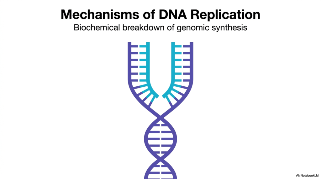
The visual presented on the opening slide serves as a fundamental primer for understanding the highly intricate mechanisms of DNA Replication. The minimalist graphic elegantly illustrates a double helix unzipping, revealing the foundational physical steps of genomic synthesis. In any functional biological system, the faithful copying of immense volumes of genetic material forms the absolute bedrock of cellular proliferation and heredity. For medical and university students, mastering this biochemical breakdown is essential for understanding how living organisms sustain themselves. The overarching process of DNA Replication involves numerous specialized enzymes and structural proteins working in seamless, highly regulated coordination to duplicate the massive genome without catastrophic structural errors.
The provided image captures the critical moment when the original double-stranded template separates, creating a distinctive and highly recognizable fork structure. This vital split represents the exact physical space where the complex enzymatic machinery of DNA Replication operates continuously. The distinct color coding on the newly synthesized strands emphasizes the precise addition of new chemical nucleotides to the existing parental blueprints. By visualizing this initial physical separation, the observer immediately grasps the sheer structural challenge the dividing cell faces. The tightly coiled helical structure must be actively unwound and mechanically stabilized, presenting a formidable thermodynamic hurdle that proteins must constantly overcome.
Understanding the full biochemical breakdown requires a rigorous, step-by-step analysis of the specific enzymes involved, which the subsequent slides systematically explore. A thorough grasp of DNA Replication is not merely an isolated academic exercise; it provides the necessary biological context for understanding severe genetic mutations, modern oncology, and targeted pharmacology. The opening slide perfectly sets the necessary stage for a comprehensive molecular exploration of genomic synthesis. It directly invites the scientific observer to dive deep into the invisible molecular choreography that makes accurate cellular division physically possible, laying the necessary groundwork for advanced biochemical and genetic studies.
Slide 2: The Biological Imperative of DNA Replication
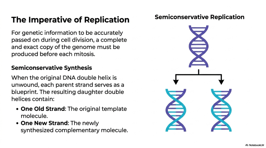
The second slide outlines the biological imperative behind DNA Replication, highlighting its crucial structural role before any physical cellular division occurs. For a complex organism to pass on its genetic information accurately during mitosis, a complete and absolutely exact genomic copy must be produced beforehand. The visual effectively demonstrates the defining concept of semiconservative synthesis. When the original double helix safely unwinds, each parent strand functions as a flawless reading matrix. The term semiconservative refers to the fact that the resulting daughter helices are not completely newly formed entities. Instead, the process of DNA Replication creates a molecular hybrid, seamlessly blending the past and the future.
This resulting structure features one strictly old strand—the original parent template—and one completely newly synthesized complementary molecule. The diagram clearly maps out the single parent molecule physically splitting into two distinct structural paths, with the newly added nucleotide sequences prominently represented in a lighter shade. This precise visual distinction helps the observer conceptualize how ultimate genetic fidelity is meticulously maintained across successive cellular generations. By utilizing the original strand as an unyielding physical guide, the machinery of DNA Replication inherently minimizes the severe risk of catastrophic genetic errors. Synthesizing entirely new double helices from scratch would invite constant biological disaster.
The fundamental biological principle of semiconservative synthesis guarantees that the delicate genetic code remains entirely stable and fully functional even as the organism grows and renews its tissues. The literal physical separation of the parent strands remains a non-negotiable prerequisite for accurately reading the immense nucleotide sequence. The slide establishes that replication is not merely a passive copying mechanism but a strictly regulated conservation strategy. Every new daughter cell successfully inherits half of its genetic material directly from the parent molecule. This fundamental structural strategy permanently secures the biological lineage of the organism, ensuring genetic continuity remains forever unbroken.
Slide 3: The Core Enzyme Powering DNA Replication
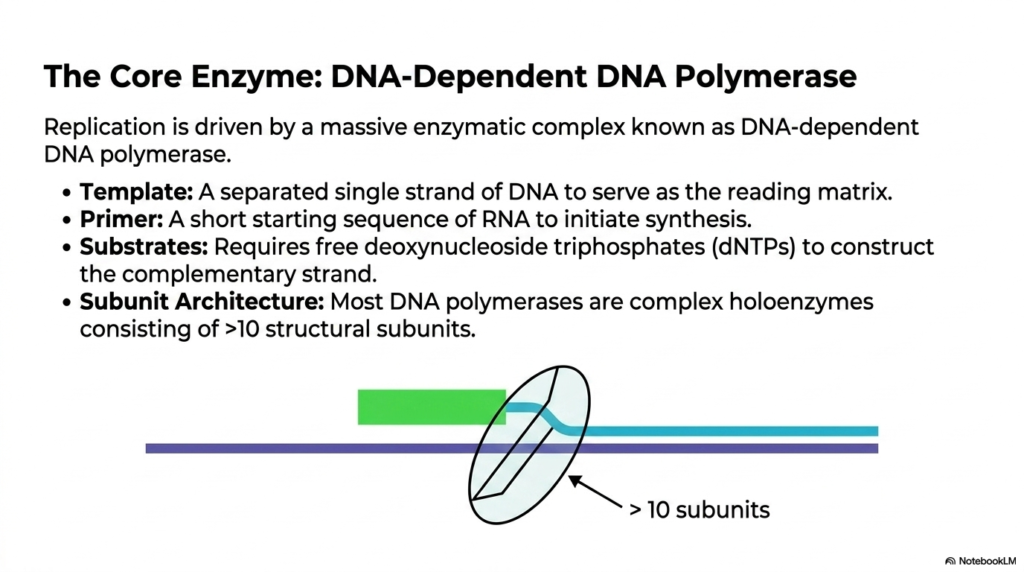
The third slide introduces the massive enzymatic complex that physically drives DNA Replication forward, formally known as the DNA-dependent DNA polymerase. This large holoenzyme operates as the primary builder, assembling the new genetic strands. To function correctly, the massive polymerase strictly requires specific structural components, beginning with a physically separated single strand of DNA to serve as the necessary reading matrix. It cannot start synthesis from absolutely nothing; it biologically mandates a short starting sequence of RNA, called a primer, to initiate the chemical sequence. The continuous progression of DNA Replication relies entirely on the constant availability of these precursors.
Beyond the template and primer, the core enzyme requires essential building blocks in the form of free deoxynucleoside triphosphates. These energetic substrates provide both the raw structural material and the necessary chemical energy to construct the complementary strand from scratch. The provided illustration showcases the impressive subunit architecture of the polymerase complex. Most DNA polymerases are not simple, singular proteins but highly complex holoenzymes consisting of more than ten distinct structural subunits. The coordinated action of these individual subunits allows DNA Replication to proceed with astonishing speed and remarkable overall accuracy, processing nucleotides in a highly systematic and controlled manner.
The visual representation of the polymerase encircling the template strand highlights the vital physical interaction between the working enzyme and the delicate genetic material. The overall complex structure ensures that the reading matrix remains stable while new bases are added sequentially. Each specific subunit within the massive holoenzyme performs a highly targeted role, from proofreading chemical errors to maintaining a firm structural grip on the elongating chain. Understanding this massive enzymatic complex is vital for advanced biochemical studies, as its continuous function dictates the overall success of the genomic copying mechanism. A structural disruption in any single subunit halts the entire procedure.
Slide 4: Substrate Alignment in DNA Replication
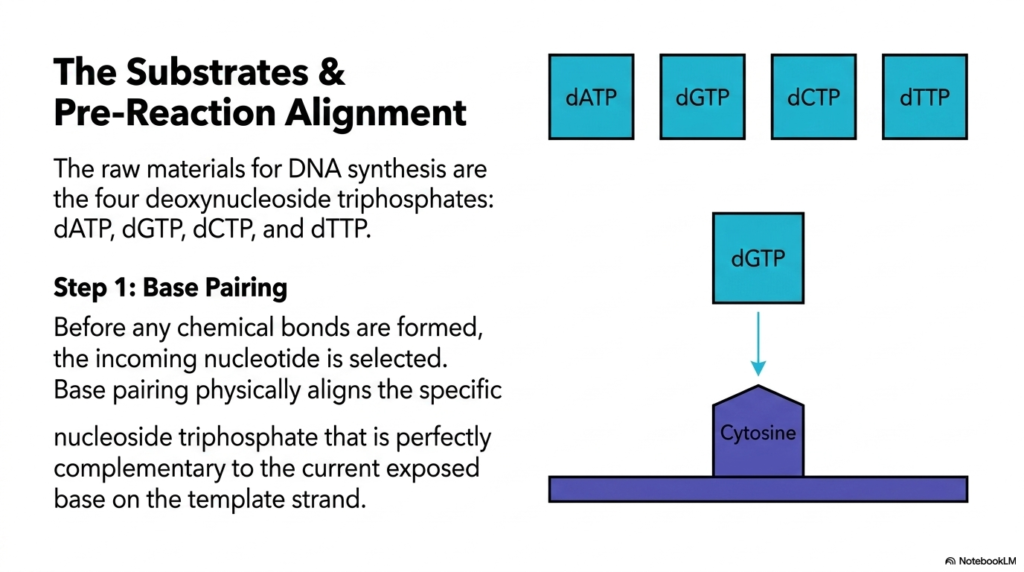
Slide four delves into the precise pre-reaction alignment required during DNA Replication, focusing heavily on the specific substrates involved. The raw molecular materials necessary for genomic synthesis are four specific deoxynucleoside triphosphates: deoxyadenosine, deoxyguanosine, deoxycytidine, and deoxythymidine triphosphates. The graphic clearly displays these essential chemical building blocks hovering directly above the exposed template strand. Before any permanent chemical bonds are established, the cell must select the exact correct incoming nucleotide. The absolute accuracy of DNA Replication depends entirely on this initial step of physical base pairing, which ensures the inherited genetic code remains intact and functional.
The diagram illustrates the passive physical alignment process, specifically showing a deoxyguanosine triphosphate molecule approaching a cytosine base located on the template strand. Base pairing relies on highly specific hydrogen-bond formation between complementary chemical structures. The incoming nucleoside triphosphate must perfectly match the currently exposed base located on the reading matrix. If an incorrect nucleotide attempts to enter the restrictive active site, the physical and chemical geometry will not align correctly. This strict complementary requirement acts as the absolute first line of defense against mutations, ensuring that the process of DNA Replication proceeds with immense fidelity.
This critical alignment is a passive physical selection process that must precede the active chemical reaction. The massive polymerase enzyme provides the optimal structural environment for this pairing to occur rapidly and efficiently without energy input. By waiting for the correct substrate to lock into place physically, the molecular machinery guarantees that the newly synthesized strand becomes a perfect mirror image of the original template. Understanding this specific pre-reaction alignment helps students appreciate the remarkable physical constraints constantly governing cellular processes. The precise geometric shape of the nitrogenous bases dictates the entire genetic transfer, ensuring perfect biological continuity.
Slide 5: The Chemical Mechanism of DNA Replication
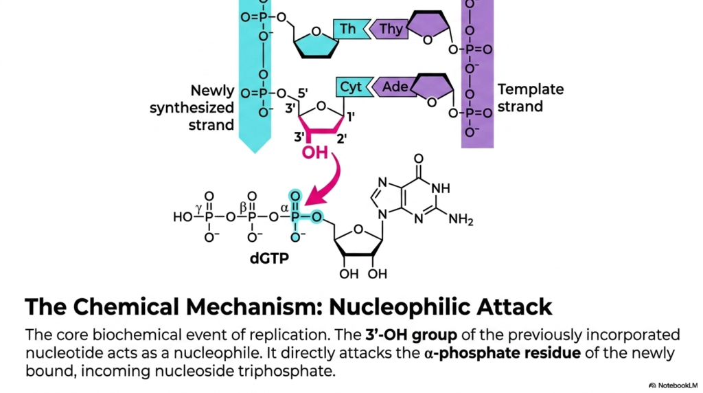
The fifth slide breaks down the core biochemical event that actively propels DNA Replication forward: the essential nucleophilic attack. This slide moves beyond mere physical substrate alignment and details the exact chemical reaction that permanently links individual nucleotides together. The crucial key player in this interaction is the exposed hydroxyl group located on the three-prime carbon of the previously incorporated nucleotide. This specific hydroxyl group acts as a powerful chemical nucleophile. The continuous structural extension of the genetic strand during DNA Replication relies entirely on this reactive site locating and engaging the next incoming building block within the active site.
The detailed chemical structure provided in the visual shows the nucleophile directly attacking the alpha-phosphate residue of the newly bound, incoming nucleoside triphosphate. This specific active phosphate is the one located closest to the sugar ring of the incoming nucleotide structure. The directed attack initiates a critical electron transfer that breaks existing weak bonds to form new, permanent chemical connections. The fundamental mechanism of DNA Replication requires this precise sequence of energetic events for every single base added to the growing genetic chain. The polymerase enzyme precisely positions both the hydroxyl group and the incoming phosphate to facilitate this exact chemical reaction.
By isolating the reactive chemical components, the slide demonstrates the sheer precision occurring millions of times per second inside the nucleus. The orientation of the interacting molecules is strictly maintained by the surrounding rigid polymerase structure. If the necessary hydroxyl group is missing or blocked by a structural analog, the nucleophilic attack cannot occur, and chain elongation stops immediately. This fundamental targeted reaction represents the exact moment when temporary physical base pairing converts into a permanent structural bond. It is the defining chemical action that physically builds the new strand, forming the vital backbone of the inherited genetic code.
Slide 6: Bond Formation During DNA Replication
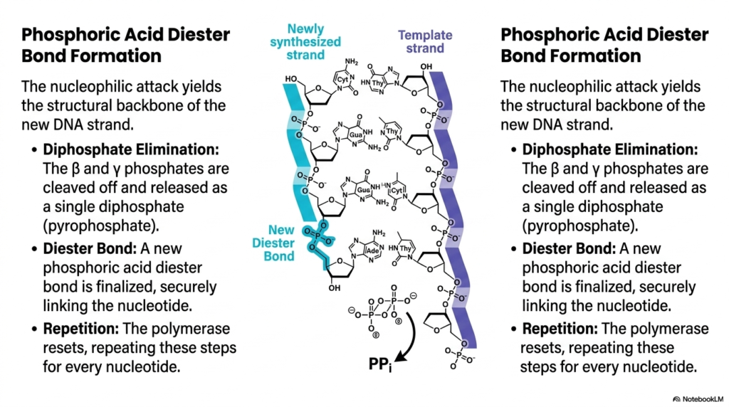
Slide six visualizes the direct structural consequence of the nucleophilic attack, highlighting the formation of the critical phosphoric acid diester bond during DNA Replication. When the targeted nucleophilic attack occurs, it fundamentally alters the structural backbone of the newly synthesizing genetic strand. The immediate chemical result of the attack on the alpha-phosphate is the rapid elimination of a diphosphate molecule. The beta and gamma phosphates from the incoming nucleotide are cleanly cleaved off and released into the surrounding cellular environment as a single pyrophosphate unit. This specific release provides the critical thermodynamic driving force necessary to propel DNA Replication forward.
Once the cleaved diphosphate leaves the active enzymatic site, a new phosphoric acid diester bond is officially finalized. This crucial chemical linkage securely connects the newly incorporated individual nucleotide to the growing macromolecular chain. The detailed diagram highlights this newly formed strong bond, demonstrating how it integrates seamlessly into the continuous sugar-phosphate backbone of the synthesized strand. The entire continuous process of DNA Replication depends on this exact bond formation to create a stable, durable molecule capable of withstanding the mechanical stresses of future cellular division. The rapid removal of the pyrophosphate ensures that the reaction remains virtually irreversible under standard physiological conditions.
Following the successful finalization of the strong diester bond, the complex polymerase enzyme must physically reset itself. It shifts forward along the rigid template matrix to precisely position the newly formed three-prime hydroxyl group for the next incoming nucleotide substrate. The biological cell repeats these exact coordinated steps—nucleophilic attack, diphosphate elimination, and diester bond formation—for every single nucleotide added to the massive sequence. This repetitive chemical cycle forms the physical basis of continuous genomic elongation. Understanding this rapid, repetitive loop allows the observer to grasp how massive genetic sequences are constructed with incredible speed and structural integrity.
Slide 7: The Absolute Rule of Directionality in DNA Replication
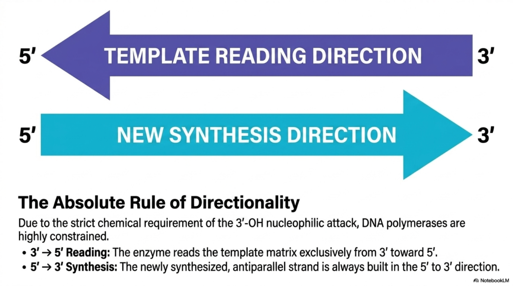
The seventh slide addresses the strict spatial constraints that govern DNA Replication, emphasizing the absolute rule of molecular directionality. Due to the highly specific chemical requirement of the nucleophilic attack detailed previously, active DNA polymerases are highly constrained in their physical movement and operation. The enzyme relies entirely on the reactive three-prime hydroxyl group to form any new chemical bonds. Therefore, the physical structural progression of DNA Replication must follow a rigid directional pathway without exception. The bold arrows on the visual slide clearly delineate the opposing flows: the template reading direction and the completely opposite new synthesis direction.
The massive polymerase enzyme reads the existing single template matrix exclusively from the three-prime end toward the five-prime end. As it moves along this strictly defined path, it constructs the newly synthesized, antiparallel genetic strand. Because of the required molecular geometry for bond formation, this new genetic strand is always built strictly in the five-prime to three-prime direction. The fundamental chemical mechanism of DNA Replication cannot be reversed or altered under any natural circumstances. The incoming energetic nucleotides can only be attached to an exposed three-prime end, meaning the newly formed chain grows in one very specific spatial orientation.
This continuous unidirectional growth creates significant mechanical challenges when copying a double-stranded molecule where the two strands physically run in opposite directions. The strict antiparallel nature of the original double helix means the replication machinery must employ specialized biological strategies to copy both sides simultaneously. The absolute rule of directionality is not merely a biological preference; it is an unyielding chemical law dictated solely by the available reactive groups on the sugar ring. Recognizing this unbreakable molecular rule provides the necessary foundation for understanding the complex mechanical paradoxes that constantly arise as the genomic duplication process continues.
Slide 8: System Architecture of DNA Replication in E. coli
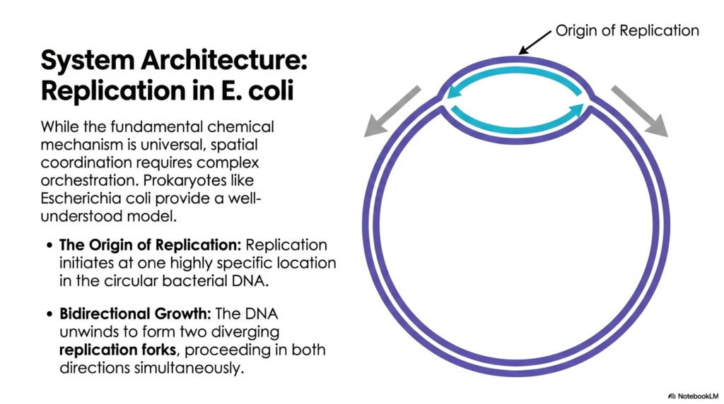
Slide eight shifts the scientific focus from universal chemical mechanisms to the spatial macroscopic coordination of DNA Replication, using the well-understood biological model of Escherichia coli. While the fundamental chemical principles of adding nucleotides remain shared across all life, orchestrating the entire process requires significant architectural planning. Prokaryotes provide a highly streamlined and accessible biological model for studying these physical dynamics. The provided diagram illustrates the circular bacterial chromosome, highlighting a specific genomic starting zone. For DNA Replication to begin properly, the process must initiate at one highly specific genetic location, formally designated as the solitary origin of replication.
Once the complex enzymatic machinery successfully binds to this origin, the tightly wound genetic material begins to unwind physically. This initial unwinding creates a distinctive physical opening within the circular genome, often referred to as a functional replication bubble. The slide strongly emphasizes the core concept of bidirectional growth during DNA Replication. The enzymatic complex does not simply move in one single direction along the circle; instead, the original separation creates two diverging active replication forks. These dual operational forks proceed away from the central origin in opposite directions simultaneously, effectively halving the total time required to copy the entire bacterial chromosome.
The distinct arrows on the visual track the steady movement of these two independent forks as they travel around the circular DNA matrix. This simultaneous, bidirectional approach is an incredibly efficient evolutionary strategy, allowing the bacterium to duplicate its entire genome rapidly in preparation for upcoming cellular division. By studying this elegant bacterial system, scientific students can clearly visualize how dense molecular coordination functions on a macro cellular scale. The circular nature of the simple prokaryotic genome ensures that the two diverging forks will eventually meet precisely opposite the origin, seamlessly completing the massive task of whole-genome duplication.
Slide 9: Unwinding the Helix for DNA Replication
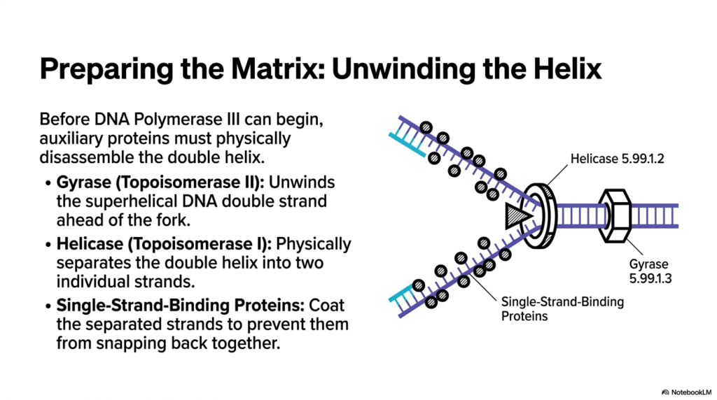
The ninth slide details the extensive mechanical preparation required before the main polymerase can begin its synthetic work during DNA Replication. Before new biological strands can be synthesized, auxiliary proteins must physically disassemble the tightly wound double helix to expose the hidden reading matrix. The diagram clearly illustrates this dynamic physical environment at the opening fork. The continuous process of DNA Replication requires a powerful enzyme known as Helicase, operating alongside the functions of topoisomerase I, to physically separate the double helix into two individual vulnerable strands. This specific enzyme operates exactly like a molecular wedge, forcefully breaking the stabilizing hydrogen bonds holding the paired bases together.
As the driving helicase forces the parent strands apart, it naturally creates severe physical torsion further down the coiled molecule. To prevent the remaining double helix from becoming overwhelmingly tangled, a distinct enzyme called Gyrase, functioning as a Topoisomerase II, actively unwinds the intense superhelical structures ahead of the advancing fork. The uninterrupted continuous progression of DNA Replication depends heavily on Gyrase relieving this intense mechanical stress. Without this critical supercoil relaxation, the mounting physical pressure would eventually halt the entire unzipping process completely, preventing the main polymerase from accessing the necessary genetic information required for copying.
Once the strands are physically forcefully separated by the helicase wedge, they are highly structurally unstable and naturally tend to snap back together. To counteract this constant threat, Single-Strand-Binding Proteins rapidly coat the newly separated nucleotide sequences. These vital stabilizing proteins ensure the exposed single templates remain open and accessible for the trailing synthetic machinery. Understanding this physical preparation phase highlights the intensely cooperative nature of cellular processes. The major building polymerases simply cannot function without this dedicated team of auxiliary proteins clearing the difficult path and managing the formidable structural stresses inherent in rapid molecular uncoiling.
Slide 10: The Leading Strand in DNA Replication
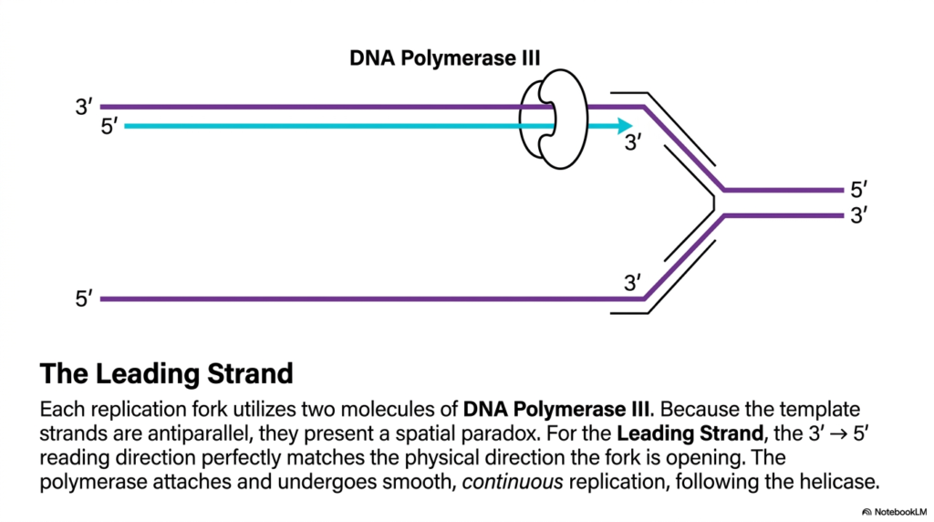
Slide ten illustrates the straightforward continuous synthesis process occurring exclusively on the leading strand during DNA Replication. As the active helicase enzyme continuously opens the moving replication fork, it immediately exposes the two antiparallel template strands, presenting a distinct spatial and mechanical paradox for the synthetic machinery. Each active replication fork universally utilizes two distinct molecules of DNA Polymerase III to handle the simultaneous molecular copying of both exposed sides. For the continuous leading strand, the physical direction of the DNA Replication process perfectly matches the unyielding chemical constraints of the polymerase enzyme. The physical reading direction allows for a smooth, uninterrupted synthesis run.
The diagram effectively maps the clear three-prime to five-prime reading direction of the leading template, which aligns exactly with the physical direction the replication fork is opening. Because the primary polymerase must synthesize the new chain in the mandatory five-prime to three-prime direction, it can simply attach to the template and follow right behind the advancing helicase. The cellular process of DNA Replication on this specific side is remarkably efficient and wholly continuous. Once the initial RNA primer is laid down, the primary polymerase complex can rapidly process thousands of nucleotides without ever needing to detach or reset its position on the matrix.
The visual explicitly shows the straight, unbroken cyan arrow representing this rapid, continuous elongation. This particular side of the replication fork perfectly satisfies the strict directional rules discussed in earlier slides without generating any mechanical complications. The leading strand represents the absolute ideal operational state for the polymerase complex, allowing it to function at maximum speed and processivity. Recognizing the structural simplicity of the continuous leading strand is crucial for students, as it provides a stark biological contrast to the highly complex, disjointed mechanisms strictly required to synthesize the opposite side of the replication fork.
Slide 11: The Lagging Strand Paradox in DNA Replication
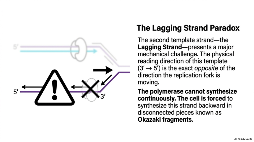
Slide eleven directly addresses the major mechanical challenge inherent in copying an antiparallel double helix, introducing the fundamental lagging strand paradox in DNA Replication. The second exposed template strand presents a formidable physical hurdle because its mandatory reading direction runs exactly opposite to the physical direction the replication fork is moving. As the driving helicase unwinds the double helix, it exposes a bare template that must be read backward relative to the overall physical progression of the fork. The unyielding strict directional rules of DNA Replication mean the primary polymerase complex simply cannot synthesize this newly exposed section in a continuous forward motion.
The diagram starkly highlights this spatial conflict with a clear warning symbol, emphasizing that the massive polymerase cannot travel forward while actively synthesizing. Instead, the biological cell is forced to compromise structurally and synthesize this specific strand backward in short, completely disconnected segments. These discontinuous DNA segments are widely known in the field of DNA Replication as Okazaki fragments. The enzyme must wait patiently for a sufficient stretch of the template to be exposed by the advancing helicase, attach itself physically, and synthesize a short genetic sequence backward until it collides directly with previously copied material. This disjointed process requires constant starting and stopping.
This paradoxical mechanism effectively solves the rigid directional constraints of the polymerase while still allowing the overall replication fork to advance in a single forward direction. The provided visual perfectly captures the backward-pointing synthesis arrows, contrasting sharply with the relentless forward motion of the overall complex. Understanding the specific lagging strand paradox is essential for grasping the sheer mechanical complexity of successful cellular division. The cell prioritizes strict chemical compliance over physical simplicity, utilizing a highly orchestrated, heavily fragmented approach to ensure the accurate structural transfer of the genetic code on the difficult antiparallel strand.
Slide 12: Initiating Fragments for DNA Replication
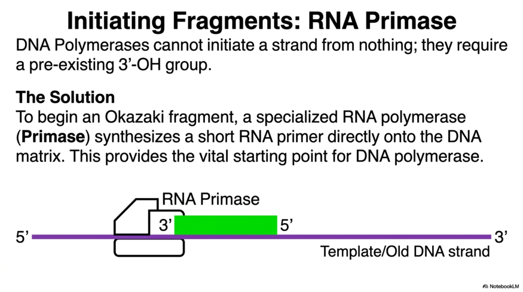
Slide twelve introduces the critical first step in actively solving the lagging strand paradox during DNA Replication, focusing entirely on the initiation of Okazaki fragments. As previously established, major DNA polymerases cannot simply pull a new strand out of thin air; they possess a strict chemical requirement for a pre-existing three-prime hydroxyl group to begin any synthesis. On the continuous leading strand, this is a simple one-time requirement, but the discontinuous lagging strand needs a completely new starting point for every single disconnected segment. The continuous overall process of DNA Replication relies on a specialized auxiliary enzyme to constantly provide these chemical anchors.
The biological solution to this massive initiation problem involves a highly specialized RNA polymerase widely known as Primase. Unlike the main DNA building enzymes, Primase has the unique structural ability to start synthesizing directly onto the bare template matrix without needing a pre-existing free end. The clear diagram illustrates this specific enzyme laying down a short RNA primer directly onto the newly exposed lagging strand. This critical enzymatic action provides the vital starting point strictly required for the main machinery of DNA Replication to physically engage. The bright green block in the visual clearly represents this short, newly constructed RNA sequence.
By rapidly synthesizing this temporary RNA primer, the Primase enzyme artificially creates the necessary three-prime hydroxyl group that DNA Polymerase III desperately requires. The primary cellular builder can now instantly recognize this structure, bind securely to the template, and begin adding the standard deoxynucleotides. The constant, repetitive enzymatic action of Primase is absolutely necessary for the creation of every single Okazaki fragment. Understanding this temporary initiation strategy allows biology students to see how biological systems utilize transient, non-permanent structures to overcome strict chemical rules, ensuring the massive genomic copying process never physically stalls due to a lack of starting material.
Slide 13: Fragment Elongation in DNA Replication
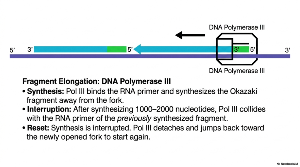
The thirteenth slide examines the active elongation phase of Okazaki fragments, detailing the repetitive physical movements constantly occurring during DNA Replication. Once the RNA Primase has successfully laid down the vital initiating primer, the primary cellular builder, DNA Polymerase III, takes over the rigorous process. The enzyme actively binds to the exposed hydroxyl group of the green RNA primer and begins synthesizing the actual DNA segment, moving directly backward away from the opening fork. This specific backward synthesis is the core defining action of DNA Replication on the lagging strand, allowing the enzyme to strictly comply with its unyielding directional constraints.
The main polymerase rapidly adds nucleotides, typically synthesizing a steady stretch of 1,000 to 2,000 bases in simple prokaryotic organisms. The illustration carefully tracks the blue newly synthesized DNA steadily extending from the green RNA primer. However, this seemingly uninterrupted run is strictly temporary. Eventually, the actively elongating polymerase collides physically with the original RNA primer belonging to the previously synthesized fragment. The continuous machinery of DNA Replication is abruptly interrupted by this direct molecular collision. Because it cannot synthesize past or structurally fuse with this existing block, the primary polymerase must halt its chemical activity entirely.
Following this absolutely necessary interruption, the massive polymerase complex must physically detach entirely from the genetic matrix. It then actively jumps backward toward the newly opened section of the advancing replication fork to locate the next freshly laid primer and start the entire synthetic cycle again. The provided slide captures this highly dynamic looping and resetting behavior, which remains essential for properly managing the discontinuous side of the fork. This exhausting biological process of binding, synthesizing, detaching, and jumping highlights the incredible mechanical agility required by the enzymatic complex to accurately duplicate an antiparallel template without massive errors.
Slide 14: Fragment Maturation in DNA Replication
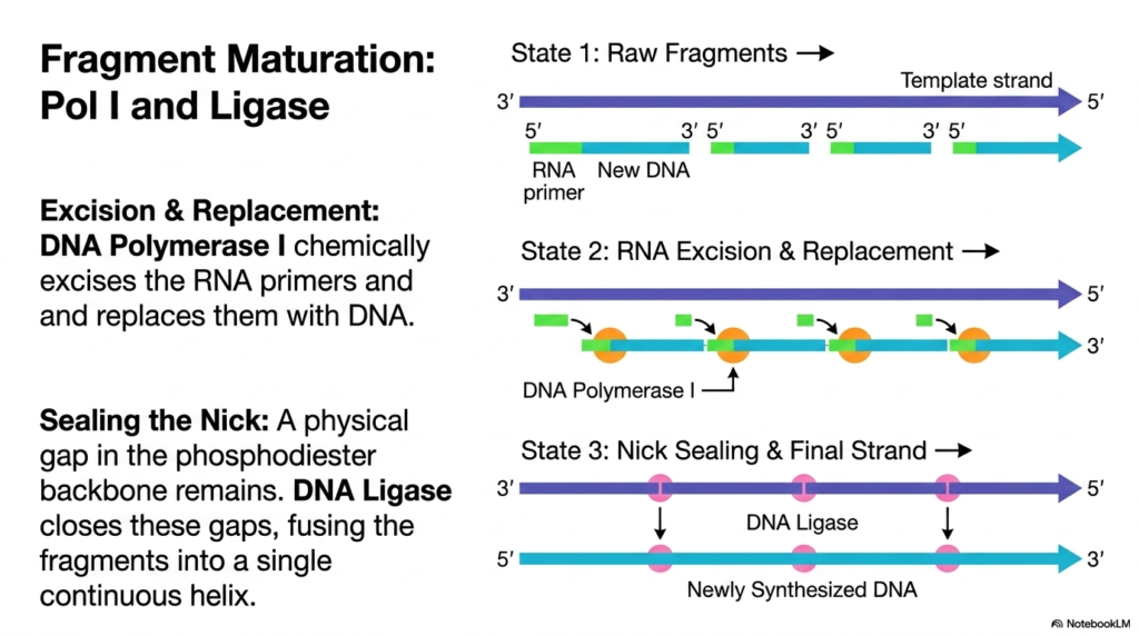
Slide fourteen details the extensive final cleanup and maturation phase absolutely required to complete the lagging strand during DNA Replication. The raw, newly synthesized genetic strand exists in a highly precarious fragmented state, littered with temporary RNA primers that must be systematically removed. An entirely new specialized enzyme, DNA Polymerase I, takes direct charge of this critical excision and replacement process. This specific polymerase recognizes the foreign RNA segments, chemically excises them from the sequence, and seamlessly replaces them with standard stable DNA nucleotides. The overall structural integrity of DNA Replication depends completely on permanently removing all traces of these temporary starting blocks.
The detailed diagram maps out this essential transitional state, showing the step-by-step molecular conversion from an unstable RNA-DNA hybrid into a pure, stable DNA sequence. However, even after Polymerase I successfully finishes replacing the nucleotides, a significant structural problem remains. A physical microscopic gap, or nick, persists in the newly formed phosphodiester backbone located precisely between the adjacent fragments. The final absolute step of DNA Replication requires a dedicated enzyme known as DNA Ligase to locate and securely seal these minute gaps. Ligase effectively welds the adjacent sections together by forming the final necessary chemical bonds, ensuring the backbone remains entirely unbroken.
The visual sequence concludes perfectly with the state three representation, showing a solid, continuous double helix entirely devoid of any dangerous nicks or RNA segments. The lagging strand, initially built backward in thousands of chaotic disconnected pieces, is finally permanently fused into a single continuous macromolecule. This maturation phase remains vital for cellular survival; if the genome were left highly fragmented, the chromosome would instantly degrade during cell division. Understanding the combined actions of Polymerase I and Ligase provides a complete picture of how chaotic, discontinuous synthesis is ultimately resolved into perfect structural stability by the cell.
Slide 15: Kinetics and Scale of DNA Replication
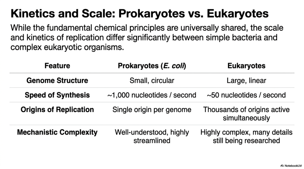
The final slide broadens the overall perspective by comparing the kinetics and massive scale of DNA Replication between simple prokaryotes and complex eukaryotic organisms. While the fundamental chemical principles—such as nucleophilic attacks and strict directional constraints—are universally shared across all domains of biological life, the physical execution differs significantly. The provided table highlights these major logistical differences, starting with genome structure. Prokaryotes, like Escherichia coli, possess a small, circular chromosome, whereas eukaryotic cells must properly manage massive, linear genomic structures. The overall physical strategy of DNA Replication must heavily adapt to these vastly different physical architectures.
The raw speed of synthesis reveals another striking biological contrast in how distinct organisms manage genomic copying. The highly streamlined prokaryotic machinery operates at a blistering pace, adding approximately one thousand nucleotides every single second. In stark contrast, eukaryotic DNA Replication proceeds much more cautiously, crawling along at a mere fifty nucleotides per second. To properly compensate for this slower enzymatic speed and the vastly larger overall genome size, eukaryotes entirely abandon the single-origin strategy. Instead of relying on one lone starting point, eukaryotic cells activate thousands of origins of replication simultaneously right across their massive linear chromosomes.
This simultaneous multi-origin approach allows highly complex organisms to copy their massive genomes within a perfectly reasonable timeframe. The detailed table also points out the severe mechanistic complexity distinguishing the two major groups. While the simple bacterial system is well-understood and highly streamlined, eukaryotic replication is immensely complex, heavily mechanistically regulated, and involves numerous intricate details that are still actively being researched by modern biochemists. Concluding the slide deck with this large-scale comparison helps advanced students appreciate that while the core molecular chemistry is elegantly simple, the evolutionary adaptations required to manage massive genomic datasets remain wonderfully complex.
Please read our Content Disclaimer Statement.
Check out our social media channels: