82. Chromatin Structure and Histone Organization: A Biochemical Overview
Imagine attempting to fit a microscopic thread stretching the length of two human adults perfectly into a tiny sphere smaller than a grain of sand. This immense physical challenge is the reality faced by every living cell. This educational guide explores the profound molecular mechanisms the cell utilizes to compress, organize, and read its genetic material. By examining the fundamental components of DNA packaging, this comprehensive post illuminates the biochemical elegance that makes complex life possible. Medical and college students alike will discover the vital structural interactions that manage and protect the human genome.
Slide 1: The Molecular Architecture of Chromatin Structure
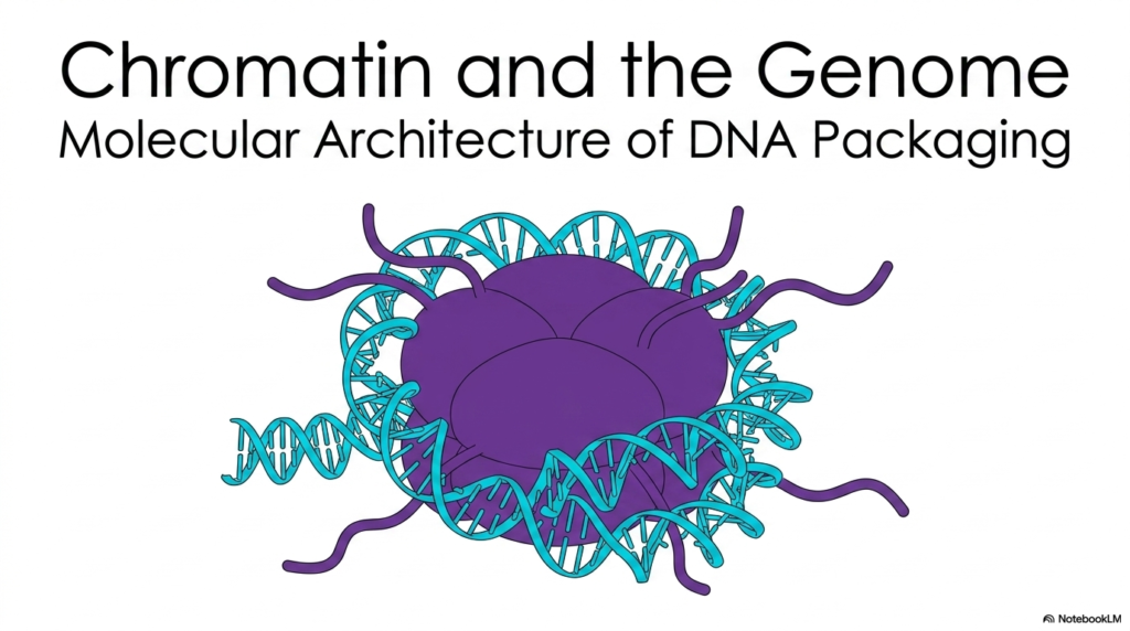
The study of modern genetics often begins with the familiar double helix, but the true marvel of cellular engineering lies in the highly organized molecular architecture known as chromatin structure. This introductory visual captures the foundational building block of DNA packaging. It displays a segment of the genetic code wrapped tightly around a robust central protein core. This core consists of specialized histone proteins, which form the functional heart of all chromatin structure within the human body.
Understanding this precise molecular arrangement is absolutely crucial for medical and college students diving into advanced biochemistry. Without this intricate wrapping mechanism, the biological cell could never manage its immense genetic blueprint. The brilliant illustration highlights the direct physical interaction between the winding nucleic acid and the central protein complex. This initial layer of dense organization represents just the very beginning of a much larger, complex geometric hierarchy.
By continuously coiling the long DNA strands around these microscopic protein hubs, the cell successfully creates a highly stable and compact chromatin structure. This basic building block is meticulously replicated millions of times across the entire global genome. It serves not just as a static biological storage solution, but as an incredibly dynamic platform for continuous genetic regulation.
This slide brilliantly sets the stage for exploring how living cells perfectly balance opposing needs. The cell must somehow tightly pack massive amounts of DNA while simultaneously maintaining the critical ability to access and read the genetic code whenever necessary. The vivid blue strands and the dense purple core perfectly embody this beautiful biological paradox.
Slide 2: The Spatial Paradox and Chromatin Structure
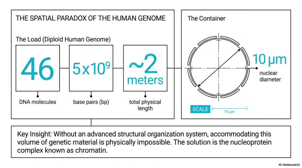
The sheer volume of genetic information contained within a single human cell presents an astonishing and seemingly impossible physical paradox. A standard diploid human genome contains forty-six distinct DNA molecules, comprising approximately five billion individual base pairs. If these microscopic molecules were laid end-to-end, they would reach a massive total physical length of nearly two meters. This immense load of genetic material must fit perfectly into a nuclear container measuring only ten micrometers in diameter.
Without a highly advanced, specialized structural organization system, accommodating this massive volume of genetic material is physically impossible. If the long DNA strands were left completely unpackaged, the resulting tangled mass would cause the cell to malfunction and perish instantly. The brilliant biological solution to this extreme spatial limitation is the highly ordered nucleoprotein complex known as chromatin. The intricate geometric nature of chromatin structure perfectly allows the cell to overcome the massive discrepancy between the genetic load and the cellular container.
By utilizing this specialized biochemical packaging system, the cell successfully compresses its two meters of genetic code by a factor of hundreds of thousands. This informative slide visually contrasts the massive physical length of the uncoiled genome against the microscopic dimensions of the nuclear diameter. The simple mathematical illustration effectively demonstrates why stable chromatin structure is a fundamental requirement for the existence of all complex lifeforms.
The visual layout strongly highlights the stark mathematical reality of this spatial paradox. It clearly emphasizes that the basic double helix cannot exist freely within the nucleus. The complex folding mechanisms provided by specialized chromatin structure ensure that the genetic code remains safely tucked away, yet perfectly organized for the biological cell to survive.
Slide 3: Defining the Composition of Chromatin Structure
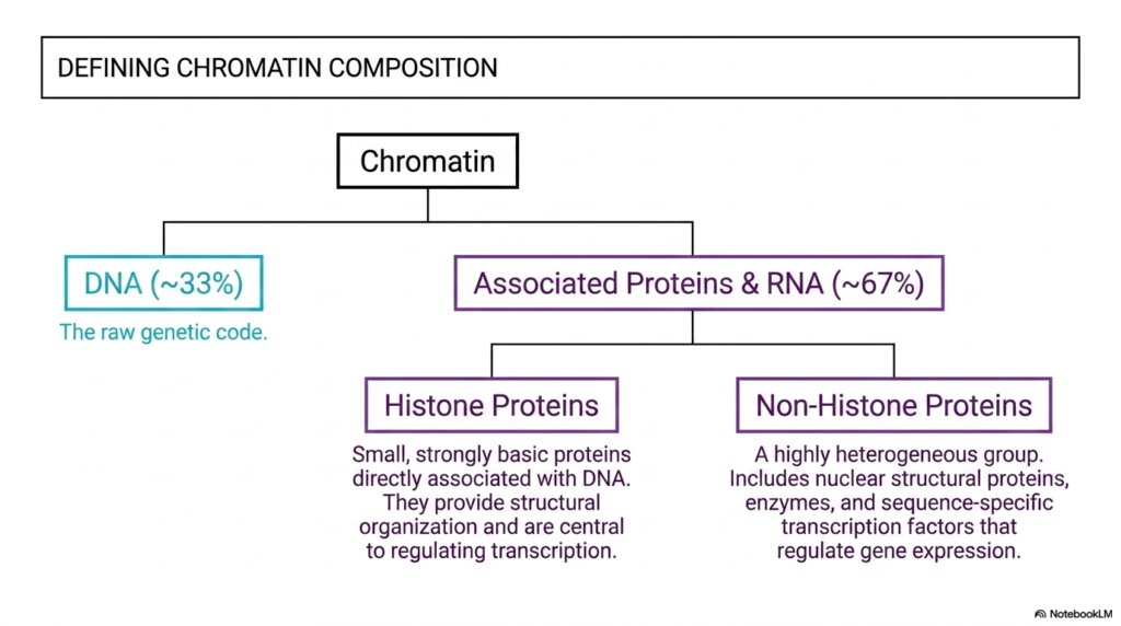
To fully comprehend how the entire genome is organized, students must examine the precise biochemical makeup of chromatin structure. The slide expertly breaks down this composition, revealing that the raw genetic code makes up only a minor fraction of the total complex. Specifically, naked DNA constitutes approximately thirty-three percent of the overall mass. The remaining sixty-seven percent is composed entirely of heavily associated proteins and essential RNA molecules.
This fascinating ratio clearly highlights that chromatin structure is predominantly a protein-based system. It emphasizes the massive structural scaffolding absolutely required to manage the lengthy DNA strand. The protein component is further categorized into two entirely distinct biological groups: histone proteins and non-histone proteins. Histones are remarkably small, strongly basic proteins that physically associate directly with the fragile DNA double helix.
These basic histone proteins provide the essential structural organization required for successful DNA packaging. They serve as the primary foundational building blocks of all chromatin structure and play a highly central role in dynamically regulating transcription. By physically restricting or granting access to the underlying code, histones control the entire cellular factory. On the other side of the biochemical spectrum lies a highly heterogeneous group known as non-histone proteins.
This diverse collection of non-histone molecules includes various nuclear structural proteins, vital cellular enzymes, and crucial sequence-specific transcription factors. While histones provide the robust foundational framework of chromatin structure, these non-histone proteins perform highly specialized tasks. They dynamically regulate active gene expression and meticulously maintain the overall structural integrity of the cellular nucleus.
Slide 4: Interphase Morphologies in Chromatin Structure
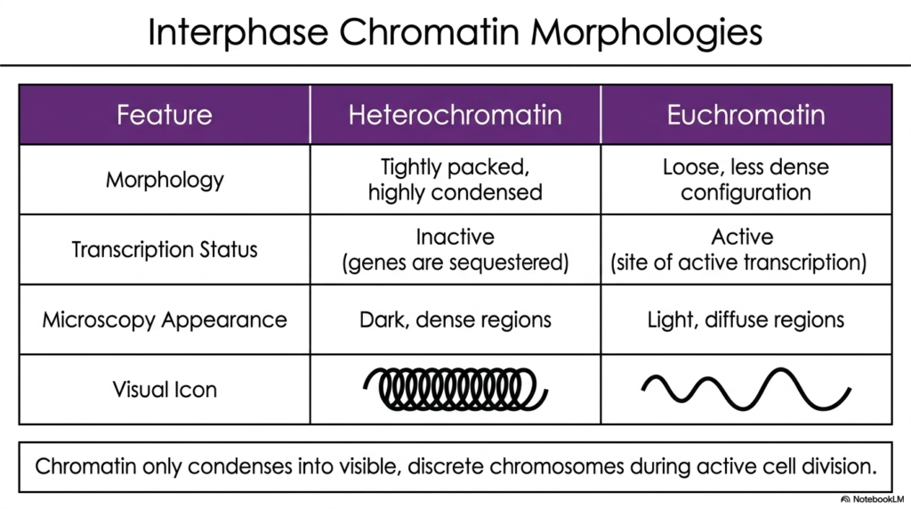
During the active interphase stage of the standard cell cycle, the vast genome continuously exists in two highly distinct morphological states. This educational slide categorizes these separate states into heterochromatin and euchromatin, detailing exactly how chromatin structure dictates overall genetic activity. Heterochromatin represents a tightly packed and highly condensed structural configuration. Because the physical arrangement is so incredibly dense, the genes located within these specific regions are completely sequestered.
Due to this restrictive compaction, these dense genetic regions remain entirely transcriptionally inactive. The incredibly tight chromatin structure successfully prevents the necessary cellular reading machinery from physically accessing the genetic code. Under an electron microscope, heterochromatin predictably appears as very dark, highly dense regions scattered within the nucleus. The slide provides a highly visual icon of a tightly coiled spring to perfectly represent this inaccessible, restrictive state.
In stark contrast, euchromatin constantly maintains a very loose, significantly less dense structural configuration. This open, highly accessible form of chromatin structure is heavily active, serving as the primary physical site for ongoing gene transcription. The relaxed physical state easily allows vital enzymes and necessary transcription factors to freely bind directly to the exposed DNA strand. Euchromatin readily appears as light, highly diffuse regions during standard microscopy.
Its corresponding visual icon resembles a relaxed, loose, wavy line, strongly emphasizing its overall biological accessibility. The constant, dynamic transition between these two critical morphologies definitively proves that overall chromatin structure is highly adaptable. It constantly shifts its physical form to meet the immediate functional demands of the living cell, condensing into discrete, visible chromosomes only during cellular division.
Slide 5: Electrostatic Mechanics Behind Chromatin Structure
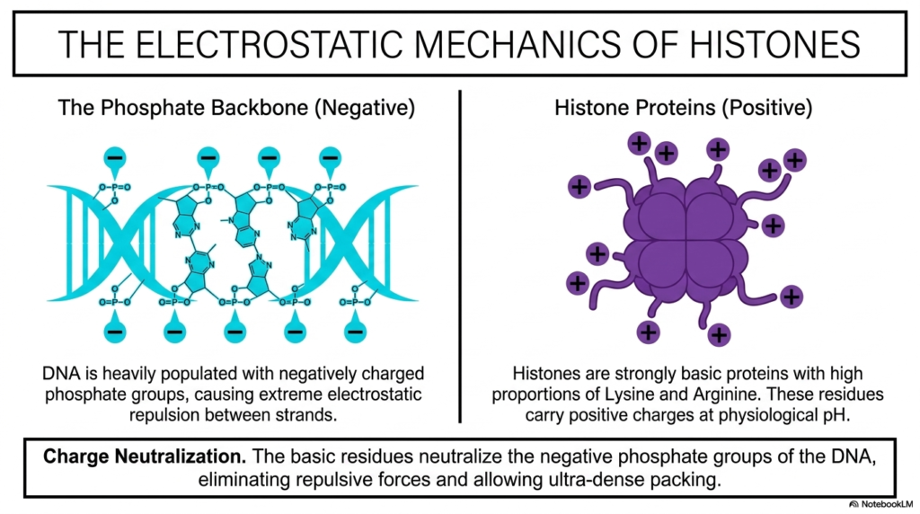
The physical compaction of the entire genome is fundamentally driven by the basic scientific principles of chemistry and atomic physics. This fascinating slide brightly illustrates the vital electrostatic mechanics that make all chromatin structure physically possible. At the absolute core of the biological problem is the long DNA phosphate backbone. The fragile DNA strand is heavily populated with highly negatively charged phosphate groups running along its entire exterior.
Because similar atomic charges constantly repel each other, these numerous groups cause extreme electrostatic repulsion between the genetic strands. This natural phenomenon actively resists any form of dense biological packaging. To completely overcome this immense repulsive force, the cell heavily employs positively charged histone proteins. Histones are strongly basic proteins naturally equipped with incredibly high proportions of specific amino acids, specifically Lysine and Arginine.
These particular amino acid residues naturally carry strong positive charges at standard physiological pH levels. The direct physical interaction between the negatively charged DNA and the positively charged histone proteins forms the crucial energetic basis of stable chromatin structure. This specific electrostatic pairing is perfectly calibrated by biological evolution to securely lock the molecules together. The incredible result of this biochemical pairing is complete charge neutralization.
The basic, positively charged residues of the histone proteins entirely neutralize the acidic, negatively charged phosphate groups of the DNA backbone. This crucial neutralization completely eliminates the repulsive forces that would naturally tear the complex apart. The ultimate physical stability of all chromatin structure relies entirely on this continuous electrostatic balancing act. It enables the ultra-dense packing required for human life.
Slide 6: The Nucleosome Core in Chromatin Structure
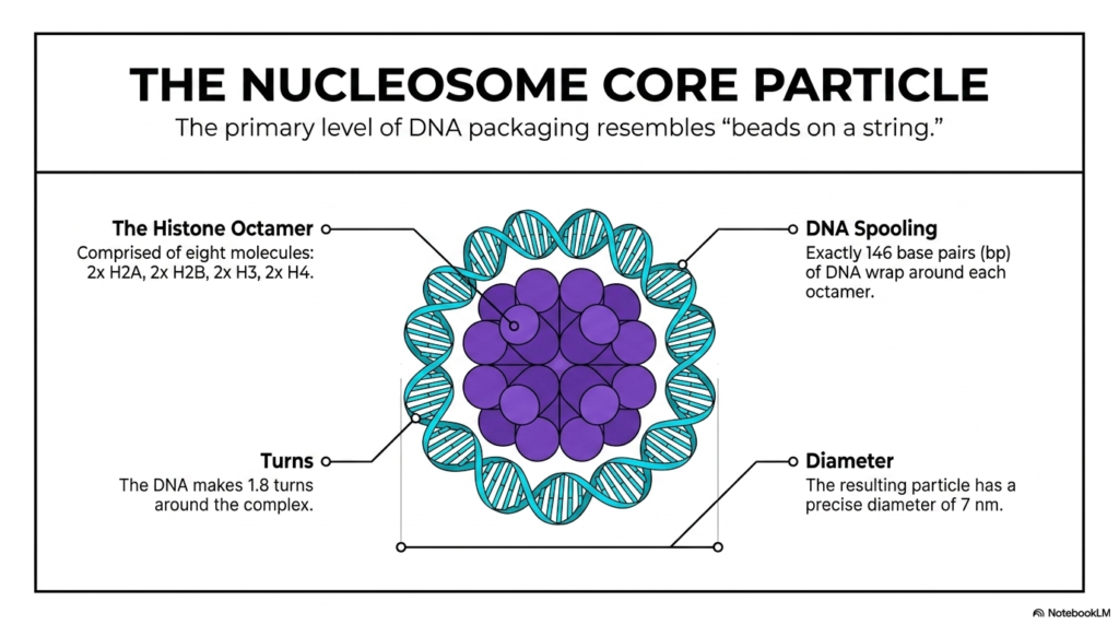
The primary foundational level of all DNA packaging naturally creates a microscopic formation that famously resembles “beads on a string.” This detailed slide expertly breaks down the vital nucleosome core particle, the fundamental repeating unit of chromatin structure. At the center of this core particle sits the robust histone octamer, a specialized complex composed of eight distinct protein molecules, perfectly locked together.
This functional octamer consists of precisely two copies each of four specific core histones: H2A, H2B, H3, and H4, forming a highly stable central spool. The biological process of DNA spooling precisely around this central octamer is highly organized. Exactly one hundred and forty-six base pairs of DNA securely wrap tightly around each specific histone core. As it gently wraps around the octamer, the genetic strand makes 1.8 highly precise turns around the protein complex.
This meticulous mathematical organization at the microscopic level beautifully demonstrates the immense physical precision continuously required by the cell. It actively maintains stable chromatin structure without ever accidentally damaging the incredibly fragile genetic code. The resulting microscopic nucleosome core particle continuously boasts a precise and uniform physical diameter of exactly seven nanometers.
This structural consistency is absolutely vital for the larger hierarchical folding of the entire genome later on. Every single nucleosome actively acts as a highly standardized building block, ensuring that the entire lengthy genetic code is compressed uniformly. Without this precise standardization at the microscopic level, the massive higher-order levels of chromatin structure would be physically impossible to construct.
Slide 7: Linker DNA, Histone H1, and Chromatin Structure
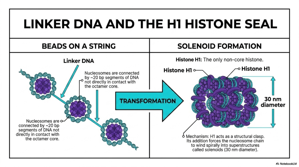
While the central nucleosome core particles form the initial loose “beads on a string” configuration, they never actually exist in total isolation. This informative slide visually explains the massive transition from individual floating beads to a highly condensed higher-order chromatin structure. The individual nucleosomes are securely connected by short segments of genetic material universally known as linker DNA.
These connecting segments consistently consist of approximately twenty base pairs of DNA that are never directly in contact with the central octamer core. They act as highly flexible biological bridges directly between the distinct protein hubs. The critical physical transformation from a loose nucleosome chain into a dense genetic fiber relies entirely on the rapid addition of Histone H1. Unlike the interior components of the central octamer, Histone H1 is the only non-core histone.
Its primary biological function is to actively act as a robust structural clasp, tightly sealing the genetic DNA as it enters and exits the specific nucleosome. The rapid integration of this specific structural protein fundamentally alters the local chromatin structure. It rapidly locks the previously loose linker DNA tightly into a highly rigid geometric position. The physical mechanism of this H1 clasp forces the entire nucleosome chain to wind spirally.
This winding action radically compresses the loose genetic material into massive superstructures scientifically called solenoids. This spiral winding heavily compresses the genetic material, rapidly creating a thick biological fiber boasting a diameter of thirty nanometers. This dense thirty-nanometer chromatin structure successfully provides the sturdy architectural foundation absolutely necessary for managing the massive genome.
Slide 8: The Full Hierarchy of Chromatin Structure
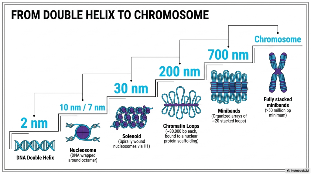
The biological journey from a simple genetic double helix to a fully condensed human chromosome is a beautiful masterclass in biological engineering. This comprehensive slide thoroughly details the strict, continuous five-level spatial hierarchy that heavily defines all modern chromatin structure. The microscopic process begins rapidly with the raw DNA double helix, measuring just two nanometers across.
It quickly upgrades to the basic nucleosome level, wrapping tightly around the specific octamer to form the microscopic seven to ten-nanometer core particles. This crucial initial step successfully sets the perfect stage for the dramatic physical compaction that directly follows. The third specific level of chromatin structure successfully occurs when the tiny nucleosomes spiral directly into the dense thirty-nanometer solenoid, heavily driven by the H1 histone clasp.
From there, the dense thirty-nanometer fiber carefully organizes into massive, winding chromatin loops. Each of these specific loops contains approximately eighty thousand base pairs and is securely anchored directly to a central nuclear protein scaffolding. This complex loop formation rapidly creates a massive two-hundred-nanometer structure, properly managing massive amounts of genetic data inside heavily organized geographic domains. The strict hierarchy continuously builds as the loops physically compress.
They compress into highly organized, stacked arrays universally called minibands. These large minibands measure roughly seven hundred nanometers across and physically represent fully stacked arrays containing roughly twenty looped sections. Finally, these minibands securely stack together to ultimately create the fully condensed chromosome. This complete chromatin structure guarantees that the immense genetic load is always managed perfectly without ever succumbing to chaotic tangles.
Slide 9: Evolutionary Perfection within Chromatin Structure
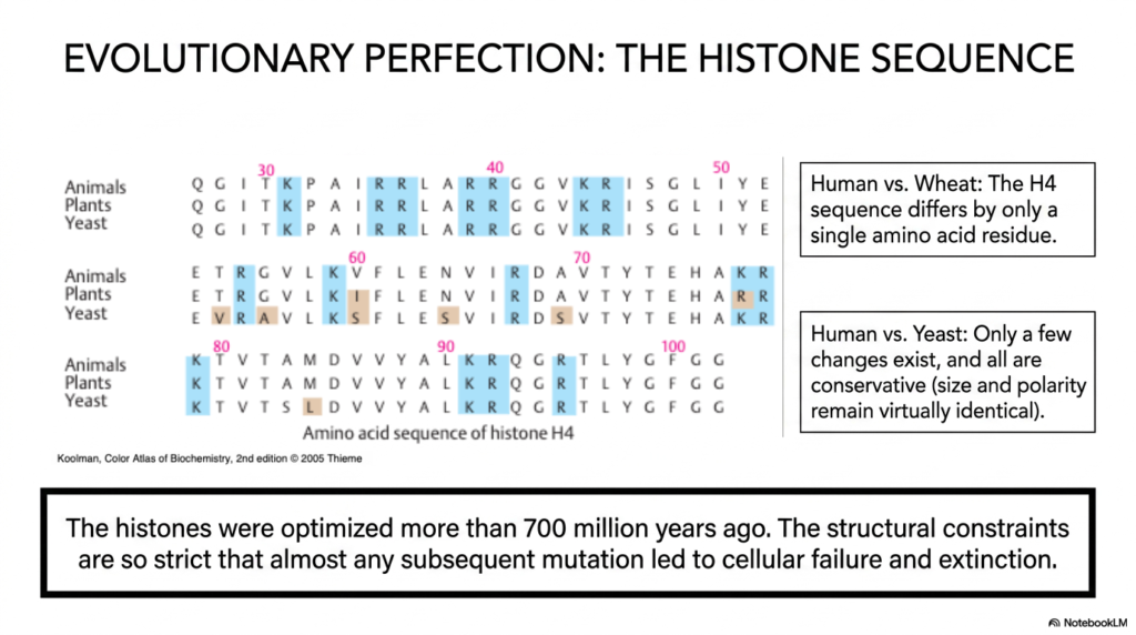
The sophisticated protein machinery entirely responsible for massive DNA packaging is not just biologically effective; it is practically flawless from a historical evolutionary standpoint. This intriguing slide thoroughly explores the profound biological conservation of the specific histone sequence. It strongly highlights the extreme biological constraints heavily placed on all global chromatin structure.
The provided comparison chart explicitly details the precise amino acid sequences of distinct animals, leafy plants, and microscopic yeast. The direct comparison readily reveals a truly shocking level of genetic preservation actively maintained across vastly different biological kingdoms over hundreds of millions of years. The slide visually points out a highly fascinating biological example involving the specific Histone H4 sequence.
When carefully comparing a modern human to a simple wheat plant, the H4 sequence differs by only a single microscopic amino acid residue. Even when comparing humans directly to yeast, only a few highly microscopic chemical changes exist. Furthermore, all of these minor alterations are highly conservative. This means the specific physical size and the vital chemical polarity of the necessary proteins successfully remain virtually identical across vastly different species.
This incredibly strict preservation ensures that the global chromatin structure stays entirely functional. The basic microscopic building blocks of chromatin structure were thoroughly optimized more than seven hundred million years ago. Because the precise structural constraints on the histones are so incredibly strict, any random mutation typically destroys the cellular machinery. Almost any alteration to the histones caused the packaging to fail, leading immediately to cellular extinction.
Slide 10: N-Terminal Tails Regulating Chromatin Structure
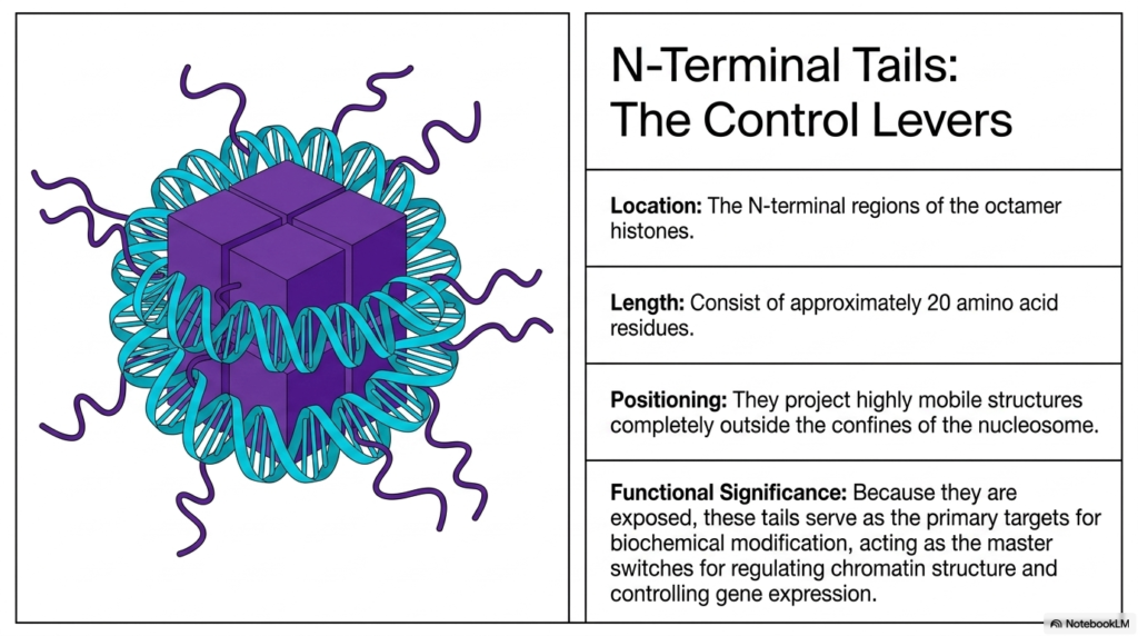
While the massive central histone octamer constantly remains locked tightly inside the genetic DNA coil, highly important functional elements deliberately protrude heavily into the cellular environment. This specific slide heavily focuses on the crucial N-terminal tails, which constantly act as the primary biological control levers for actively manipulating global chromatin structure.
These critical flexible regions are constantly located at the extreme N-terminal ends of the specific octamer histones. They typically consist of relatively short polypeptide chains, usually measuring approximately twenty amino acid residues in total length. The exact physical positioning of these tails is absolutely vital to their overall biochemical function. Rather than being tucked safely away, they visibly project out as highly mobile microscopic structures.
They project completely outside the strict physical confines of the wrapped nucleosome. Because they heavily extend completely beyond the wrapped DNA, these loose tails are fully chemically exposed to the surrounding nucleoplasm. This brilliant architectural feature flawlessly ensures that the rigid chromatin structure can effortlessly communicate directly with the numerous reading enzymes widely floating throughout the cellular nucleus. The true functional significance of these exposed residues completely dictates genetic life.
Because they are readily available to external molecules, these specific N-terminal tails serve as the absolute primary targets for critical biochemical modification. By chemically altering these distinct tails, the biological cell essentially flips the tiny master switches for actively regulating overall chromatin structure. They absolutely represent the perfect biological interface seamlessly connecting rigid DNA storage and dynamic genetic expression.
Slide 11: Epigenetic Modifications Altering Chromatin Structure
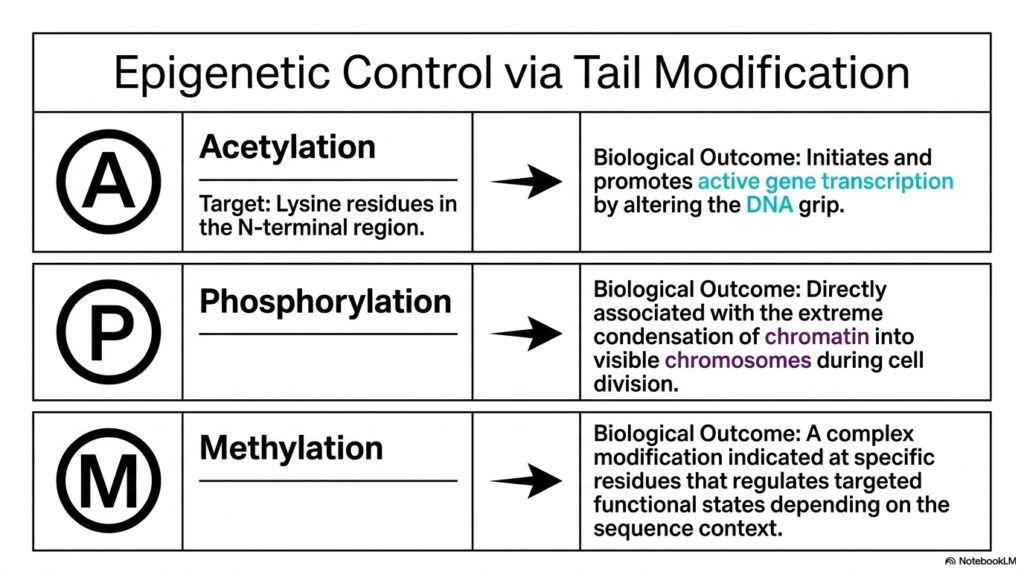
The biological cell constantly manages its heavily guarded genetic blueprints by actively chemically altering the highly protruding histone tails. This slide explicitly outlines the primary epigenetic controls that physically alter local chromatin structure on demand. The absolute first major chemical modification is Acetylation.
This vital process specifically heavily targets the microscopic lysine residues naturally located within the exposed N-terminal region. By actively chemically neutralizing the strong positive charge of the specific lysine, acetylation fundamentally successfully alters the biological grip the massive histone has on the surrounding genetic strand. The biological outcome of this acetylation is highly profound for the cell. By effectively loosening the electrostatic grip, the cell physically opens the specific local chromatin structure.
This structural relaxation strongly initiates and promotes active gene transcription, allowing the cellular reading machinery to access the previously securely hidden genetic code. Conversely, the slide explicitly details Phosphorylation, a crucial modification directly functionally associated with the extreme global condensation of the genetic material into visible chromosomes. The third major chemical modification heavily discussed is targeted Methylation.
Unlike rapid acetylation, methylation is a highly complex chemical modification that carefully occurs at highly specific exposed residues. Depending heavily on the exact sequence context, it specifically regulates targeted functional states. Methylation can selectively tighten or loosen the distinct local chromatin structure. By constantly deploying specific cellular enzymes, the cell flawlessly gains absolute epigenetic control over its massive genome.
Slide 12: The Structure-Function Paradigm of Chromatin Structure
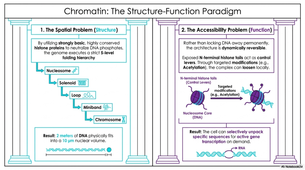
The incredible architectural design of the massive human genome perfectly delicately balances two entirely opposing necessary biological requirements. This excellent final slide expertly summarizes the ultimate, profound structure-function paradigm of all chromatin structure. On the far left side, the living cell constantly faces the massive Spatial Problem.
It absolutely must rapidly neutralize the heavily repelling DNA phosphates using highly conserved, strongly basic histone proteins. This allows it to successfully execute a strict five-level spatial folding hierarchy. This incredibly dense microscopic packaging ultimately forces two full meters of vital genetic material to physically comfortably fit inside a microscopic ten-micrometer volume. However, this extreme compaction instantly creates a severe secondary biological issue: the Accessibility Problem.
If the vital DNA is permanently tightly locked away in a highly rigid chromatin structure, the cell absolutely cannot transcribe the necessary genes to survive. Therefore, the biological architecture absolutely must be highly dynamically reversible. Rather than entirely permanently sealing the genetic code, the smart cell happily utilizes the widely exposed N-terminal histone tails as dynamic biological control levers.
Through highly targeted biochemical modifications, the microscopic complex can smoothly physically loosen on a highly localized level. This localized relaxation of the massive chromatin structure seamlessly permits the vital machinery of transcription to easily access and read the exact required genes. The beautiful structural dichotomy represents the absolute pinnacle of cellular engineering. The massive rigid folding flawlessly handles the immense spatial burden, while the flexible histone tails perfectly handle the massive functional demands of complex human life.
Please read our Content Disclaimer Statement.
Check out our social media channels: