119. The Biochemistry of Bile Acids: Synthesis, Function, and Enterohepatic Circulation
Have you ever wondered how your body digests greasy meals without clogging its aqueous pathways? The secret lies in a fascinating biochemical transformation in the liver. This blog post explores a slide deck that breaks down the molecular pathway converting rigid cholesterol into powerful digestive detergent molecules. Understanding this pathway reveals how human metabolism maintains cholesterol balance and absorbs vital nutrients.
Slide 1: Synthesis, Circulation, and Function of Bile Acids
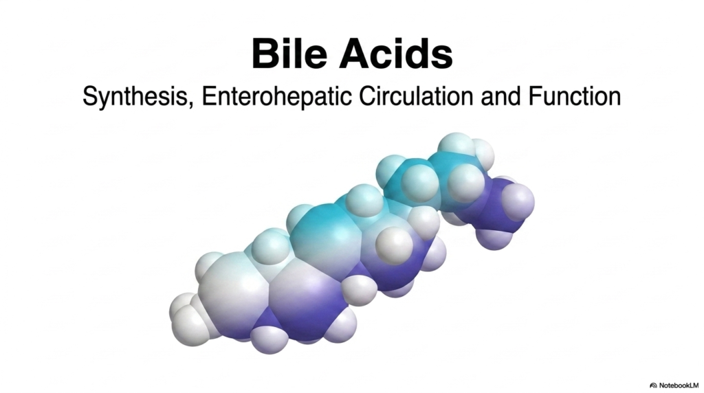
Metabolism relies on specialized chemical tools to solve physical problems in physiological fluid environments. Bile Acids serve as the body’s primary biological detergents, fulfilling a vital role in lipid digestion, nutrient absorption, and cholesterol elimination. Synthesized in hepatocytes, Bile Acids undergo structural alterations that turn a nonpolar lipid into an effective emulsifying agent. Once produced, these molecules move through the biliary system into the intestinal lumen, where they break down dietary fats into digestible particles.
The life cycle of these biochemical tools extends far beyond simple fat breakdown. A process called enterohepatic circulation continuously recycles Bile Acids between the liver and the gastrointestinal tract. This circuit conserves resources by reabsorbing over ninety percent of secreted molecules from the terminal ileum. Returning molecules travel through the portal bloodstream back to hepatocytes, minimizing the need for constant new synthesis from precursor cholesterol pools.
Beyond their mechanical roles in digestion, these compounds also act as crucial signaling molecules that regulate metabolic homeostasis. By binding to specific cellular receptors throughout the body, they influence lipid turnover, glucose metabolism, and overall energy balance. Disruptions in their synthesis or circulation can lead to severe clinical disorders, such as gallstones or malabsorption. Studying these structures reveals how hepatocytes engineer simple lipids into multifunctional regulators essential for survival.
This introductory slide sets the foundation for our deep dive into hepatic pathways. By examining molecular geometry and enzymatic steps, students can appreciate how structural adjustments unlock crucial digestive capabilities. Throughout this presentation, we will track the journey of these compounds from their steroid origins to their final functional forms in human physiology.
Slide 2: Cholesterol as the Precursor to Bile Acids
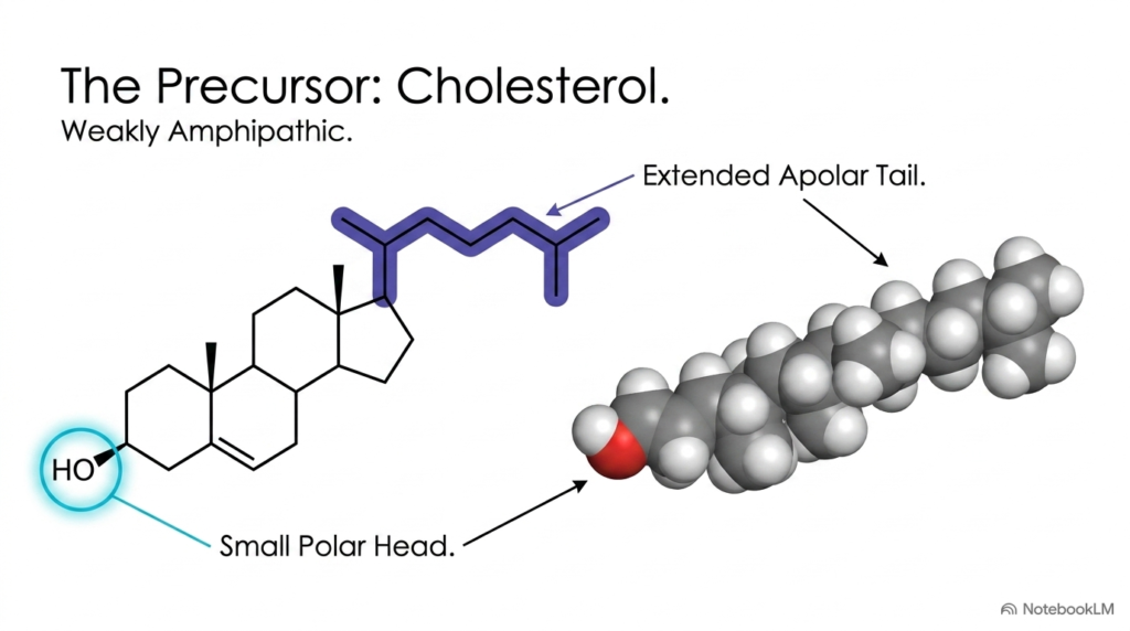
Every bile acid molecule originates from cholesterol, a sterol lipid essential for cellular membrane integrity and hormone production. Cholesterol’s molecular structure features a rigid four-ring hydrocarbon core known as the cyclopentanoperhydrophenanthrene, or sterane, framework. A single hydroxyl group attached to C-3 of this steroid framework forms a tiny polar head on an otherwise hydrophobic molecule. An extended apolar hydrocarbon tail attaches to the opposite end at carbon C-17.
Because cholesterol possesses only one small hydroxyl group against a massive hydrophobic steroid skeleton, it is extremely insoluble in water. Scientists classify cholesterol as weakly amphipathic due to this extreme imbalance between its tiny polar head and large apolar body. In aqueous blood plasma, cholesterol cannot float freely or emulsify fats on its own. To serve as digestive agents, raw cholesterol molecules must undergo significant structural redesign by liver enzymes to become soluble Bile Acids.
The liver faces a major biochemical challenge when transforming cholesterol into active Bile Acids. Hepatocytes must add hydrophilic regions to the hydrophobic ring system while shortening the hydrocarbon tail. This conversion process increases water solubility, allowing the resulting compounds to interact effectively with water and dietary lipids simultaneously in the small intestine.
Understanding cholesterol’s physical limitations highlights why hepatic transformation is necessary. Without these enzyme-driven changes, the body could not eliminate surplus cholesterol or absorb dietary fats efficiently. The starting material has the basic steroid ring structure, but it lacks the polarity needed for lipid emulsification. By modifying this precursor, hepatocytes create specialized Bile Acids tailored for digestion in the intestinal environment.
Slide 3: Hepatic Transformation Steps in Creating Bile Acids
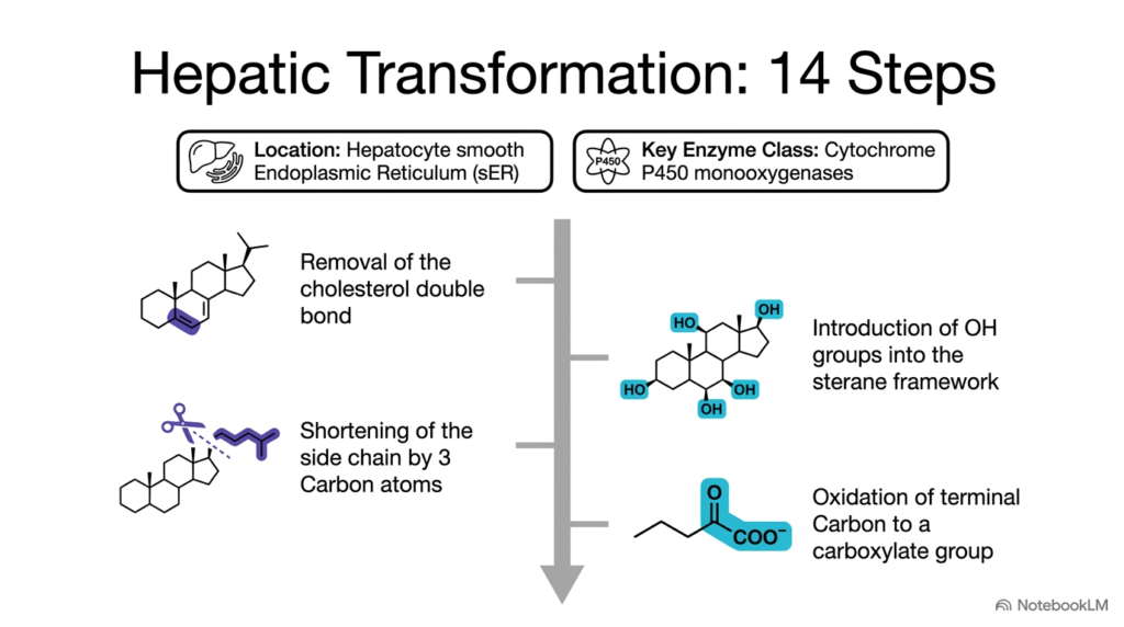
Converting cholesterol into functional molecules requires a complex pathway consisting of fourteen distinct enzymatic steps inside liver cells. This extensive transformation occurs within the smooth endoplasmic reticulum of hepatocytes. A specialized group of enzymes known as cytochrome P450 monooxygenases carries out these oxidation reactions. These enzymes systematically alter the hydrophobic steroid nucleus, adding polar hydroxyl groups while modifying the hydrocarbon side chain to generate amphipathic Bile Acids.
The conversion begins with key chemical modifications to the steroid ring system. First, enzymes remove the double bond located between carbon atoms C-5 and C-6 in the cholesterol molecule. Next, enzymes add hydroxyl groups at specific positions along the sterane framework, primarily at C-7 and C-12. These added oxygen atoms introduce hydrophilic regions, significantly boosting the overall polarity of the molecule so that newly formed Bile Acids can operate in watery digestive fluids.
At the same time, liver enzymes alter the hydrocarbon side chain attached to carbon C-17. The enzymatic machinery shortens this apolar tail by three carbon atoms through a series of cleavage reactions. After this shortening, the terminal carbon atom is oxidized to form a carboxylate group. This newly added carboxyl group carries a negative charge at physiological pH, dramatically increasing water solubility and completing the baseline structure of primary Bile Acids.
This multi-step hepatic process demonstrates remarkable precision in molecular engineering. By systematically altering double bonds, adding hydroxyl groups, and oxidizing the side chain, hepatocytes convert an insoluble sterol into versatile metabolic tools. Each enzymatic step moves the molecule closer to its functional role, ensuring proper formation of bile acids needed for human digestion.
Slide 4: Geometric Shift from Trans to Cis in Bile Acids
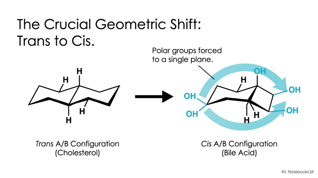
Beyond adding polar chemical groups, the liver alters the spatial geometry of the steroid ring system during synthesis. In precursor cholesterol, the junction between ring A and ring B exists in a trans configuration. This planar arrangement creates a relatively flat, rigid steroid skeleton where polar and nonpolar regions are distributed evenly along the molecule. To convert this flat structure into efficient digestive agents, liver enzymes convert this planar layout into a bent configuration found in Bile Acids.
Enzymatic reduction of the C-5 double bond alters the spatial orientation of the A/B ring junction from trans to cis. This single stereochemical change forces ring A to fold downward relative to the rest of the steroid nucleus. Consequently, the molecule loses its flat shape and adopts a distinct bucket-like or curved conformation. This structural shift reorganizes how functional groups are oriented across the surface of synthesized Bile Acids.
Because of this cis orientation, all newly added hydroxyl groups on rings A, B, and C are forced onto the same concave face of the molecule. Meanwhile, hydrophobic methyl groups and ring hydrogens project outward from the opposite convex face. This spatial alignment creates a clear separation between polar and nonpolar surfaces, a defining physical property that allows functional Bile Acids to act as detergent molecules.
This geometric shift highlights the critical relationship between molecular shape and biological function. Without the trans-to-cis conversion, polar hydroxyl groups would remain scattered on opposing sides of the ring system. By forcing all hydrophilic groups onto a single plane, hepatocytes construct amphipathic structures optimized for lipid emulsification in the digestive tract.
Slide 5: The Amphipathic Disc Structure of Bile Acids
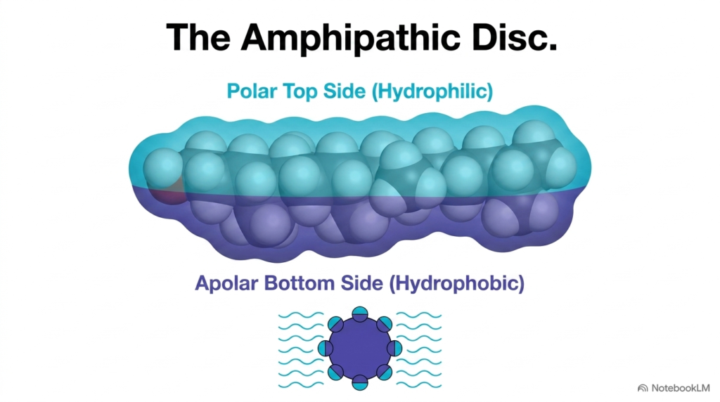
The cis ring junction produces a unique amphipathic disc structure. Rather than having a polar head and nonpolar tail like classic phospholipids, these molecules possess two distinct physical faces. The concave top face contains multiple hydroxyl groups and the terminal carboxylate group, creating a polar hydrophilic surface. Conversely, the convex bottom face consists exclusively of the hydrophobic hydrocarbon sterane skeleton, forming an apolar surface on Bile Acids.
This planar polarity allows these amphipathic molecules to function as powerful facial detergents in aqueous intestinal fluids. When mixed with dietary lipids, the apolar bottom face interacts directly with hydrophobic triglyceride droplets, while the polar top face interacts with surrounding water molecules. This orientation reduces surface tension at the lipid-water interface, breaking large lipid drops into smaller droplets through emulsification, a process facilitated by Bile Acids.
As emulsification proceeds, these specialized structures arrange themselves around lipid fragments to form mixed micelles. In these microscopic aggregates, hydrophobic lipid components remain trapped inside the core, shielded by the polar surfaces facing outward toward the aqueous intestinal contents. This arrangement allows pancreatic lipases to access and digest dietary fats efficiently. Without these organized discs, fat digestion would stall because lipids are poorly soluble in water.
The amphipathic disc geometry represents a remarkable biochemical solution to lipid transport in fluid environments. By separating polar and nonpolar properties onto opposite sides of the steroid nucleus, hepatocytes create optimal tools for nutrient handling. These physical mechanisms highlight how bile acid structure directly enables efficient fat digestion and absorption throughout the intestinal tract.
Slide 6: Primary Bile Acids Formed in the Liver
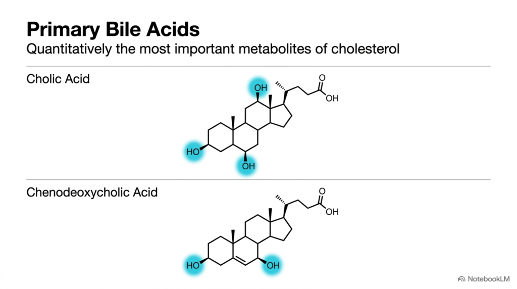
Hepatocytes directly synthesize two major molecules known as primary bile acids, which represent the quantitatively most important metabolites of cholesterol. The most abundant of these synthesized compounds is cholic acid. Cholic acid features three hydroxyl groups positioned at carbon atoms C-3, C-7, and C-12 along the steroid skeleton. This trihydroxy structure gives cholic acid high polarity and excellent water solubility, making it an effective detergent for emulsifying dietary lipids in intestinal fluids.
The second primary compound synthesized by the liver is chenodeoxycholic acid. Unlike cholic acid, chenodeoxycholic acid contains only two hydroxyl groups, located at carbon atoms C-3 and C-7. Lacking a hydroxyl group at C-12, this dihydroxy compound is slightly less polar than cholic acid. Nevertheless, both compounds serve as essential components of human bile, working together to maintain lipid solubility during digestion as primary Bile Acids.
Liver enzymes carefully regulate the production ratio of cholic acid and chenodeoxycholic acid according to metabolic needs. The enzyme 12-alpha-hydroxylase determines whether the pathway yields the trihydroxy or dihydroxy variant. Both primary molecules undergo further chemical modifications before secretion into the biliary tree, ensuring they remain fully dissolved in digestive secretions as active Bile Acids.
Primary products synthesized from cholesterol fulfill essential homeostatic functions in human physiology. By converting excess cholesterol into cholic acid and chenodeoxycholic acid, the liver prevents toxic lipid accumulation in tissues while manufacturing necessary digestive fluid components. These synthesized primary Bile Acids establish the baseline pool required for ongoing fat digestion and metabolic balance.
Slide 7: Activation and Conjugation to Form Bile Salts from Bile Acids
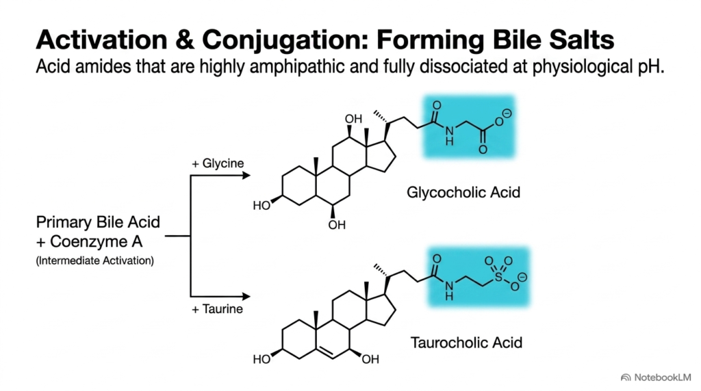
Before primary molecules leave hepatocytes, they undergo activation and conjugation that transforms them into functional bile salts. Unconjugated molecules have a pKa around five, meaning they exist partially in an uncharged, protonated state in acidic intestinal environments. To prevent premature reabsorption across cell membranes, the liver attaches polar amino acids to lower the pKa value, creating fully ionized molecules known as conjugated Bile Acids.
Conjugation begins when liver enzymes activate primary molecules by attaching Coenzyme A to the terminal carboxyl group. Next, specific acyltransferase enzymes link this activated intermediate to either glycine or taurine through an amide bond. Conjugation with glycine yields glycocholic acid, while conjugation with taurine produces taurocholic acid. This enzymatic step drastically lowers the pKa to around two or three for glycine conjugates and below two for taurine conjugates, ensuring complete ionization as Bile Acids.
Because conjugated forms carry a full negative charge at normal intestinal pH, they remain completely ionized as bile salts paired with sodium or potassium cations. This complete ionization makes them impermeable to cell membranes in the upper intestine, keeping them in the intestinal lumen where they can continuously emulsify lipids. This property enhances their stability and detergent efficiency in digestive fluid containing Bile Acids.
The formation of conjugated salts represents a vital adaptation that optimizes digestive function. By appending glycine or taurine, hepatocytes create highly amphipathic molecules that resist passive diffusion until they reach the terminal ileum. This chemical adjustment maximizes lipid emulsification efficiency while ensuring controlled absorption down the intestinal tract.
Slide 8: Hydroxylation Matrix of Primary and Secondary Bile Acids
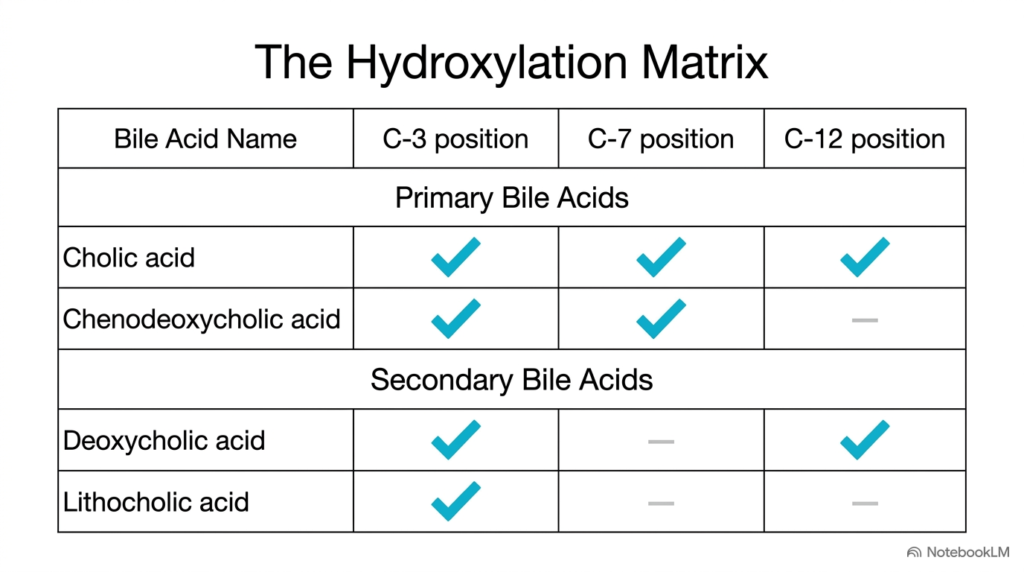
The chemical diversity among digestive steroids can be understood by analyzing their hydroxylation patterns at carbons C-3, C-7, and C-12. A hydroxylation matrix categorizes primary and secondary forms by where oxygen atoms attach to the sterane core. Every major variant retains a hydroxyl group at carbon C-3, inherited directly from precursor cholesterol. However, variations at carbons C-7 and C-12 alter the overall solubility of individual Bile Acids.
Primary forms synthesized by hepatocytes feature hydroxyl groups at multiple positions. Cholic acid contains hydroxyl groups at C-3, C-7, and C-12, making it the most polar variant in the matrix. Chenodeoxycholic acid possesses hydroxyl groups at C-3 and C-7, lacking hydroxyl modification at carbon C-12. These specific oxygen additions create the hydrophilic surfaces required for primary Bile Acids to perform emulsification.
Secondary forms generated by bacterial modifications in the intestine show reduced hydroxylation. Intestinal microflora remove the C-7 hydroxyl group from primary structures through dehydroxylation reactions. Consequently, deoxycholic acid retains hydroxyl groups at C-3 and C-12, while lithocholic acid retains only a single hydroxyl group at C-3. This loss of oxygen atoms reduces overall polarity in secondary Bile Acids.
Comparing these structures in a matrix illustrates how subtle atomic changes alter physiological behavior. Molecules with more hydroxyl groups display higher water solubility and detergent efficiency. Conversely, molecules with fewer hydroxyl groups are less polar and can be membrane-disruptive if they accumulate. Hepatocytes and gut microbes continually modulate this matrix to maintain metabolic balance throughout the digestive system.
Slide 9: Regulation of Synthesis by Feedback on Bile Acids
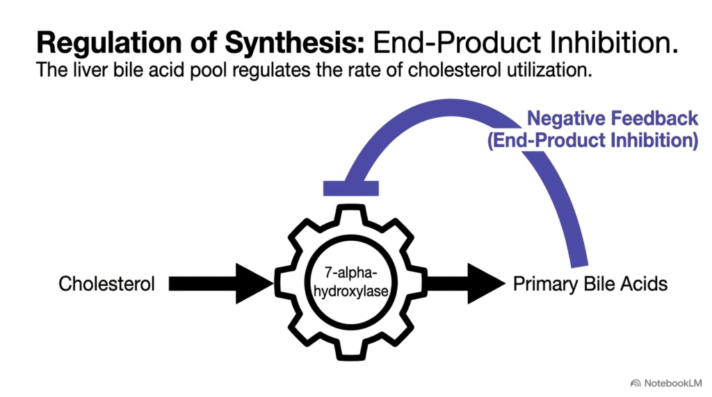
Maintaining appropriate pool sizes requires precise homeostatic regulation of liver synthesis pathways. The rate-limiting enzyme that converts cholesterol into primary bile acids is cholesterol 7-alpha-hydroxylase, encoded by the CYP7A1 gene. This microsomal enzyme catalyzes the addition of a hydroxyl group at carbon C-7 of the cholesterol ring. Because this step dictates overall pathway throughput, controlling 7-alpha-hydroxylase activity regulates production rates of all Bile Acids.
Regulation occurs through an elegant end-product negative feedback mechanism that prevents overproduction. When portal blood returns an abundant supply of these compounds to the liver, they bind to nuclear receptors inside hepatocytes, specifically the Farnesoid X Receptor. Receptor activation suppresses transcription of the 7-alpha-hydroxylase gene, slowing cholesterol conversion when returning bile acid levels are high.
Conversely, if intestinal absorption decreases or biliary loss increases, reduced portal return relieves this transcriptional repression. Lower intrahepatic concentrations upregulate 7-alpha-hydroxylase expression. This enzymatic boost accelerates the conversion of liver cholesterol into new primary molecules, restoring pool size. This feedback loop links daily cholesterol utilization directly to circulating bile acid levels.
This sensitive feedback system protects hepatocytes from accumulating excessive concentrations of amphipathic detergent molecules, which could damage cellular membranes. Furthermore, it regulates systemic cholesterol balance by coordinating cholesterol conversion with physiological demand. By continuously sensing returning concentrations, the liver precisely tunes synthesis rates to match daily metabolic needs, demonstrating tight homeostatic regulation of synthesized Bile Acids.
Slide 10: Intestinal Modifications Generating Secondary Bile Acids
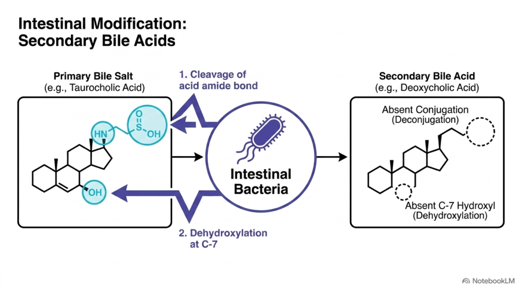
After conjugated bile salts perform their emulsification duties in the upper small intestine, they travel down into the distal ileum and colon. Here, they encounter dense populations of anaerobic intestinal bacteria. These gut microbes have specialized enzymes that chemically modify primary bile acids, transforming them into secondary variants. Through two main enzymatic steps, bacterial action alters the polarity and absorption characteristics of circulating Bile Acids.
The first bacterial modification involves deconjugation, catalyzed by bacterial bile salt hydrolases. These microbial enzymes cleave the amide bond connecting glycine or taurine to the steroid nucleus. Removing these amino acid conjugates releases free primary compounds and restores a higher pKa value. Deconjugation reduces water solubility and prepares the steroid skeleton for subsequent bacterial modifications that generate modified Bile Acids.
The second bacterial modification is 7-alpha-dehydroxylation, carried out by microbial 7-alpha-dehydroxylase enzymes. This reaction removes the hydroxyl group attached to carbon C-7 on the steroid nucleus. Converting cholic acid through this pathway produces deoxycholic acid, whereas converting chenodeoxycholic acid yields lithocholic acid. These resulting secondary structures are significantly less polar and more lipophilic than their parent compounds, altering the overall pool of circulating Bile Acids.
Intestinal microbial modifications highlight the dynamic metabolic partnership between human host biochemistry and the gut microbiome. While deoxycholic acid is efficiently reabsorbed and returned to the liver, lithocholic acid is largely insoluble and mostly excreted in feces. This bacterial transformation plays a major role in determining which compounds cycle back through portal blood and which are permanently eliminated, shaping the composition of circulating Bile Acids.
Slide 11: The Enterohepatic Circulation of Bile Acids
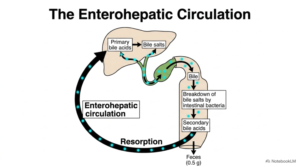
The continuous movement of digestive steroids between the liver and intestines is known as enterohepatic circulation. This recycling pathway begins when hepatocytes secrete bile containing conjugated salts into bile canaliculi. Stored and concentrated within the gallbladder, bile is released into the duodenum following meal ingestion. As bile travels through the intestinal lumen, it facilitates lipid digestion before reaching the terminal ileum, where specialized transporters reabsorb returning Bile Acids.
Reabsorption in the terminal ileum occurs primarily through active transport mediated by the apical sodium-dependent transporter. This highly efficient transport system retrieves roughly ninety-five percent of all secreted molecules from the intestinal lumen. Once inside ileal enterocytes, these reabsorbed compounds cross the basolateral membrane into portal venous blood, traveling directly back toward the liver to preserve the total pool of Bile Acids.
Upon reaching liver sinusoids, hepatocytes extract these returning molecules from portal blood using specialized membrane transporters like sodium-taurocholate cotransporting polypeptide. Liver cells re-conjugate any deconjugated molecules and re-secrete them into bile alongside newly synthesized primary compounds. Meanwhile, the unabsorbed five percent passes into the colon, undergoes bacterial modification, and is excreted in feces, representing the primary pathway for eliminating excess Bile Acids.
Enterohepatic recycling is an exceptionally efficient physiological conservation mechanism in humans. By recycling the existing pool two to three times during a single meal, the body avoids the massive energetic cost of synthesizing fresh molecules from cholesterol from scratch. This continuous circulatory loop maintains high digestive capacity in the intestine while enabling precise metabolic management of circulating Bile Acids.
Slide 12: Mass Circulation Versus Daily Loss of Bile Acids
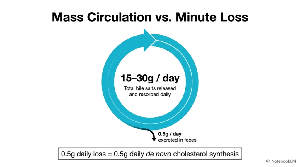
Quantitative analysis of enterohepatic recycling reveals a remarkable contrast between total daily flux and actual fecal loss. The total pool size in an adult human averages around two to four grams. However, because this pool recycles six to ten times daily through the intestine, the liver secretes and reabsorbs an enormous total flux of fifteen to thirty grams per day of circulating Bile Acids.
Despite this massive daily circulation through portal blood, intestinal reabsorption mechanisms are so efficient that only about half a gram (0.5 grams) escapes into feces each day. This tiny daily loss represents only one to two percent of the total circulating flux. Fecal excretion of this unabsorbed fraction constitutes the major physiological pathway by which the human body eliminates excess steroid ring structures, balancing synthesized Bile Acids.
To maintain a stable pool size over time, hepatic synthesis must precisely match this daily fecal excretion. Under normal homeostatic conditions, hepatocytes synthesize exactly 0.5 grams of new primary molecules per day from precursor cholesterol. This de novo synthesis replaces excreted amounts without expanding or shrinking the total pool, linking daily hepatic cholesterol turnover directly to excreted Bile Acids.
Understanding this quantitative balance explains why medical interventions targeting intestinal reabsorption can effectively lower blood cholesterol levels. Pharmacological sequestrant medications bind these molecules within the intestinal lumen, preventing ileal transport and increasing fecal excretion. To compensate for the loss, the liver upregulates conversion pathways and consumes circulating LDL cholesterol, showing how mass circulation controls overall metabolic balance through Bile Acids.
Slide 13: Clinical Disruption and Gallstone Formation Involving Bile Acids
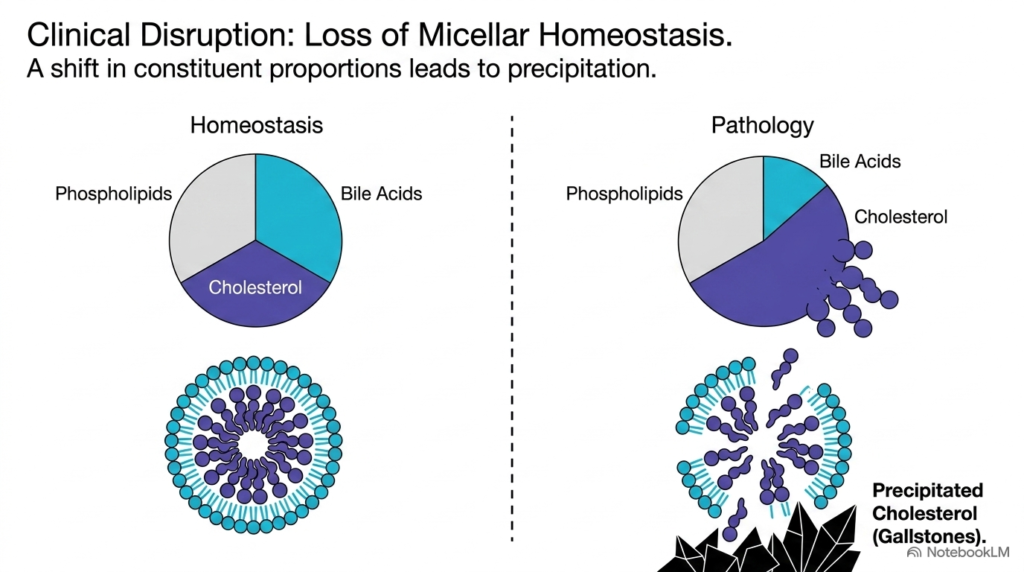
Normal digestive function depends on maintaining precise micellar homeostasis within gallbladder bile. Bile consists of three primary lipid components dissolved in an aqueous fluid: bile salts, phospholipids (predominantly phosphatidylcholine), and cholesterol. Under normal physiological conditions, amphipathic bile salts and phospholipids form stable mixed micelles that solubilize highly hydrophobic cholesterol, keeping it completely dissolved in solution alongside native Bile Acids.
If the relative proportions of these three key lipid constituents shift out of balance, micellar solubilization mechanisms fail. Clinical pathology develops when cholesterol concentration exceeds the solubilizing capacity of available mixed micelles, creating biliary supersaturation. Supersaturation occurs either when hepatocytes hypersecrete cholesterol into bile or when liver synthesis or intestinal reabsorption yields insufficient quantities of protective Bile Acids.
When supersaturation occurs, excess cholesterol can no longer remain trapped inside the hydrophobic cores of mixed micelles. Unsolubilized cholesterol molecules precipitate out of solution and nucleate into solid microcrystals within the gallbladder lumen. Over time, these precipitating crystals aggregate into solid gallstones, causing clinical cholelithiasis. This pathological transition demonstrates how disrupted micellar ratios lead to stone formation from altered Bile Acids.
Restoring proper lipid proportions represents a central clinical goal in preventing and managing gallstone pathology. Understanding the delicate triangular equilibrium among cholesterol, phospholipids, and amphipathic detergent molecules allows healthcare professionals to assess stone-formation risk in patients. By maintaining adequate synthesis, conjugation, and secretion of these specialized detergent molecules, the liver preserves micellar stability and prevents painful clinical disruptions, highlighting the physiological importance of balanced Bile Acids.
Please read our Content Disclaimer Statement.
Check out our social media channels: