118. Hepatic Lipid Metabolism: Balancing Energy Storage, Ketogenesis, and Systemic Homeostasis
Have you ever wondered how your body stays powered when you skip a meal or fast for days? The secret lies inside the liver, our master metabolic furnace. This organ seamlessly shifts gears between storing excess energy from meals and generating alternative fuel sources during hunger. Understanding hepatic lipid metabolism is essential for mastering human physiology and clinical medicine. This slide deck breaks down the core biochemical pathways, from fatty acid and cholesterol synthesis to the step-by-step production of ketone bodies and the clinical dangers of ketoacidosis.
Slide 1: Overview of Hepatic Lipid Metabolism and Systemic Fuel Distribution
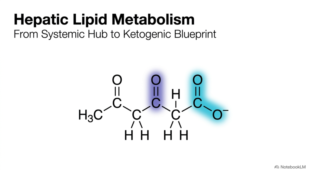
The liver serves as the body’s primary metabolic engine, directing energy distribution and cellular fuel choices across every major organ system. At the center of this organ’s physiological role is hepatic lipid metabolism, which governs how fats are synthesized, stored, packaged, and broken down under constantly changing dietary conditions. As energy availability shifts between well-fed states and periods of hunger, the liver rapidly adjusts its pathways to ensure skeletal muscle, cardiac tissue, and the central nervous system receive continuous metabolic support. Mastering hepatic lipid metabolism provides students with a foundational framework for analyzing systemic homeostasis, fuel selection, and cellular bioenergetics.
This opening slide introduces the structure of ketone bodies, highlighting the molecular configurations that allow lipid-derived energy to travel freely through aqueous blood plasma. During extended periods of carbohydrate restriction, the liver activates mitochondrial enzymes to condense acetyl-CoA units into compact energy carriers. Through hepatic lipid metabolism, bulky, insoluble fatty acids are converted into acetoacetate and 3-hydroxybutyrate, compounds that diffuse easily through the bloodstream without specialized apolipoprotein transport packages or cellular carrier proteins.
By examining these chemical structures, students can appreciate how key functional groups dictate solubility, transport efficiency, and downstream tissue utilization. The structural transformation of lipids into small, water-soluble carboxylates represents a masterclass in bioenergetic flexibility. As we progress through this slide deck, we will trace the complete journey of hepatic lipid metabolism from liver substrate processing to systemic fuel delivery, while highlighting the clinical consequences that arise when these tightly regulated biochemical pathways become overwhelmed.
Slide 2: The Liver as the Primary Hub for Hepatic Lipid Metabolism
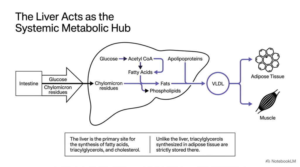
In this second slide, we examine how the liver serves as the body’s central metabolic hub. Central to this anatomical role is hepatic lipid metabolism, which processes incoming dietary nutrients and redistributes them according to systemic demands. Following a meal, dietary glucose and chylomicron remnants enter hepatocytes from the intestinal circulation. The liver processes these substrates through glycolysis to generate acetyl-CoA, which then feeds into de novo lipogenesis. Understanding hepatic lipid metabolism at this level shows how dietary carbohydrates are efficiently converted into stored lipid reserves.
Once hepatocytes synthesize fatty acids, they combine them with glycerol to form triacylglycerols or convert them into structural phospholipids for cell membranes. A defining feature of hepatic lipid metabolism is packaging these neutral fats with specialized apolipoproteins to assemble Very Low-Density Lipoproteins (VLDLs). While adipose tissue synthesizes triacylglycerols strictly for internal storage, the liver actively exports VLDLs into the bloodstream. These circulating lipoprotein particles deliver essential fatty acid cargo to peripheral tissues, including adipose tissue for storage and skeletal muscle for immediate ATP generation through oxidation.
This export mechanism highlights the selfless nature of liver physiology during energy abundance. Rather than hoarding synthesized lipids internally, the organ utilizes hepatic lipid metabolism to sustain extrahepatic tissue function and maintain global energy balance. By continuously monitoring circulating nutrient signals, hepatocytes determine whether to store, convert, or export lipid substrates. This coordinated interplay between gut absorption, hepatic processing, and peripheral delivery forms the baseline framework for studying fuel selection in health and disease.
Slide 3: Endogenous Cholesterol Synthesis and Transport in Hepatic Lipid Metabolism
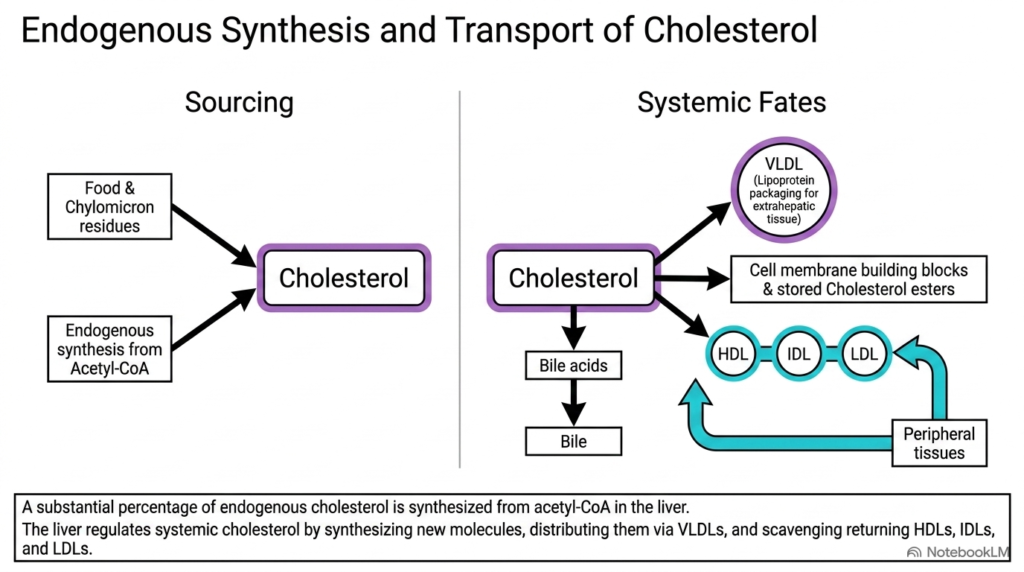
Slide 3 shifts our attention to cholesterol homeostasis, a highly regulated pathway within overall hepatic lipid metabolism. Cholesterol is an essential structural component of cellular membranes and a molecular precursor for steroid hormones, vitamin D, and bile acids. The liver acquires cholesterol through two primary inputs: direct clearance of dietary chylomicron remnants from intestinal absorption and de novo biosynthesis from cytosolic acetyl-CoA. Through tightly regulated hepatic lipid metabolism, hepatocytes continuously balance internal enzymatic synthesis with dietary intake to keep systemic cholesterol levels tightly controlled across varying nutrient conditions.
Once cholesterol enters hepatocytes, it faces several distinct metabolic fates that shape global lipid distribution. A fraction is incorporated directly into liver cell membranes or esterified for intracellular storage, while another major portion is converted into bile salts and secreted into bile to aid fat digestion. Crucially, hepatic lipid metabolism packages cholesterol alongside triacylglycerols into VLDL particles for export. As VLDLs circulate through systemic capillaries, tissues extract neutral fats, converting these particles into intermediate-density lipoproteins and low-density lipoproteins, or LDLs.
To prevent dangerous cholesterol buildup in blood vessel walls, high-density lipoproteins, or HDLs, collect excess cholesterol from peripheral cell membranes and return it to the liver via reverse cholesterol transport. Hepatocytes then clear or excrete this returned cargo through bile, completing a vital systemic loop. This continuous shuttle highlights how hepatic lipid metabolism protects blood vessels while supplying necessary structural lipids. Mastering these lipoprotein pathways enables students to understand the development of atherosclerosis, gallstones, and complex dyslipidemias in clinical practice.
Slide 4: Metabolic State Transitions in Hepatic Lipid Metabolism
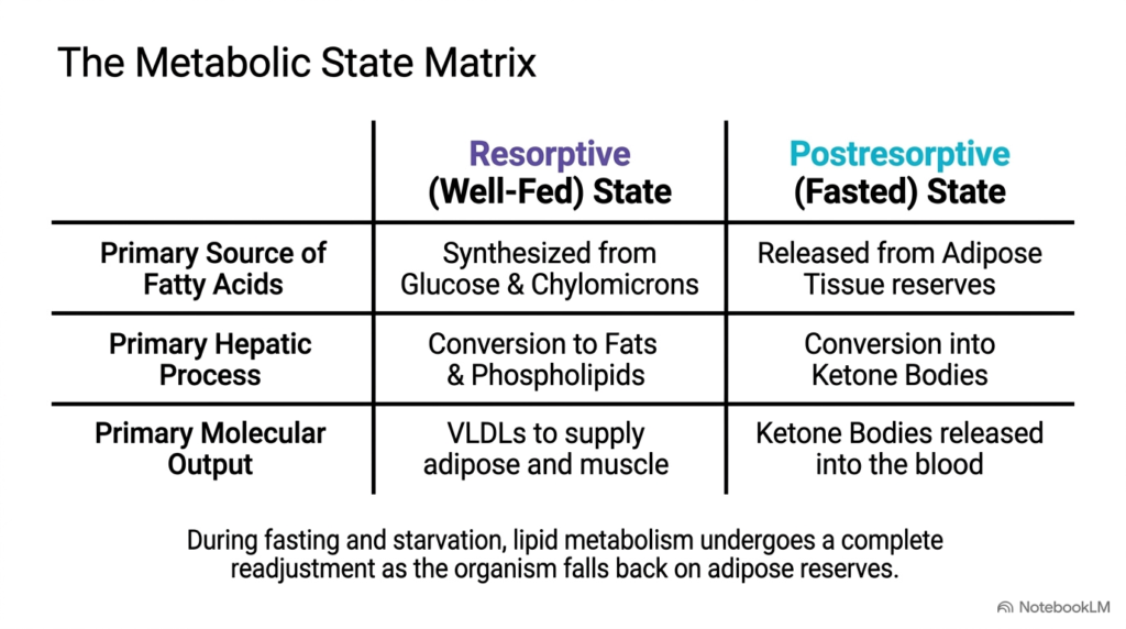
Slide 4 introduces the metabolic state matrix, contrasting how nutritional status radically alters liver function. Physiological adaptation relies entirely on the flexibility of hepatic lipid metabolism, which toggles between fuel storage and fuel release based on endocrine signals like insulin and glucagon. In the resorptive, or well-fed state, elevated blood glucose and abundant dietary chylomicrons drive de novo lipogenesis. The liver converts excess carbohydrates into fatty acids and triacylglycerols, ensuring that surplus energy is safely stored or exported. Studying hepatic lipid metabolism in this nourished state reveals how the body prepares for future fasting intervals.
In stark contrast, the postabsorptive, or fasted state, triggers a total reorganization of liver pathways. As blood glucose levels decline, insulin drops and glucagon rises, signaling adipose tissue to hydrolyze stored triacylglycerols and release free fatty acids into circulation. Through hepatic lipid metabolism, the liver absorbs these circulating fatty acids and shifts its primary internal process toward mitochondrial beta-oxidation. Rather than synthesizing new fats, hepatocytes convert acetyl-CoA into ketone bodies, providing a vital alternative fuel source for extrahepatic tissues during prolonged fasting.
This state-dependent matrix highlights the liver’s operational outputs under opposing metabolic demands. While the well-fed liver releases VLDLs to deliver triacylglycerols to muscle and fat cells, the fasting liver secretes water-soluble ketone bodies into the blood. This functional shift demonstrates how hepatic lipid metabolism preserves plasma glucose for glucose-dependent cells while fueling the rest of the body. Grasping this metabolic transition is crucial for medical students analyzing physiological fasting, ketogenic diets, and uncontrolled diabetes.
Slide 5: Chemical Structures of Ketone Bodies in Hepatic Lipid Metabolism
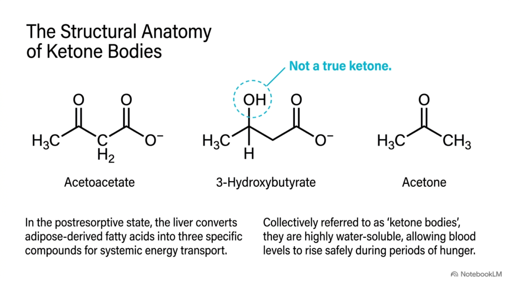
Slide 5 breaks down the exact chemical structures of the three primary molecules collectively referred to as ketone bodies. These compounds are the principal water-soluble end-products of hepatic lipid metabolism during prolonged fasting. When adipose tissue releases free fatty acids into the blood, liver mitochondria process them into acetoacetate, 3-hydroxybutyrate, and acetone. Although these three molecules fall under a single clinical heading, their distinct chemical structures give rise to different functional properties that shape their biological roles. Understanding hepatic lipid metabolism requires a close look at how these molecular structures function as circulating energy carriers.
Acetoacetate is the foundational four-carbon ketoacid produced directly within the hepatic mitochondrial matrix. Through further enzymatic reduction in hepatic lipid metabolism, acetoacetate is converted to 3-hydroxybutyrate. Biochemistry students should note the key chemical detail on this slide: 3-hydroxybutyrate is technically not a true ketone because its carbonyl oxygen is reduced to a hydroxyl group. Nevertheless, it functions as the predominant circulating ketone body in plasma, carrying high-energy electrons as reduced NADH equivalents to power energy-deprived peripheral organs such as the heart and brain.
The third compound, acetone, is a volatile three-carbon byproduct formed through the spontaneous, non-enzymatic decarboxylation of acetoacetate. Unlike its precursor molecules, acetone cannot be metabolized for energy and is exhaled through the lungs, producing the characteristic fruity breath odor seen in ketotic patients. The high water solubility of these carboxylate molecules allows hepatic lipid metabolism to transport massive quantities of lipid-derived fuel through blood plasma without requiring lipoprotein carriers.
Slide 6: Reaction 1 – Acetyl-CoA Condensation in Hepatic Lipid Metabolism
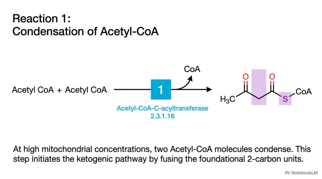
Slide 6 details the inaugural step of ketogenesis, marking the vital transition from general fatty acid breakdown to specialized ketone body synthesis. This enzymatic reaction occurs in the liver mitochondrial matrix, where hepatic lipid metabolism manages very high concentrations of acetyl-CoA generated by intensive beta-oxidation. When acetyl-CoA production exceeds the tricarboxylic acid cycle’s processing capacity, the liver redirects these two-carbon building blocks into ketone synthesis. Understanding hepatic lipid metabolism at this enzymatic junction explains how hepatocytes prevent mitochondrial congestion while recycling essential coenzyme A pools for ongoing fat oxidation.
In Reaction 1, two acetyl-CoA molecules undergo a Claisen-type condensation reaction catalyzed by the enzyme acetyl-CoA-C-acyltransferase, commonly known as thiolase. This enzyme joins two-carbon acetyl groups to form a four-carbon intermediate, acetoacetyl-CoA, while releasing one molecule of free coenzyme A. Within hepatic lipid metabolism, this reversible condensation reaction acts as an essential enzymatic gateway. The forward reaction is driven primarily by the sheer abundance of acetyl-CoA substrate accumulating inside liver mitochondria during active fasting, starvation, or carbohydrate restriction.
Thiolase also operates in reverse during the final cleavage step of beta-oxidation. However, under ketogenic conditions in liver tissue, massive substrate accumulation pushes the chemical equilibrium toward acetoacetyl-CoA formation. By joining two acetyl units, hepatic lipid metabolism constructs the four-carbon backbone needed to build circulating ketone bodies. Students should appreciate that this initial condensation step establishes the molecular foundation for the entire pathway, linking intracellular fat breakdown directly to systemic ketone generation.
Slide 7: Reaction 2 – HMG-CoA Synthesis in Hepatic Lipid Metabolism
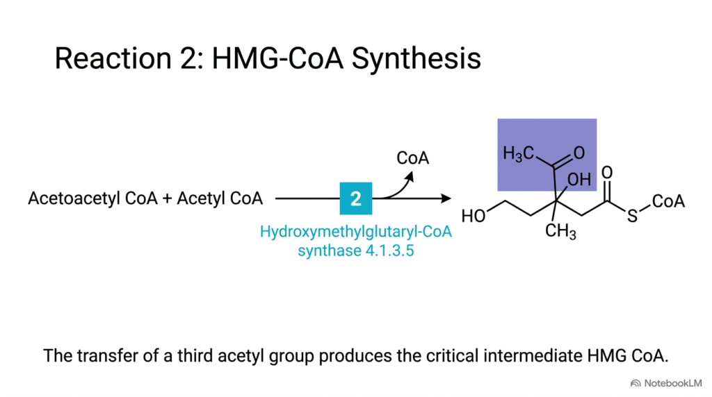
Slide 7 illustrates Reaction 2 of the ketogenic pathway, where a third acetyl group is added to synthesize 3-hydroxy-3-methylglutaryl-CoA, commonly abbreviated as HMG-CoA. This step is a cornerstone of hepatic lipid metabolism, as mitochondrial HMG-CoA serves as the rate-limiting intermediate for ketone body production. In this condensation reaction, the four-carbon acetoacetyl-CoA produced in Reaction 1 condenses with a third acetyl-CoA molecule and water. Understanding hepatic lipid metabolism at this stage highlights how the liver commits carbon skeletons specifically to ketone synthesis during periods of nutrient deprivation.
The reaction is catalyzed by mitochondrial Hydroxymethylglutaryl-CoA synthase, or HMG-CoA synthase. This enzyme attaches the acetyl group to acetoacetyl-CoA, releasing another free coenzyme A molecule and forming the branched six-carbon compound HMG-CoA. In hepatic lipid metabolism, mitochondrial HMG-CoA synthase is the primary rate-limiting enzyme of ketogenesis. Fasting, high circulating fatty acid levels, and glucagon strongly induce its gene expression and enzymatic activity, while insulin suppresses them. This precise hormonal regulation ensures that ketone production occurs only when systemic glucose supplies are scarce.
Biochemistry students must carefully distinguish mitochondrial HMG-CoA synthase from its cytosolic isoenzyme. While the cytosolic enzyme utilizes HMG-CoA for cholesterol biosynthesis, the mitochondrial enzyme channels HMG-CoA exclusively into ketone body synthesis. This spatial compartmentalization represents a fundamental regulatory feature of hepatic lipid metabolism. By physically separating cytosolic cholesterol synthesis from mitochondrial ketone production, liver cells can independently regulate these two major metabolic pathways based on the body’s overarching physiological demands.
Slide 8: Reaction 3 – The Lynen Cycle and Acidosis in Hepatic Lipid Metabolism
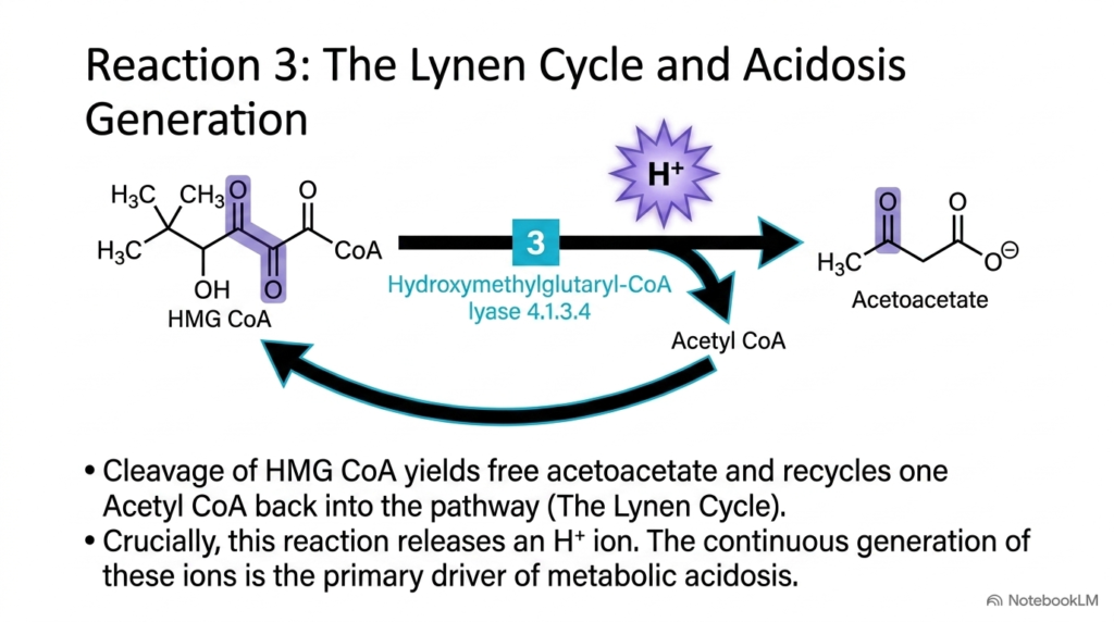
Slide 8 brings us to Reaction 3, the pivotal cleavage step that yields the first free ketone body and establishes the Lynen cycle. Here, hepatic lipid metabolism processes HMG-CoA through the mitochondrial enzyme Hydroxymethylglutaryl-CoA lyase, or HMG-CoA lyase. This cleavage reaction breaks the six-carbon HMG-CoA molecule into a four-carbon acetoacetate molecule and a two-carbon acetyl-CoA molecule. By recycling one acetyl-CoA back into the mitochondrial pool, hepatic lipid metabolism maintains a continuous catalytic loop that permits ongoing fatty acid oxidation without depleting essential coenzyme A reserves.
Beyond generating acetoacetate, Reaction 3 has immense clinical significance because of its chemical byproducts. As HMG-CoA lyase cleaves the substrate, the reaction releases a free hydrogen ion, or proton, into the mitochondrial matrix. In hepatic lipid metabolism, steady acetoacetate synthesis is stoichiometrically coupled to proton generation. At normal physiological rates, physiological blood buffering systems easily neutralize these free protons. However, when ketone production accelerates drastically during prolonged fasting or insulin deficiency, proton accumulation rapidly overwhelms plasma bicarbonate buffers, driving down systemic arterial blood pH.
This continuous proton release explains the direct biochemical mechanism linking excessive ketone synthesis to life-threatening metabolic acidosis. Medical students should appreciate how hepatic lipid metabolism shifts from a benevolent physiological adaptation into a dangerous clinical condition when ketone production becomes uncontrolled. Cleavage of HMG-CoA via the Lynen cycle shows how an essential energy pathway can cause severe acid-base disturbances. Mastering this proton release mechanism is fundamental for diagnosing and treating diabetic ketoacidosis and starvation ketoacidosis in clinical practice.
Slide 9: Reactions 4 and 5 – Fates of Acetoacetate in Hepatic Lipid Metabolism
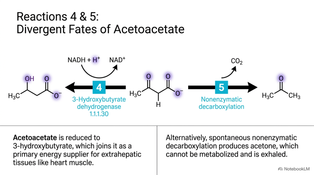
Slide 9 illustrates Reactions 4 and 5, detailing the two divergent biochemical fates of newly synthesized acetoacetate. Once acetoacetate forms inside liver mitochondria, hepatic lipid metabolism processes it through two distinct pathways, depending on local redox state and chemical kinetics. In Reaction 4, acetoacetate undergoes reversible reduction to 3-hydroxybutyrate, catalyzed by 3-hydroxybutyrate dehydrogenase. Understanding hepatic lipid metabolism at this enzymatic step highlights how mitochondrial electron ratios directly influence the composition and energy content of circulating ketone bodies.
Reaction 4 is a reversible, NADH-dependent reduction. When mitochondrial NADH levels rise due to rapid beta-oxidation, the equilibrium strongly favors converting acetoacetate to 3-hydroxybutyrate. Through hepatic lipid metabolism, 3-hydroxybutyrate becomes the predominant circulating ketone body, storing reducing equivalents that yield significant ATP when oxidized in peripheral tissues such as cardiac muscle, renal cortex, and skeletal muscle. The plasma ratio of 3-hydroxybutyrate to acetoacetate reflects the internal mitochondrial NADH-to-NAD+ ratio in hepatocytes and serves as a valuable metabolic biomarker.
Reaction 5 represents a secondary, non-enzymatic pathway where acetoacetate undergoes spontaneous decarboxylation to yield acetone and carbon dioxide. Because human tissues lack the metabolic pathways to convert acetone back into usable acetyl-CoA, it is exhaled through the lungs. Within hepatic lipid metabolism, acetone production increases proportionally alongside rising acetoacetate concentrations. This volatile byproduct causes the classic sweet, fruity breath odor seen in patients with severe ketoacidosis, serving as an immediate diagnostic clue for clinicians evaluating metabolic emergencies in hospital settings.
Slide 10: Extrahepatic Utilization of Ketone Bodies in Hepatic Lipid Metabolism
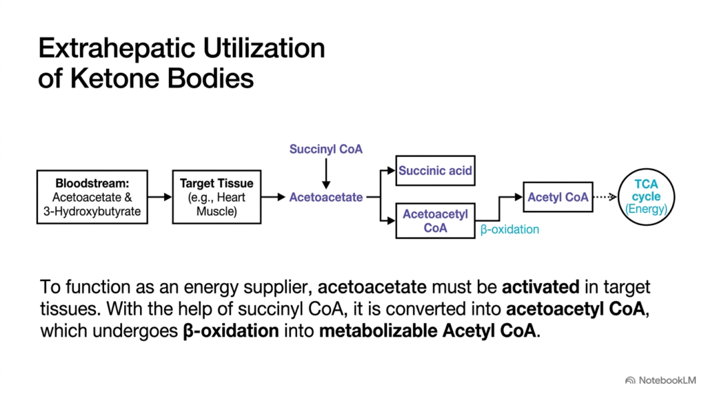
Slide 10 traces the ultimate fate of circulating ketone bodies as extrahepatic tissues consume them for cellular energy production. Although the liver is the sole organ that synthesizes ketone bodies, it cannot use them for its own energy needs. Instead, hepatic lipid metabolism exports acetoacetate and 3-hydroxybutyrate into the bloodstream to power high-demand organs such as cardiac muscle, skeletal muscle, renal cortex, and brain during fasting. Studying hepatic lipid metabolism alongside extrahepatic ketolysis illustrates the cooperative energetic relationship between the liver and peripheral tissues.
Once acetoacetate enters a target tissue cell, it must undergo enzymatic activation before its carbon skeleton can enter mitochondrial energy pathways. Activation requires the enzyme succinyl-CoA:3-ketoacid CoA-transferase, commonly known as thiophorase. Thiophorase transfers a coenzyme A group from succinyl-CoA to acetoacetate, generating acetoacetyl-CoA and succinate. Within hepatic lipid metabolism, hepatocytes completely lack thiophorase enzyme activity. This intentional physiological deficiency prevents the liver from consuming the ketone bodies it produces, ensuring that all exported ketone fuels remain available for extrahepatic tissue survival.
Following activation, acetoacetyl-CoA is cleaved by thiolase into two acetyl-CoA molecules, which enter the citric acid cycle to generate ATP through oxidative phosphorylation. When 3-hydroxybutyrate is utilized, it is first oxidized back to acetoacetate, generating NADH for additional ATP synthesis. Through this coordinated inter-organ axis, hepatic lipid metabolism transforms stored adipose fats into a universal, glucose-sparing fuel source. Understanding extrahepatic ketolysis enables students to appreciate how peripheral organs adapt to prolonged carbohydrate deprivation without compromising metabolic performance.
Slide 11: The Pathological Cascade of Ketoacidosis in Hepatic Lipid Metabolism
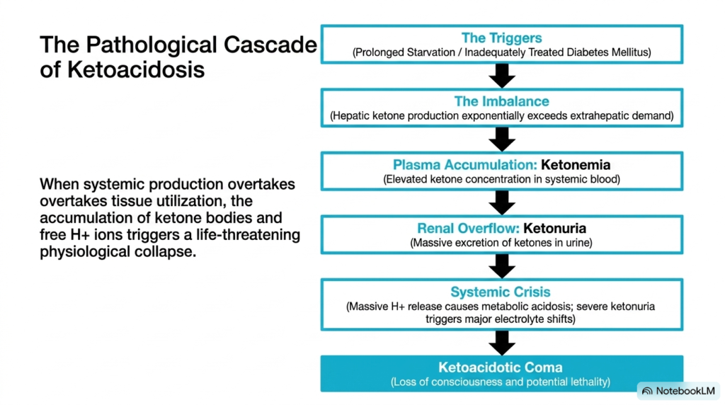
Slide 11 details the clinical cascade of ketoacidosis, illustrating what happens when physiological ketone production transitions into an uncontrolled pathological state. Under normal fasting conditions, hepatic lipid metabolism produces ketone bodies at rates that match extrahepatic energy consumption. However, clinical triggers such as type 1 diabetes mellitus or prolonged starvation break this homeostatic equilibrium. In uncontrolled diabetes, absolute insulin deficiency combined with uninhibited glucagon signaling accelerates adipose lipolysis. Understanding hepatic lipid metabolism under these conditions reveals how massive substrate overload floods the liver with free fatty acids.
This massive fatty acid delivery forces liver mitochondria into hyperactive beta-oxidation, driving exponential ketone production that far exceeds peripheral tissues’ oxidative capacity. As ketone bodies overwhelm peripheral clearance mechanisms, they accumulate in the bloodstream, producing ketonemia. Because ketone bodies are small organic acids, their rapid accumulation floods plasma with acetoacetate, 3-hydroxybutyrate, and free protons. Through hepatic lipid metabolism, uncontrolled synthesis elevates blood ketone levels from normal micromolar concentrations into dangerous millimolar ranges, overwhelming the body’s bicarbonate buffering capacity.
As blood levels rise further, the kidney’s reabsorptive threshold for ketones is exceeded, resulting in ketonuria. Excretion of negatively charged ketone anions in urine pulls positively charged cations like sodium and potassium, along with water, causing severe osmotic diuresis, dehydration, and electrolyte imbalances. Coupled with uncompensated metabolic acidosis, this systemic crisis impairs cellular function, leads to altered mental status, and eventually induces ketoacidotic coma or death. This cascade demonstrates why medical students must understand hepatic lipid metabolism to recognize and treat metabolic emergencies effectively.
Slide 12: From Molecular Cleavage to Clinical Crisis in Hepatic Lipid Metabolism
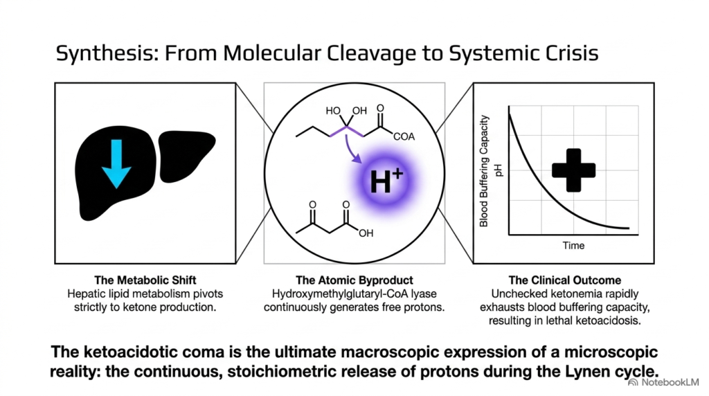
Slide 12 provides a comprehensive synthesis of the slide deck, bridging the gap between microscopic enzymatic cleavage and macroscopic clinical crisis. At the core of this summary is hepatic lipid metabolism, which undergoes a complete functional shift when gluconeogenesis depletes oxaloacetate reserves. Deprived of oxaloacetate, acetyl-CoA produced from intensive fat breakdown cannot enter the citric acid cycle. Consequently, hepatocytes redirect acetyl-CoA into ketogenesis, converting liver mitochondria into specialized ketone factories. Analyzing hepatic lipid metabolism at this systemic level reveals the precise molecular events that precede metabolic collapse.
The critical molecular culprit behind diabetic ketoacidosis is the atomic byproduct generated during Reaction 3 of the Lynen cycle. Every time HMG-CoA lyase cleaves HMG-CoA into acetoacetate and acetyl-CoA, it releases a free proton. Within hepatic lipid metabolism, this stoichiometric release means that for every mole of acetoacetate generated, a mole of hydrogen ions enters the systemic circulation. When hepatic ketone production accelerates uncontrollably, millions of protons flood the bloodstream per second, rapidly depleting plasma bicarbonate buffers and causing arterial pH to plummet below physiological limits.
This precipitous drop in blood pH destabilizes protein structures, impairs myocardial contractility, and disrupts central nervous system function. Ultimately, ketoacidotic coma represents the macroscopic manifestation of an unbuffered microscopic reaction happening continuously inside liver mitochondria. By connecting single-enzyme mechanics to whole-body physiology, studying hepatic lipid metabolism emphasizes why molecular details matter in clinical medicine. Medical and biochemistry students who master these fundamental pathways will be fully equipped to diagnose acid-base disturbances, interpret lab values, and save lives in critical care settings.
Please read our Content Disclaimer Statement.
Check out our social media channels: