117. Carbohydrate Metabolism: Hepatic Glucose Control and Biochemical Pathways
Have you ever wondered how the human body maintains stable energy levels whether a person feasts or fasts? The answer lies in hepatic sugar regulation. The liver functions as a central biochemical hub, managing fuel distribution across every tissue and organ. This slide deck breaks down the essential biochemical pathways that govern glucose balance, alternative hexose assimilation, and systemic inter-organ cooperation. Designed for college and medical students, this guide simplifies complex molecular pathways into intuitive biochemical concepts for advanced learning.
Slide 01: Introduction to Hepatic Carbohydrate Metabolism
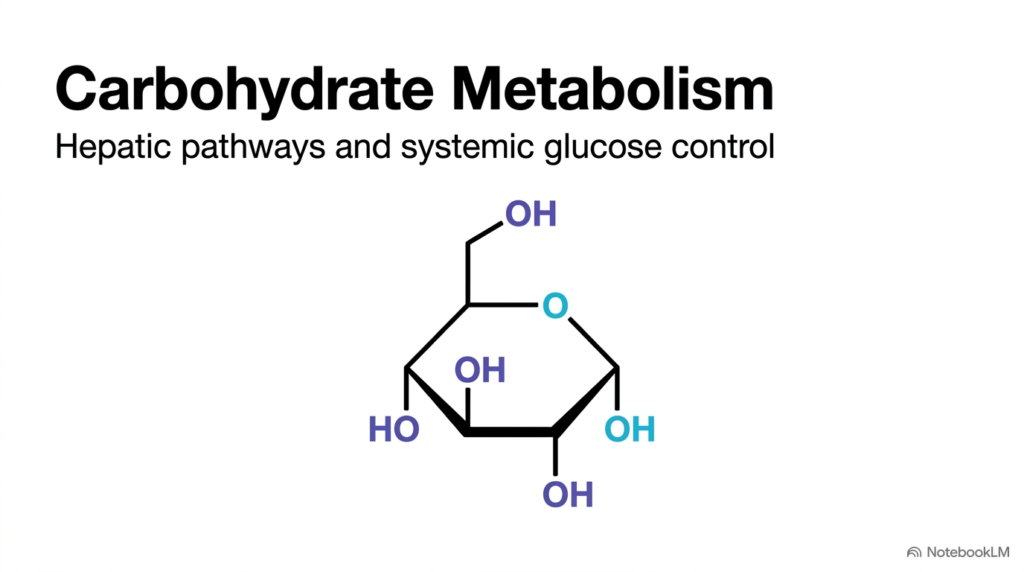
Carbohydrate metabolism is the fundamental chemical engine powering human physiology. At the molecular foundation of this dynamic system sits glucose, shown here in its classic cyclic Haworth projection. Glucose acts as an essential energy source for cellular function across virtually all human tissues. However, circulating blood sugar levels cannot remain unmonitored or fluctuate wildly without causing severe pathological consequences. The human body relies heavily on the liver as a central regulatory organ to manage Carbohydrate Metabolism, ensuring a steady and reliable energy supply during periods of feeding and fasting alike.
Inside hepatocytes, specialized enzymatic pathways process incoming dietary monosaccharides to maintain systemic energy balance. When dietary carbohydrates are abundant after meals, hepatocytes store extra glucose as glycogen or convert it into fatty acids for long-term energy storage. Conversely, when circulating blood glucose levels drop during starvation or vigorous exercise, the liver mobilizes stored glycogen reserves and actively synthesizes new sugar molecules. These tightly regulated biochemical responses prevent dangerous drops in blood sugar that could otherwise severely compromise brain and red blood cell function.
Beyond processing glucose, hepatic Carbohydrate Metabolism integrates other dietary sugars into a unified metabolic pipeline. Fructose derived from fruits and galactose derived from dairy products are funneled directly into central energy pathways within hepatocyte cytoplasm. As an organ-level metabolic clearinghouse, the liver transforms diverse dietary inputs into common, usable cellular energy intermediates. This introductory slide establishes the liver as the master regulator of systemic fuel distribution, setting the foundation for exploring specific metabolic pathways, enzymatic mechanisms, and inter-organ cycles.
Slide 02: Systemic Control and Transporters in Carbohydrate Metabolism
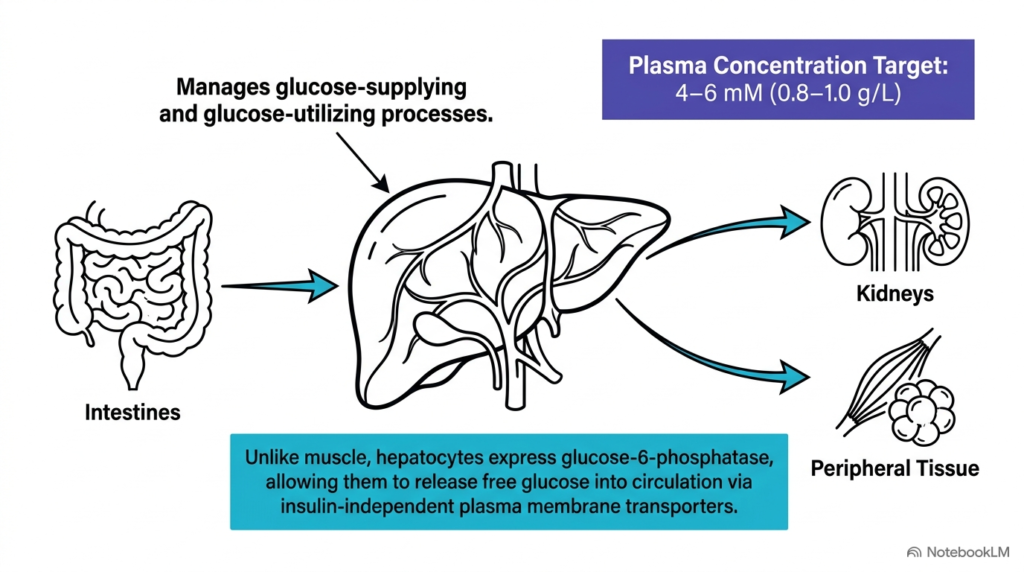
Maintaining plasma glucose within the strict physiological target range of 4 to 6 mM, or 0.8 to 1.0 g/L, is a cornerstone objective of human Carbohydrate Metabolism. Following a meal, dietary carbohydrates are absorbed through the intestinal lining into the bloodstream and travel directly to the liver via the hepatic portal vein. The liver acts as an indispensable physiological buffer, balancing glucose-supplying and glucose-utilizing pathways to protect the body against severe postprandial glycemic spikes and dangerous hypoglycemic drops. By managing these nutrient fluxes, the liver maintains constant energy delivery to peripheral tissues throughout the day.
A key molecular feature that distinguishes hepatic Carbohydrate Metabolism from muscle tissue is the abundant expression of the enzyme glucose-6-phosphatase. Skeletal muscle lacks this crucial enzyme, so muscle cells trap phosphorylated glucose internally to fuel localized contraction. In sharp contrast, hepatocytes express abundant glucose-6-phosphatase, which strips the phosphate group from glucose-6-phosphate. This key enzymatic reaction generates free, unphosphorylated glucose molecules that readily exit the hepatocyte to supply distant organs like the kidneys, brain, and peripheral tissues with vital metabolic fuel.
Furthermore, hepatic Carbohydrate Metabolism relies on specialized GLUT2 transporters embedded within the hepatocyte plasma membrane. Unlike the insulin-dependent GLUT4 transporters found in skeletal muscle and adipose tissue, GLUT2 operates independently of circulating insulin levels. GLUT2 allows rapid, bidirectional glucose flux along concentration gradients across the plasma membrane. Consequently, hepatocytes absorb large glucose loads during postprandial states and release glucose efficiently into the circulation during fasting, safeguarding systemic metabolic stability around the clock.
Slide 03: Substrates and Hormonal Control in Gluconeogenesis and Carbohydrate Metabolism
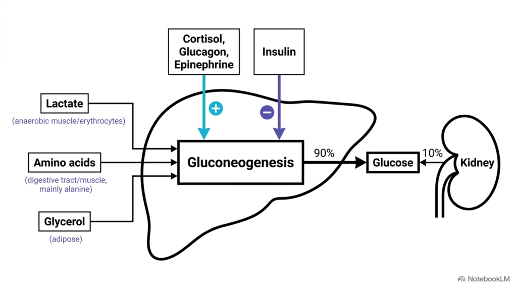
When dietary carbohydrates are unavailable during starvation, the liver uses gluconeogenesis to sustain human Carbohydrate Metabolism. This critical anabolic pathway generates fresh glucose from non-carbohydrate precursors during extended fasting, intense physical exercise, or strict dietary carbohydrate restriction. The principal substrates feeding into hepatic gluconeogenesis include lactate produced by anaerobic glycolysis in skeletal muscle and erythrocytes, glucogenic amino acids like alanine derived from tissue protein breakdown, and glycerol released from adipose tissue triacylglycerols. Together, these peripheral precursors supply the carbon skeletons needed to build new glucose molecules in the liver.
Hormonal signaling tightly coordinates gluconeogenic activity to match real-time physiological demands in carbohydrate metabolism. Stress and fasting hormones, including glucagon, epinephrine, and cortisol, strongly stimulate gluconeogenesis by upregulating key rate-limiting enzymes like phosphoenolpyruvate carboxykinase and fructose-1,6-bisphosphatase. In contrast, insulin acts as a potent metabolic inhibitor, rapidly shutting down hepatic glucose production when blood sugar levels rise after meals. Under normal fasting conditions, the liver performs approximately 90% of total body gluconeogenesis, while the renal cortex contributes the remaining 10% to systemic circulation.
This division of labor underscores the liver’s primary role in maintaining systemic glucose balance. By synthesizing glucose from diverse peripheral metabolic waste products, the liver protects vital glucose-dependent organs during prolonged starvation. Understanding this substrate flow and hormonal control reveals how the body maintains continuous energy production when external food supplies are completely depleted, illustrating the remarkable adaptability and resilience of hepatic Carbohydrate Metabolism across various physiological challenges, environmental conditions, and metabolic stresses.
Slide 04: The Metabolic Blockade in Fatty Acid Use and Carbohydrate Metabolism
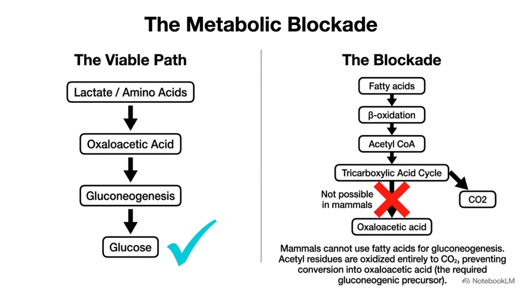
A fascinating structural constraint in mammalian Carbohydrate Metabolism is known as the metabolic blockade. While non-carbohydrate precursors like lactate and glucogenic amino acids readily convert into oxaloacetate to yield new glucose, fatty acids cannot serve as a net source of glucose in mammals. Mammalian tissues break down fatty acids through beta-oxidation to produce acetyl-CoA inside mitochondria. However, converting acetyl-CoA back into oxaloacetate is biochemically impossible in mammalian cells because they lack glyoxylate cycle enzymes such as isocitrate lyase and malate synthase.
Within mammalian Carbohydrate Metabolism, the pyruvate dehydrogenase reaction that converts pyruvate into acetyl-CoA is completely irreversible under physiological conditions. When acetyl-CoA enters the tricarboxylic acid cycle, its two carbon atoms are fully oxidized into two molecules of carbon dioxide before the cycle regenerates oxaloacetate. Because two carbons enter as acetyl-CoA and two carbons leave as carbon dioxide, there is zero net gain of carbon skeletons to fuel gluconeogenic glucose production from fatty acid degradation in mammalian liver tissue during fasting states.
This biochemical limitation means that even extensive adipose fat breakdown cannot supply carbon skeletons for glucose synthesis during extended starvation. Mammals must rely exclusively on glucogenic amino acids, lactate, and glycerol to maintain plasma glucose levels. The metabolic blockade emphasizes the critical physiological need to preserve lean muscle tissue during prolonged fasting, as muscle amino acids represent the largest available reservoir of gluconeogenic carbon in human Carbohydrate Metabolism during extended starvation conditions, ensuring survival when external food resources are completely unavailable for energy production.
Slide 05: Alternative Sugar Assimilation in Hepatic Carbohydrate Metabolism
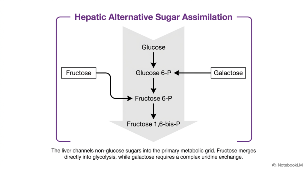
The liver excels at integrating non-glucose sugars into central Carbohydrate Metabolism through alternative sugar assimilation pathways. Human diets supply significant quantities of fructose and galactose alongside standard glucose. Rather than maintaining separate, isolated metabolic machinery for each monosaccharide, the liver channels these alternative hexoses directly into the primary glycolytic pathway. This unified entry strategy maximizes cellular energy extraction while maintaining high metabolic efficiency in hepatocyte cytoplasm, preventing unprocessed dietary sugars from accumulating in systemic circulation after digestion. Consequently, hepatocytes efficiently assimilate complex dietary mixtures containing multiple monosaccharides without cellular stress.
As shown in the pathway diagram, galactose enters hepatic Carbohydrate Metabolism near the top of the glycolytic pathway. Specific hepatic enzymes convert galactose to glucose-6-phosphate, allowing it to merge seamlessly into glycogen synthesis or glycolytic breakdown depending on real-time cellular energy status. In contrast, fructose enters downstream at the level of fructose-6-phosphate or triose phosphate intermediates. By bypassing early regulatory checkpoints like hexokinase and phosphofructokinase-1, fructose rapidly feeds into downstream metabolic cascades within hepatocyte cytoplasm to generate energy or lipid precursors. This structural organization allows the liver to prioritize glucose production while simultaneously processing secondary dietary sugars.
This assimilation framework shows how the liver acts as a highly flexible sugar-processing plant in human Carbohydrate Metabolism. Whether an individual consumes dairy products containing galactose or fruits and sweeteners containing fructose, hepatic Carbohydrate Metabolism transforms these distinct sugars into common phosphorylated intermediates. Understanding these specific entry points provides essential context for analyzing how different dietary sugars influence liver health, lipid synthesis, and overall systemic metabolic regulation across various nutritional states, dietary habits, and clinical conditions in modern nutritional science and biochemistry. Ultimately, this integrated pathway design minimizes metabolic friction and prevents pathological accumulation of unphosphorylated hexoses.
Slide 06: Fructose Pathway and Kinase Steps in Carbohydrate Metabolism
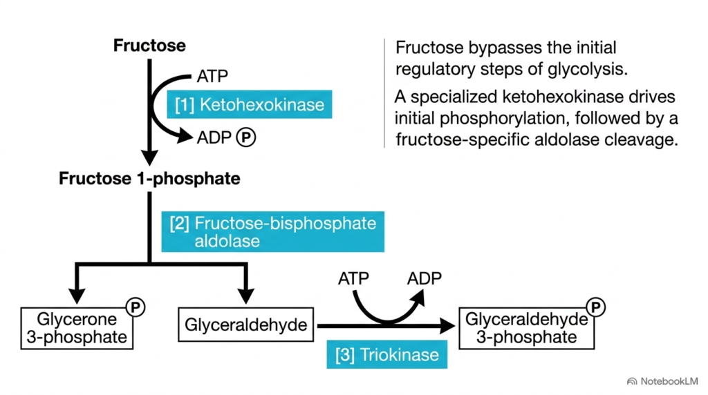
Fructose processing represents a unique metabolic pathway within hepatic Carbohydrate Metabolism that bypasses major regulatory checkpoints. Unlike glucose, which requires hexokinase or glucokinase and is tightly controlled by phosphofructokinase-1, fructose enters glycolysis through a specialized three-step enzymatic cascade inside liver cells. First, the enzyme ketohexokinase phosphorylates fructose using ATP to produce fructose 1-phosphate. This initial phosphorylation traps fructose inside hepatocytes and commits it to rapid hepatic processing, without feedback inhibition from cellular energy levels or intracellular ATP concentrations. This high affinity for fructose ensures that dietary fructose is cleared almost entirely during its first pass through the portal circulation.
Next, a specific isoenzyme called fructose-bisphosphate aldolase cleaves fructose 1-phosphate into two distinct three-carbon molecules: glycerone phosphate, also known as dihydroxyacetone phosphate, and glyceraldehyde. In the final step of this specialized pathway within Carbohydrate Metabolism, the enzyme triokinase phosphorylates glyceraldehyde using ATP to yield glyceraldehyde 3-phosphate. Both resulting triose phosphate intermediates can then directly enter the main glycolytic stream to produce pyruvate and acetyl-CoA within the hepatocyte cytoplasm for energy production. The production of dihydroxyacetone phosphate and glyceraldehyde allows hepatocytes to generate both glycolytic fuel and glycerol backbones for lipid synthesis.
Because ketohexokinase and triokinase bypass the rate-limiting phosphofructokinase-1 checkpoint, fructose metabolism is not restricted by cellular energy charge or high intracellular ATP levels. As a result, high fructose intake floods hepatocytes with massive amounts of triose phosphate intermediates. This unrestrained carbon flux can overwhelm downstream mitochondrial oxidation, driving excess acetyl-CoA into de novo lipogenesis. Consequently, understanding fructose handling in Carbohydrate Metabolism clarifies why high dietary fructose consumption strongly correlates with hepatic steatosis, hypertriglyceridemia, visceral fat accumulation, insulin resistance, and systemic metabolic dysfunction in clinical populations. Therefore, medical students must recognize how unregulated fructose flux alters liver metabolism differently than regulated glucose pathways.
Slide 07: The Alcohol Connection and Redox Balance in Carbohydrate Metabolism
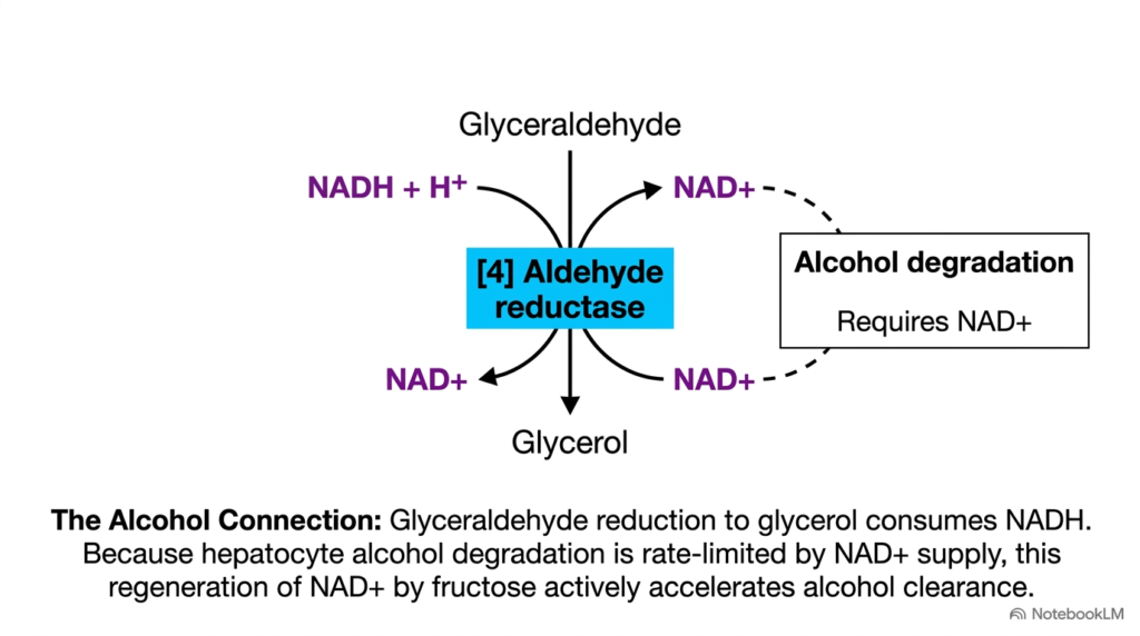
An intriguing biochemical cross-talk exists between fructose handling and ethanol clearance within hepatic Carbohydrate Metabolism. During fructose processing, the intermediate glyceraldehyde is reduced to glycerol by the enzyme aldehyde reductase. This reduction reaction consumes cytosolic NADH and regenerates oxidized NAD+. The resulting increase in available cytosolic NAD+ helps modulate other hepatic oxidation pathways that depend heavily on coenzyme availability for their catalytic cycles in the hepatocyte cytoplasm during active metabolism. This coenzyme regeneration mechanism highlights how metabolic pathways maintain intracellular redox homeostasis during nutrient breakdown.
Ethanol degradation in hepatocytes relies primarily on alcohol dehydrogenase and aldehyde dehydrogenase enzymes, both of which require NAD+ as an essential electron acceptor. Because intracellular NAD+ supply is relatively limited inside liver cells, ethanol clearance is naturally rate-limited by the speed of cytosolic NAD+ regeneration. By driving the enzymatic reduction of glyceraldehyde, fructose processing within Carbohydrate Metabolism rapidly replenishes cytosolic NAD+ pools, thereby accelerating the overall rate of alcohol breakdown and clearance in human liver tissue. Without adequate NAD+ supply, hepatic ethanol oxidation halts, leading to toxic acetaldehyde accumulation in liver cells.
This metabolic connection explains why fructose administration clinically enhances ethanol elimination rates in human subjects. It provides a striking example of how distinct dietary substrates interact through shared coenzyme pools in Carbohydrate Metabolism. By examining these redox interactions, students gain a deeper appreciation for how hepatic pathways operate as an interconnected biochemical network rather than isolated chemical reactions, illustrating the dynamic nature of hepatic Carbohydrate Metabolism, coenzyme recycling, and overall metabolic regulation in health, clinical toxicology, and disease states. Thus, understanding redox coupling in Carbohydrate Metabolism provides valuable insights for managing acute metabolic disturbances in clinical settings.
Slide 08: Galactose Epimerization Pathway in Carbohydrate Metabolism
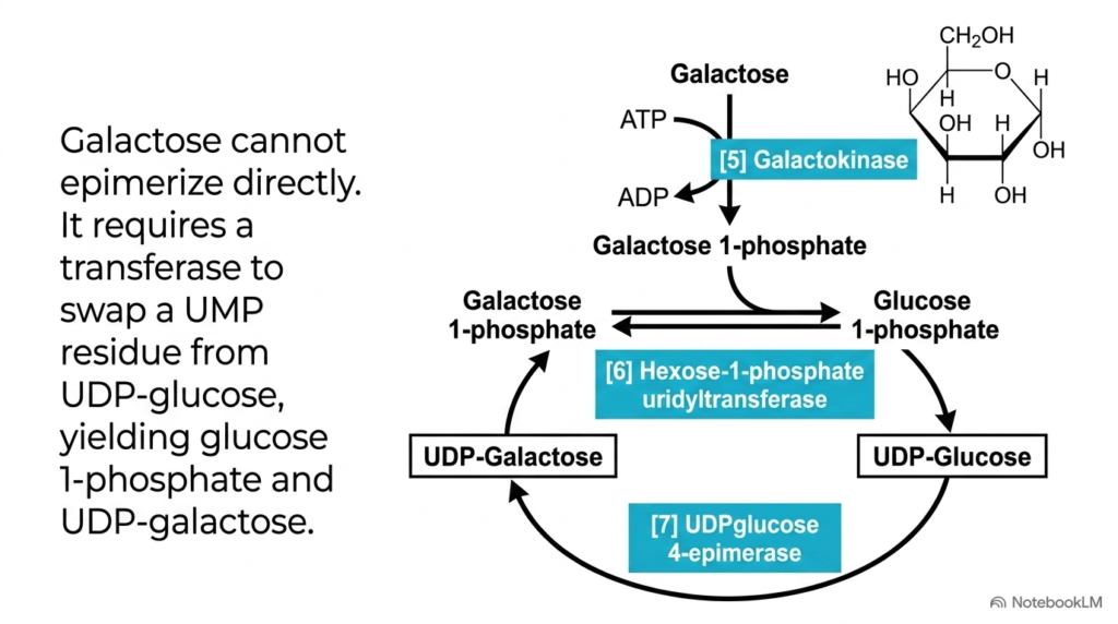
Galactose processing highlights the remarkable stereochemical specificity required in human Carbohydrate Metabolism. Galactose differs from glucose only in the spatial orientation of the hydroxyl group at carbon-4, making them C4 epimers. Because human cells cannot directly epimerize free galactose, hepatocytes use a specialized pathway involving nucleotide-sugar intermediates to convert galactose into a usable form of glucose that can readily enter central energy pathways in the liver cell cytoplasm for cellular use. This mechanism ensures that dietary galactose is rapidly integrated into central energy pathways without disturbing osmotic balance.
The pathway begins when galactokinase uses ATP to phosphorylate galactose, forming galactose 1-phosphate. Next, the crucial enzyme hexose-1-phosphate uridylyltransferase catalyzes a swap reaction. It transfers a uridine monophosphate residue from UDP-glucose onto galactose 1-phosphate, yielding glucose 1-phosphate and UDP-galactose. Finally, UDP-glucose 4-epimerase flips the C4 hydroxyl group of UDP-galactose to regenerate UDP-glucose, maintaining a continuous catalytic cycle within carbohydrate metabolism and sustaining sugar conversion without depleting nucleotide carrier pools required for other vital metabolic processes. The reversible nature of epimerization also allows hepatocytes to synthesize galactose from glucose when dietary intake of dairy products is absent.
Phosphoglucomutase then converts the resulting glucose 1-phosphate to glucose 6-phosphate, allowing it to enter glycolysis or glycogen synthesis directly. This elegant pathway shows how carbohydrate metabolism uses activated nucleotide carriers to perform challenging stereochemical inversions without wasting cellular energy. Through this multi-step exchange mechanism, the liver efficiently converts dietary lactose and milk sugars into central cellular energy reserves during postprandial states in human Carbohydrate Metabolism, ensuring seamless fuel integration for systemic energy needs across the organism. In summary, nucleotide sugar activation represents a sophisticated biochemical strategy for interconverting structural epimers in human cells.
Slide 09: Galactosemia Clinical Correlations in Carbohydrate Metabolism
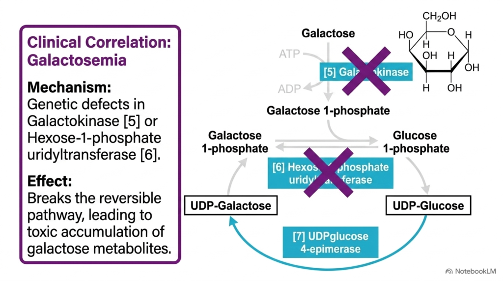
Disruptions in the enzymatic machinery of galactose processing lead to significant clinical disorders in carbohydrate metabolism. Galactosemia is an inherited autosomal recessive condition caused by genetic mutations in key galactose-metabolizing enzymes. As shown in the pathway diagram, defects in galactokinase or hexose-1-phosphate uridylyltransferase disrupt the reversible conversion cycle, preventing hepatocytes from processing dietary galactose normally and leading to toxic metabolite accumulation in tissues and organ systems throughout the body. Genetic defects in these enzymes disrupt cellular energy balance and cause severe metabolic toxicity in neonates after milk feeding.
When hexose-1-phosphate uridylyltransferase is deficient, classic galactosemia develops. In this severe form, galactose-1-phosphate accumulates inside tissues, exerting toxic effects on the liver, kidneys, and central nervous system. Alternatively, when galactose processing is blocked, alternative pathways convert excess galactose into galactitol via the enzyme aldose reductase. Galactitol accumulation in the lens of the eye creates osmotic swelling, rapidly leading to infantile cataract formation in human galactosemia disorders if left untreated. The osmotic gradient generated by galactitol accumulation pulls water into lens fibers, disrupting transparency and causing permanent damage.
Clinical management of galactosemia requires immediate dietary intervention following newborn screening. Infants diagnosed with this metabolic defect must strictly eliminate lactose and galactose from their diet to prevent irreversible liver failure, cataracts, and intellectual disability. This clinical correlation highlights the profound medical importance of understanding Carbohydrate Metabolism, showing how a single inherited enzymatic defect can cause severe multisystemic pathology in patients with carbohydrate metabolism disorders across pediatric clinical settings worldwide. Early diagnosis through routine newborn screening allows immediate dietary modification, enabling affected children to develop normally and stay healthy.
Slide 10: Comparative Analysis of Fructose and Galactose in Carbohydrate Metabolism
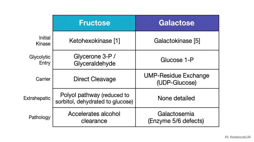
A comparative analysis of fructose and galactose pathways clarifies the diverse strategies employed in hepatic Carbohydrate Metabolism. Although both hexoses are processed primarily by the liver, their enzymatic machinery, entry points, and pathological risks differ significantly. Fructose utilizes ketohexokinase for initial phosphorylation and enters glycolysis at the triose level, whereas galactose relies on galactokinase and merges upstream at glucose-1-phosphate following nucleotide exchange in human Carbohydrate Metabolism across various physiological conditions. These enzymatic differences explain why fructose and galactose cause distinct clinical manifestations when metabolic pathways are overloaded or genetically impaired.
Furthermore, carrier requirements and extrahepatic fates diverge across these sugar pathways in carbohydrate metabolism. Fructose processing involves direct aldolase cleavage without nucleotide carriers and can utilize the polyol pathway in extrahepatic tissues. In contrast, galactose processing strictly requires a UMP-residue exchange via UDP-glucose. Clinically, excess fructose accelerates ethanol clearance but promotes lipogenesis, while defective galactose processing causes toxic metabolite accumulation and classic galactosemia in affected pediatric patients with inborn errors of metabolism. Understanding carrier requirements helps clinicians evaluate why galactosemia presents early in infancy whereas fructose toxicity manifests as long-term metabolic dysfunction.
Comparing these sugar pathways reinforces essential principles of hepatic Carbohydrate Metabolism. While fructose bypasses key glycolytic controls and can drive fatty liver development, galactose depends on careful nucleotide exchange and is highly sensitive to genetic enzyme defects. Mastering these differences helps biochemistry students analyze how dietary sugar composition affects cellular energetics, liver metabolic health, and clinical pathology across diverse patient populations in human Carbohydrate Metabolism, bridging basic science, nutritional biochemistry, and clinical medicine. As a result, comparative biochemistry gives medical students an intuitive framework for diagnosing and managing inherited or diet-induced metabolic disorders.
Slide 11: The Unified Inter-Organ Network in Carbohydrate Metabolism
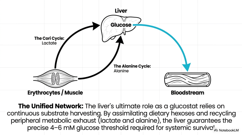
Systemic energy homeostasis relies on an interconnected inter-organ network within human Carbohydrate Metabolism. The liver serves as a central glucostat by continuously harvesting metabolic substrates from peripheral tissues and redistributing glucose into circulation. Two major metabolic cycles demonstrate this inter-organ cooperation: the Cori cycle and the alanine cycle. Both cycles transport carbon skeletons from peripheral tissues back to the liver for gluconeogenic glucose synthesis during various metabolic demands. This inter-organ coordination illustrates how distant tissues cooperate biochemically to maintain whole-body energy balance under varying workload conditions.
In the Cori cycle, red blood cells and exercising skeletal muscle generate lactate during anaerobic glycolysis. This lactate enters the bloodstream, travels to the liver, and undergoes conversion back into glucose via gluconeogenesis. Similarly, the alanine cycle transports amino nitrogen and pyruvate from active muscle to the liver as alanine. The liver transaminates alanine to pyruvate for gluconeogenesis while routing the nitrogen into urea synthesis, reinforcing Carbohydrate Metabolism across multiple tissues, organs, and systemic physiological networks. Furthermore, the alanine cycle allows muscle tissue to safely export toxic amino nitrogen to the liver while preserving carbon skeletons for glucose synthesis.
By assimilating dietary hexoses and recycling peripheral metabolic exhaust, the liver maintains circulating blood glucose within the critical 4 to 6 mM threshold. This continuous inter-organ communication ensures an uninterrupted energy supply to glucose-dependent tissues like the brain. Ultimately, this unified network illustrates the profound integration of human Carbohydrate Metabolism, showing how systemic survival depends on liver-centered metabolic harmony across all physiological states in Carbohydrate Metabolism and inter-organ physiology. In conclusion, inter-organ metabolic cycles demonstrate that systemic energy regulation is a collective effort centered on hepatic biochemical activity.
Please read our Content Disclaimer Statement.
Check out our social media channels: