105. Fundamentals of Human Iron Metabolism: A Biochemical Analysis
Have students ever wondered how a single metal dictates human life and breath? Iron is the unsung hero of human physiology, driving everything from oxygen transport to cellular energy production. This comprehensive slide deck explores the intricate biochemical dance of molecular iron. Its core purpose is to break down the complex mechanisms of human iron handling for medical and college students. It provides a visual and scientific roadmap for understanding structural frameworks, transport pathways, and critical clinical pathologies associated with this essential element.
Slide 1: Molecular Iron Metabolism: Why the Body Depends on a Single Element
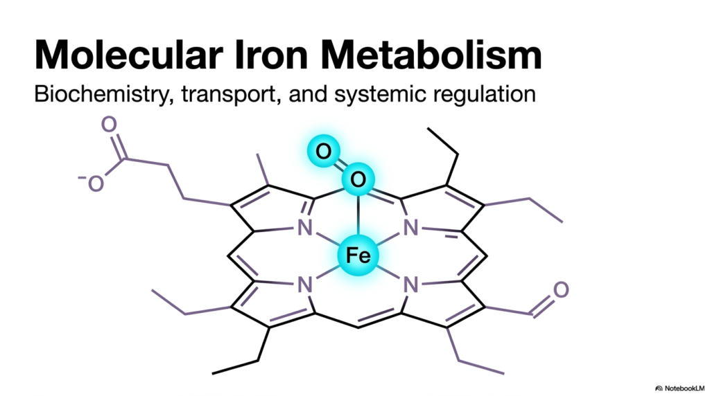
The study of biochemistry often reveals that the most crucial life processes rely on deceptively simple elements. At the heart of human energy and respiration sits a single transition metal, meticulously managed by intricate biological systems. This foundational slide introduces the sweeping concept of molecular Iron Metabolism, setting the stage for understanding its biochemistry, transport, and systemic regulation. A profound grasp of Iron Metabolism is essential for future medical professionals, as it bridges basic molecular structure with macroscopic human health. The body dedicates vast resources to managing this element safely and efficiently.
The illustration highlights the iconic tetrapyrrole ring, the structural cradle that holds molecular iron in biological systems. This specialized organic framework cages the metal, allowing it to safely interact with other molecules without causing uncontrolled oxidative damage. Within the context of Iron Metabolism, this specific arrangement is the cornerstone of life-sustaining oxygen transport. The central iron atom, displayed in its bivalent state, serves as the active docking site for diatomic oxygen. This precise molecular geometry enables reversible binding, allowing oxygen to be picked up in the lungs and released in tissues.
Understanding this central biochemical structure reveals why the body goes to such great lengths to acquire and protect its reserves. Free iron is chemically volatile and dangerous in biological tissues, capable of generating destructive free radicals. Therefore, systemic regulation involves constructing specialized organic cages to safely harness its immense chemical potential. By studying these molecular frameworks, students gain deep insights into the evolutionary elegance of cellular design. The transport and regulation of this metal dictate the very limits of human physical endurance and overall metabolic capacity.
As the presentation unfolds, this foundational molecular structure will appear repeatedly across different physiological contexts. Recognizing how the central metal interacts with its surrounding organic ring is the first step toward mastering complex physiological pathways. The systemic mastery of this element ultimately determines human vitality and survival.
Slide 2: Systemic Distribution in Iron Metabolism: Where the Body Stores Its Treasures
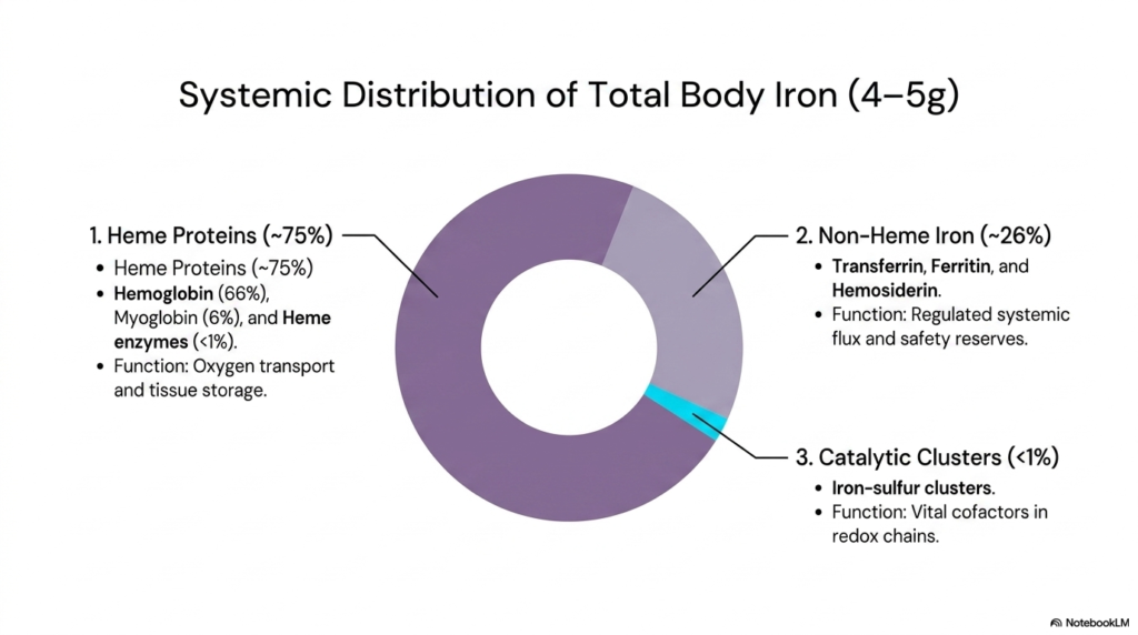
A healthy human adult contains roughly four to five grams of total body iron, a surprisingly small amount for such a critical element. This slide offers a comprehensive macro-level view of how these precious grams are allocated throughout the body. The systemic distribution of iron within Iron Metabolism is highly regulated, ensuring that every tissue receives exactly what it needs without risking toxicity. For students of medicine, visualizing this distribution is critical for understanding where metabolic priorities lie. The body treats this metal like a precious currency, investing it where it yields the highest physiological return.
The vast majority of this element, approximately seventy-five percent, is dedicated to heme proteins. The primary consumer is hemoglobin, which accounts for 66% of total reserves and functions as the vital oxygen transporter in erythrocytes. Myoglobin in muscle tissue accounts for another six percent, while various heme enzymes account for less than one percent. This dominant allocation underscores the absolute necessity of oxygen transport in human Iron Metabolism. The biological imperative to keep tissues oxygenated drives the immense demand for this specific protein family, making it the largest reservoir in the human system.
Beyond the functional heme pool, the body maintains a strategic reserve of non-heme iron, accounting for roughly 26% of the total. This fraction is primarily managed by specialized proteins like transferrin, ferritin, and hemosiderin. These molecules act as a safety buffer, regulating systemic flux and protecting tissues from oxidative stress. This non-heme compartment is a dynamic storage system that expands and contracts in response to dietary intake and physiological demand. A deep understanding of Iron Metabolism requires recognizing how these storage forms are mobilized during periods of nutritional scarcity or acute blood loss.
Finally, less than one percent of total reserves is allocated to catalytic clusters, specifically iron-sulfur complexes. Though tiny in overall mass, this fraction is biochemically irreplaceable. These clusters act as vital cofactors in essential redox chains, driving fundamental energy production at the cellular level.
Slide 3: The Heme Framework in Iron Metabolism: Engineering the Oxygen Carrier
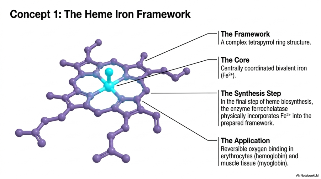
Delving deeper into the molecular architecture, this slide isolates the intricate heme framework, the most vital structural component in human oxygen transport. The structural elegance of this molecule lies in its complex tetrapyrrole ring, a specialized organic scaffold designed specifically to house a single transition metal. In the broader scope of Iron Metabolism, this framework represents the ultimate biochemical achievement in safe and effective molecular transport. Students must recognize this structure as the foundation of respiration, enabling red blood cells to ferry life-sustaining oxygen from the lungs to distant peripheral tissues.
At the center of this ring sits the core: a centrally coordinated bivalent metal atom. This bivalent state is non-negotiable for physiological function, as it possesses the exact electronic configuration required for reversible oxygen binding. The biological synthesis of this structure involves a crucial final assembly step mediated by the enzyme ferrochelatase. This remarkable enzyme physically incorporates the bivalent core into the prepared tetrapyrrole framework. The study of Iron Metabolism places heavy emphasis on this specific enzymatic step, as any genetic or chemical disruption at this step halts the production of functional hemoglobin entirely.
The practical application of this complex organic engineering is immediately evident in human physiology. The primary role is reversible oxygen binding, predominantly performed by hemoglobin in erythrocytes and myoglobin in muscle tissue. The term ‘reversible’ is the key functional concept; the molecule must grip oxygen tightly enough to transport it, but loosely enough to release it into oxygen-deprived tissues. This delicate chemical balance is the defining feature of efficient Iron Metabolism. Without this precise structural calibration, the systemic delivery of oxygen would catastrophically fail, leading to rapid cellular death.
The intricate design of the tetrapyrrole ring prevents the central core from undergoing irreversible oxidation. By shielding the reactive metal, the framework ensures a long functional lifespan for circulating red blood cells. This molecular stability is essential for sustaining the high energy demands of complex organisms.
Slide 4: Iron-Sulfur Clusters in Iron Metabolism: Powering Cellular Energy
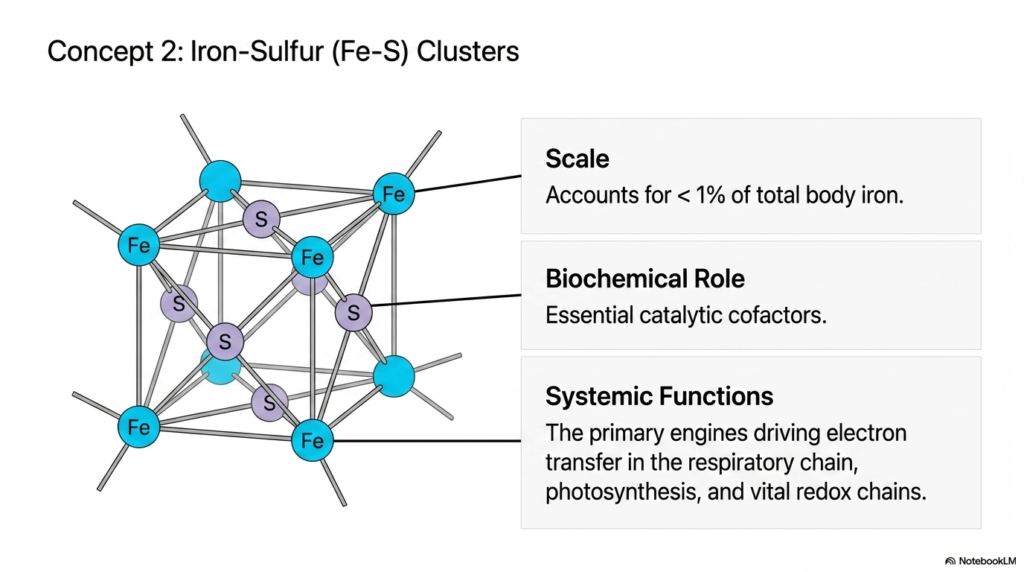
While massive quantities of resources are dedicated to oxygen transport, a microscopic fraction plays an equally vital role deep within cellular mitochondria. This slide shifts focus to iron-sulfur clusters, fascinating structural arrangements that account for less than one percent of total body reserves. Despite their minuscule scale within total Iron Metabolism, their biochemical role is absolutely monumental. These clusters serve as essential catalytic cofactors, driving the fundamental chemical reactions that keep cells alive. For biochemistry students, understanding these minute structures is key to unlocking the secrets of cellular respiration and energy production.
Structurally, these clusters feature multiple metal atoms bridged by sulfur molecules, forming a complex, cube-like lattice. This unique geometric arrangement allows the cluster to rapidly accept and donate electrons. In the grand scheme of Iron Metabolism, these structures act as molecular wires, facilitating rapid electron transfer within proteins. They are the primary engines driving electron transfer in the respiratory chain, the intricate pathway that ultimately synthesizes adenosine triphosphate. Without these specialized clusters rapidly shuttling electrons, the cellular powerhouses would immediately shut down, halting all biological activity.
Beyond human respiration, these clusters share a deep evolutionary history and play vital roles in fundamental processes such as plant photosynthesis and ancient biological redox chains. The systemic functions of these tiny structures highlight the incredible versatility of this transition metal. A thorough mastery of Iron Metabolism demands an appreciation for how a single element can simultaneously carry oxygen in the blood and drive electrical currents inside mitochondria. These diverse applications demonstrate the remarkable efficiency of biological systems in repurposing a single chemical resource.
Medical pathology often overlooks these clusters, yet molecular defects in their assembly lead to severe metabolic diseases. Recognizing their importance broadens the academic perspective beyond simple anemia, highlighting the element’s profound influence on fundamental cellular thermodynamics and systemic survival.
Slide 5: Transferrin and Plasma Transport in Iron Metabolism: The High-Speed Transit System
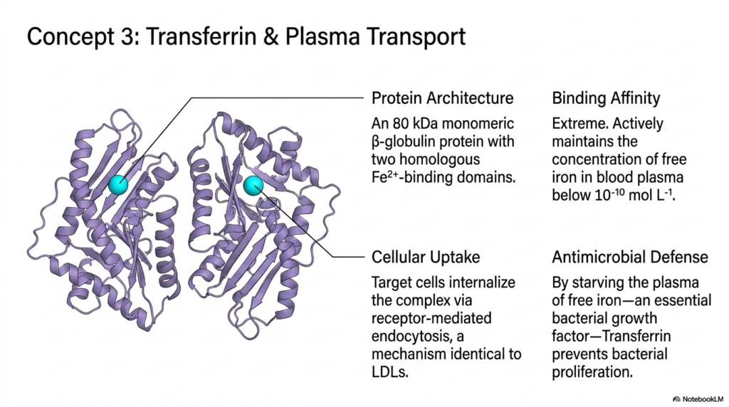
Moving from cellular structures to systemic circulation, the biological narrative shifts to the highly regulated transport mechanisms found in blood plasma. Because free reactive metals are incredibly toxic and insoluble in human blood, the body requires a specialized transit system. This slide details the protein architecture of transferrin, the undisputed primary carrier molecule in Iron Metabolism. Transferrin functions as a dedicated molecular taxi, safely ferrying its volatile cargo through the bloodstream to supply demanding tissues like the bone marrow and liver.
The protein architecture of transferrin is highly specific; it is an eighty kilodalton monomeric beta-globulin protein featuring two homologous binding domains. Its binding affinity for its target is extraordinarily high, actively maintaining the concentration of free reactive metal in blood plasma well below dangerous toxicity thresholds. Proper Iron Metabolism relies on this extreme affinity to prevent uncontrolled systemic oxidative damage. Target cells acquire this precious cargo by internalizing the entire protein complex via receptor-mediated endocytosis. Interestingly, this cellular uptake mechanism is biochemically identical to the mechanism by which cells absorb low-density lipoproteins.
Beyond simple transport, this circulating protein serves a fascinating secondary role in innate immunity and antimicrobial defense. By tightly binding and starving the blood plasma of free reserves, transferrin actively prevents bacterial proliferation. Many pathogenic bacteria require this specific element as an essential growth factor to colonize a host. By hiding the metal within a bulky protein cage, human Iron Metabolism effectively starves invading microbes. This dual function of safe transport and active immunological defense makes transferrin an incredibly elegant biological molecule.
Clinical assessments heavily rely on monitoring this specific transport protein to evaluate nutritional status, overall metabolic health, and systemic disease. Saturation levels provide physicians with a highly accurate, real-time window into circulating systemic reserves, making this vital transport molecule a truly critical biomarker in modern diagnostic medicine and everyday clinical practice.
Slide 6: Ferritin and Intracellular Storage in Iron Metabolism: The Microscopic Vault
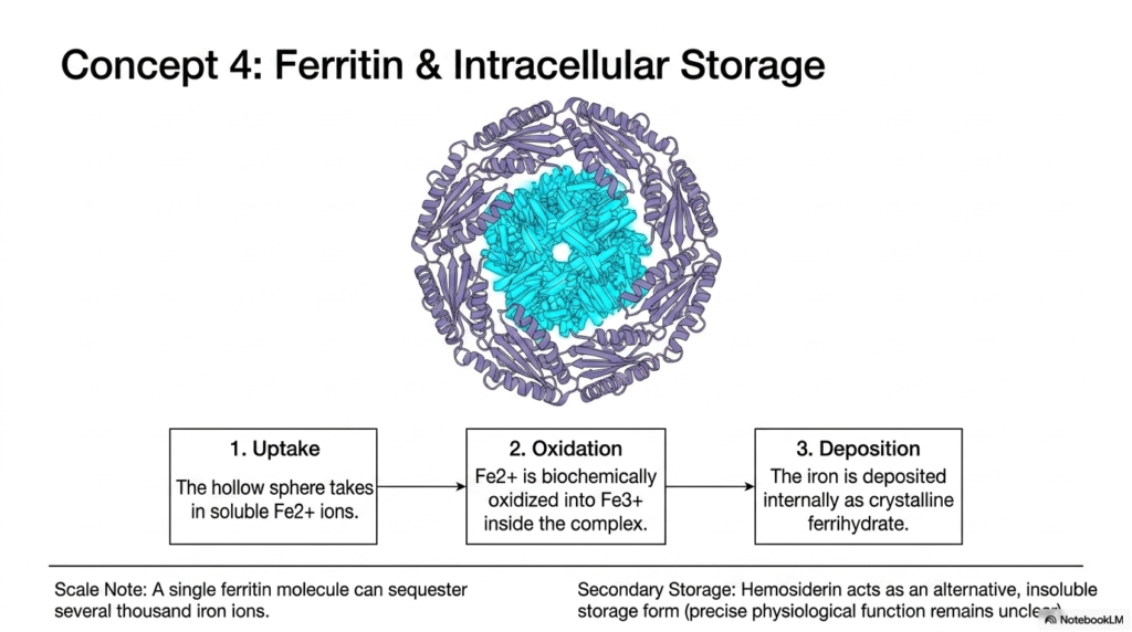
While transferrin facilitates active transport, the body requires a reliable mechanism for long-term intracellular sequestration and storage. This slide introduces ferritin, the primary storage molecule that safeguards cellular integrity against toxic metal accumulation. This massive protein complex functions as a microscopic vault, safely locking away volatile elements until physiological demands rise. A foundational pillar of healthy Iron Metabolism is the ability to store vast reserves without triggering cellular oxidation or widespread tissue damage. Without this storage capacity, the body would constantly fluctuate between lethal toxicity and severe deficiency.
The structural design of ferritin is remarkably elegant, forming a large hollow sphere assembled from multiple protein subunits. This hollow sphere takes in soluble bivalent ions from the surrounding cellular cytoplasm. Once inside, a critical biochemical transformation occurs: the bivalent metal is enzymatically oxidized into a trivalent state inside the complex. This oxidation step is essential for safe Iron Metabolism, as it prepares the metal for dense, stable packing. Following oxidation, the metal is deposited internally as a dense crystalline structure known as ferrihydrate.
The storage capacity of this single-molecule vault is staggering; a single ferritin molecule can sequester several thousand individual atoms. This high-density packing allows cells, particularly in the liver, to safely stockpile massive nutritional reserves. Additionally, the slide notes a secondary storage form called hemosiderin. Hemosiderin acts as an alternative, insoluble storage form within the broader scope of Iron Metabolism, though its precise physiological function remains somewhat unclear. It typically accumulates during states of pathological overload, serving as a biological backup system when primary vaults become overwhelmed.
By utilizing these sophisticated protein vaults, the human biological system maintains a delicate balance, preserving a vital metabolic resource while completely neutralizing its dangerous chemical reactivity. Understanding this sequestration process is fundamental for students analyzing cellular toxicity and nutritional biochemistry.
Slide 7: Intestinal Absorption Mechanics in Iron Metabolism: Crossing the Enterocyte Barrier
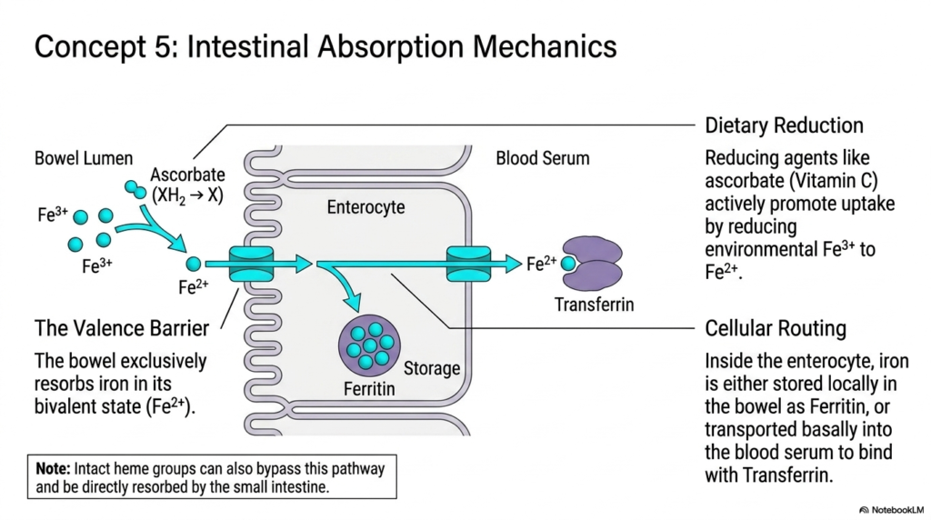
The acquisition of essential trace minerals from the external environment presents a significant biological challenge. This slide examines the complex mechanics of intestinal absorption, the highly regulated gateway that governs how external nutrients enter the systemic circulation. The enterocytes lining the bowel face a difficult task: they must selectively absorb nutrients while excluding harmful toxins. The regulation of this intestinal gateway is the primary control point for entire systemic Iron Metabolism. Because the human body cannot actively excrete this metal, meticulous control at the site of absorption is of biological paramount importance.
The absorption process encounters a strict biochemical hurdle known as the valence barrier. The human bowel almost exclusively absorbs this element in its divalent state, yet most dietary sources present it in its trivalent state. To overcome this, dietary reduction is actively promoted by reducing agents such as ascorbate, commonly known as Vitamin C. Ascorbate functions by chemically reducing environmental trivalent molecules to the absorbable bivalent form. This interaction highlights why dietary pairings are a frequent topic of discussion in Iron Metabolism, as certain vitamins drastically enhance mineral uptake.
Once the reduced mineral crosses the apical membrane into the enterocyte, cellular routing determines its ultimate biological fate. Inside the bowel cell, the mineral is either stored locally within a ferritin vault or transported basally into the bloodstream to bind to transferrin. This routing decision relies on systemic signals that reflect the body’s current nutritional status. A fascinating exception in Iron Metabolism is the intact heme group, which can bypass these traditional valence barriers entirely and be directly resorbed by the small intestine, providing a highly efficient dietary source.
This intricate cellular choreography ensures that the biological system absorbs exactly what it requires to maintain physiological homeostasis. Any disruption at this cellular gateway quickly cascades into significant systemic issues, making it a primary focus for pharmacological interventions.
Slide 8: Erythropoiesis and Macrophage Recycling in Iron Metabolism: The Biological Circular Economy
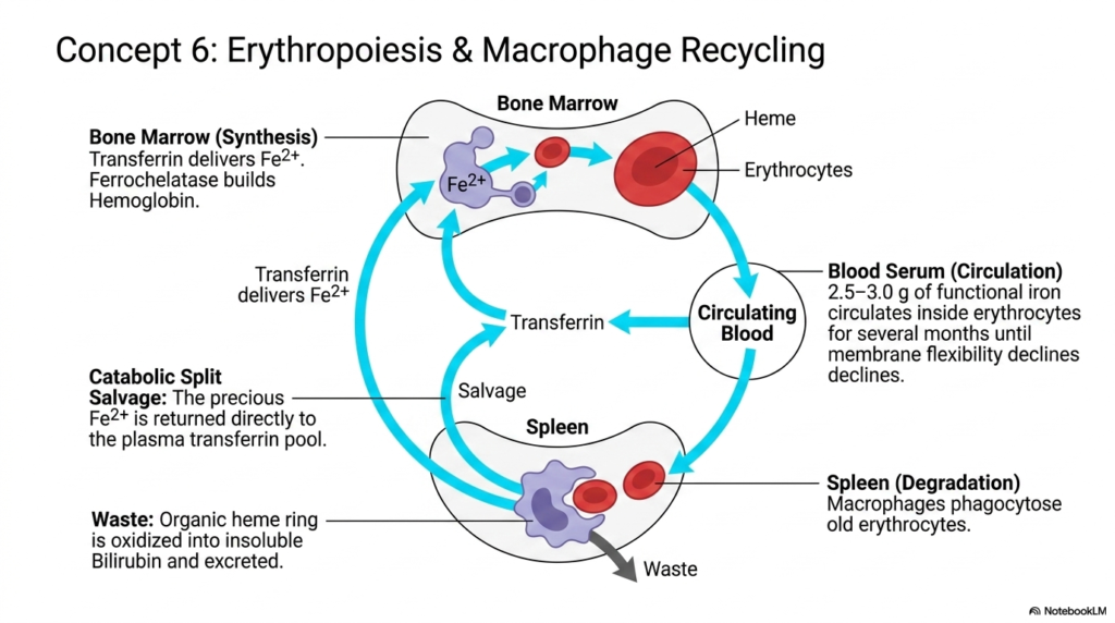
Generating fresh red blood cells requires a massive, continuous supply of biological resources that dietary intake alone simply cannot sustain. This slide illustrates the dynamic processes of erythropoiesis and macrophage recycling, showcasing the remarkable biological circular economy at work. To meet immense systemic demands, Iron Metabolism relies heavily on internal salvaging rather than continuous external acquisition. This highly efficient internal recycling system ensures a steady supply of structural materials for the bone marrow, enabling the continuous daily production of millions of new erythrocytes without depleting bodily reserves.
The cycle begins in the bone marrow, the primary site of biological synthesis. Here, circulating transferrin delivers its precious bivalent cargo, allowing the enzyme ferrochelatase to build fresh hemoglobin for new erythrocytes. These cells then enter the circulating blood serum, where roughly two and a half to three grams of functional metal circulate safely for several months. Eventually, as erythrocyte membrane flexibility declines with age, older cells are targeted for degradation. This continuous turnover is a fundamental feature of healthy Iron Metabolism, constantly refreshing the oxygen transport fleet.
The degradation phase occurs primarily in the spleen, where specialized macrophages phagocytose the senescent erythrocytes. Inside the macrophage, a critical catabolic split occurs, separating cellular waste from precious salvageable materials. The organic tetrapyrrole ring is oxidized into insoluble bilirubin and excreted as physiological waste. However, the precious bivalent metal is aggressively salvaged and returned directly to the plasma transferrin pool. This highly efficient salvage pathway is the backbone of human Iron Metabolism, preventing massive nutritional losses whenever an aging red blood cell is naturally destroyed.
By ruthlessly recycling this vital resource, the human system achieves remarkable physiological independence from constant dietary feeding. Understanding this elegant circular pathway helps medical students appreciate the resilience of human blood production and the spleen’s immense workload.
Slide 9: Systemic Flux in Iron Metabolism: Balancing the Internal Engine
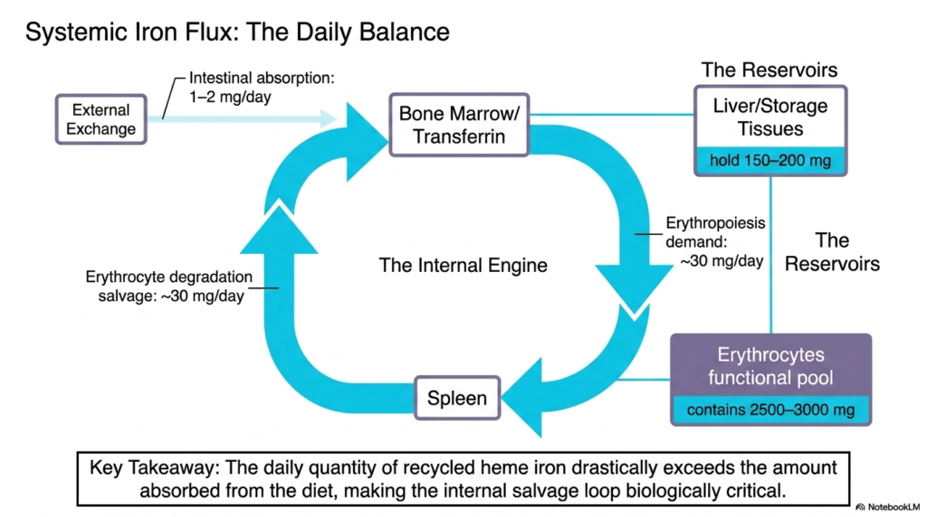
Returning to a macroscopic view, this slide quantifies the daily balance of systemic flux, mapping the precise physiological mathematics of mineral management. Maintaining homeostasis requires a delicate equilibrium between minor external inputs and massive internal biological demands. This numerical overview of Iron Metabolism perfectly illustrates just how heavily the human body relies on its own internal recycling infrastructure. Students can easily visualize the disparity between what is consumed through the diet and what is actually required to keep the internal biological engine running at optimal capacity.
The external exchange pathway provides surprisingly little material, with daily intestinal absorption yielding a mere one to two milligrams. In stark contrast, the internal demand for new erythropoiesis is massive, requiring approximately thirty milligrams every single day. This glaring numerical deficit is resolved entirely by the internal engine of erythrocyte degradation and salvage, which perfectly matches the thirty-milligram daily demand. This mathematical reality dictates that Iron Metabolism is essentially a closed-loop system, in which internal conservation takes absolute precedence over new dietary absorption to sustain standard daily operations.
The systemic reservoirs, primarily located in the liver and specialized storage tissues, hold an accessible backup supply of roughly one hundred and fifty to two hundred milligrams. Meanwhile, the massive functional pool circulating within erythrocytes contains up to three thousand milligrams. The key takeaway regarding overall Iron Metabolism is profound: the daily quantity of recycled material drastically exceeds the amount absorbed from the external environment. This makes the internal salvage loop biologically critical for human survival, completely dwarfing the contribution of a standard daily diet.
Recognizing these physiological mathematics is absolutely essential for correctly diagnosing nutritional deficiencies in a clinical setting. Medical professionals must always remember that a disruption in the internal salvage loop often causes far more rapid systemic collapse than a simple dietary shortage.
Slide 10: Clinical Pathology in Iron Metabolism: When the System Fails
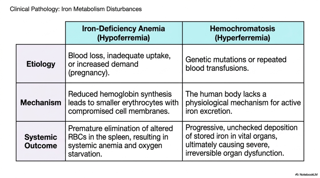
Even the most elegant biological systems occasionally break down, leading to distinct physiological consequences. The final slide shifts focus to clinical pathology, exploring what happens when the delicate balance of these biochemical pathways is fundamentally disrupted. Studying the disturbances in Iron Metabolism is highly practical for medical students, directly bridging basic molecular science with common clinical diagnostics. The presentation contrasts two diametrically opposed pathological states: systemic depletion resulting in hypoferremia, and unchecked toxic accumulation resulting in hyperferremia. Both conditions severely compromise patient health through entirely different biological mechanisms.
The first major disturbance is iron-deficiency anemia, a widespread condition characterized by a severe lack of resources. The etiology typically involves chronic blood loss, inadequate dietary uptake, or an exponentially increased physiological demand, such as during pregnancy. The fundamental mechanism involves reduced hemoglobin synthesis, which leads the body to produce smaller, compromised erythrocytes. Ultimately, the systemic outcome in this branch of Iron Metabolism dysfunction is the premature elimination of these altered cells in the spleen. This results in profound systemic anemia, leaving peripheral tissues suffering from chronic oxygen starvation.
Conversely, hemochromatosis represents a dangerous state of hyperferremia. Its etiology is frequently rooted in inherited genetic mutations or in repeated, frequent medical blood transfusions. The underlying danger lies in a glaring biological flaw: the human body completely lacks a physiological mechanism for active mineral excretion. When Iron Metabolism pathways are overloaded, the body cannot simply vent the excess material. The systemic outcome is the progressive, unchecked deposition of stored ferrihydrate crystals directly into vital organs, ultimately causing severe tissue oxidation and completely irreversible organ dysfunction.
Contrasting these two diseases provides a comprehensive understanding of why precise biochemical regulation is an absolute necessity for sustaining human life. Both extremes perfectly illustrate the catastrophic clinical consequences that arise whenever this vital transition metal escapes its strict biological constraints.
Please read our Content Disclaimer Statement.
Check out our social media channels: