116. Mastering the Buffer Function in Organ Metabolism
How does the human body maintain a steady energy supply despite erratic eating habits? The answer lies in metabolic flexibility. Humans alternate between periods of nutrient abundance and temporary starvation throughout the day. To survive these unpredictable fluctuations, specialized organs dynamically manage metabolic pathways. This slide deck breaks down the biochemical shifts that maintain continuous systemic energy homeostasis and explains how organ interactions buffer nutrient availability.
Slide 1: Overview of the Buffer Function in Organ Metabolism
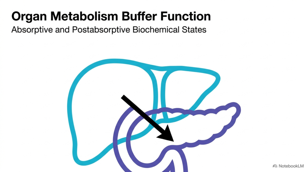
Human survival depends on maintaining a steady, uninterrupted supply of energy substrates to crucial tissues throughout the body. However, nutrient intake through food is inherently irregular and highly variable throughout the day. To bridge the vast physiological gap between intermittent meals and constant cellular energy demands, the human body relies on sophisticated organ coordination. This dynamic regulatory framework serves as a buffer in organ metabolism, continuously balancing periodic nutrient abundance against ongoing energetic deficits across systemic circulation.
In this integrated metabolic architecture, the liver and adipose tissue serve as massive biological buffers for the blood plasma. During periods of nutrient surplus following a meal, these specialized organs actively absorb circulating energy substrates and convert them into dense storage molecules. Conversely, when external nutrient supply halts during fasting, these same tissues mobilize their stored reserves to nourish peripheral organs. Understanding the buffer function in organ metabolism provides students with a unifying framework for human bioenergetics and metabolic integration.
The operational continuum of human metabolism revolves around two primary physiological states: the absorptive state and the postabsorptive state. The absorptive state occurs immediately after food ingestion, when active digestion floods the blood plasma with fresh substrates. In contrast, the postabsorptive state emerges during fasting, requiring internal mobilization of stored energy reserves.
Through these continuous metabolic transitions, organs protect sensitive tissues from metabolic stress. The coordinated action of enzymes, transporters, and regulatory hormones ensures steady energy delivery. Ultimately, the buffer function in organ metabolism ensures that metabolic rate and cellular survival remain completely uncompromised regardless of changing dietary schedules.
Slide 2: Balancing Energy Supply and Demand via the Buffer Function in Organ Metabolism
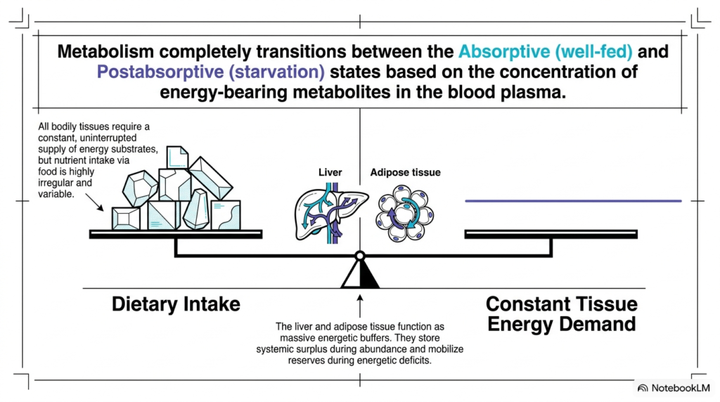
A central challenge in human biochemistry is the stark physiological mismatch between irregular dietary intake and constant cellular energy demand. Every organ system, from the contracting cardiac muscle to the metabolically demanding central nervous system, requires a continuous, uninterrupted flow of chemical energy to sustain vital cellular processes. The buffer function in organ metabolism acts as a biological balance scale, perfectly compensating for timing differences between meal ingestion and cellular fuel consumption.
When digested food enters the gastrointestinal tract, plasma concentrations of energy-bearing metabolites rise dramatically over a short period. Without robust buffering mechanisms, this sudden nutrient influx would trigger severe osmotic disturbances and dangerous hyperglycemia throughout the vascular network. Here, the buffer function in organ metabolism protects the internal circulatory environment by rapidly clearing excess glucose, lipids, and amino acids from the bloodstream and sequestering them into organized intracellular storage pools within designated metabolic organs.
During starvation or extended intervals between meals, the metabolic balance scale tilts in the opposite direction. Plasma metabolite concentrations begin to decrease rapidly, threatening the energetic stability of peripheral tissues that lack significant internal energy reserves. Biochemical pathways must respond instantly to support systemic energy balance across every vascular bed.
To counter this energetic decline, the buffer function in organ metabolism reverses its biochemical pathways, stimulating the systematic breakdown and release of stored substrates back into circulation. This dynamic interplay between substrate storage and mobilization ensures systemic energy homeostasis remains maintained across highly variable dietary conditions and activity levels. Furthermore, inter-organ metabolic cross-talk coordinates substrate availability with tissue metabolic rate. By continuously adjusting flux through key biochemical pathways, organs prevent acute nutrient toxicity and severe depletion.
Slide 3: Endocrine Regulators of the Buffer Function in Organ Metabolism
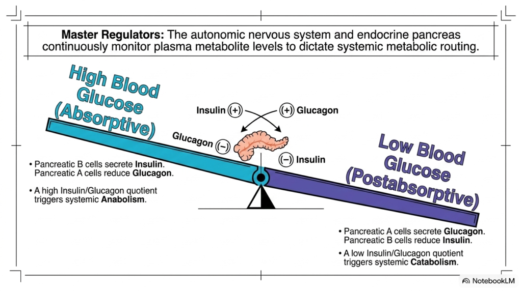
Systemic metabolic routing is tightly governed by the endocrine pancreas and autonomic nervous system. These master regulatory centers continuously monitor plasma metabolite concentrations to direct buffering in organ metabolism. Pancreatic endocrine cells act as exquisite metabolic sensors, dynamically adjusting hormone secretion into the bloodstream in direct response to circulating glucose levels in blood plasma.
Following a carbohydrate-rich meal, elevated blood glucose levels stimulate pancreatic beta cells to secrete insulin into the bloodstream, while simultaneously suppressing glucagon release from pancreatic alpha cells. A high insulin-to-glucagon ratio signifies systemic nutrient wealth, driving body-wide anabolism. Under these favorable hormonal conditions, the buffer function in organ metabolism activates glycogenesis, lipogenesis, and protein synthesis across the liver, skeletal muscle, and adipose tissues.
Conversely, when blood glucose concentrations drop during fasting intervals, pancreatic alpha cells release glucagon while insulin secretion declines significantly. A low insulin-to-glucagon ratio signals energetic scarcity, immediately flipping the systemic metabolic switch toward catabolism across all peripheral organs. Autonomic neural signals also modulate insulin and glucagon secretion to fine-tune metabolic responses. The endocrine regulation of metabolic switches is a cornerstone concept for medical biochemistry students. This delicate hormonal orchestration allows tissues to respond rapidly to changing metabolic demands.
This crucial hormonal shift engages the buffer function in organ metabolism to initiate glycogenolysis, lipolysis, and gluconeogenesis. By carefully modulating enzyme phosphorylation cascades, the endocrine system mobilizes internal energy stores, maintaining blood glucose within narrow physiological limits to sustain cellular life across every organ system. This reciprocal hormonal balance prevents simultaneous activation of opposing metabolic pathways. Understanding endocrine regulation provides students with essential clinical insight into metabolic homeostasis.
Slide 4: Systemic Anabolism and the Buffer Function in Organ Metabolism
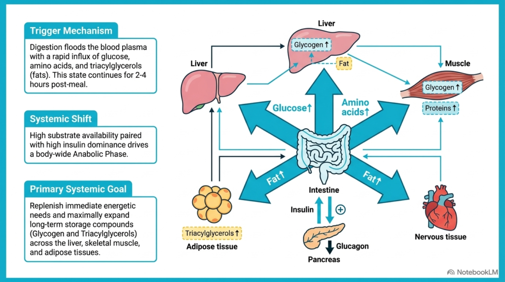
The absorptive state begins during food ingestion and typically lasts two to four hours after a complete meal. Digestive processes break down complex dietary macronutrients into basic chemical monomers, flooding the hepatic portal vein and systemic circulation with glucose, amino acids, and triacylglycerols. This massive influx of nutrient substrates serves as the primary biochemical trigger for the body’s buffering function in organ metabolism.
High substrate availability, paired with insulin dominance, shifts the entire organism into a body-wide anabolic phase. Tissues throughout the body absorb incoming plasma nutrients to satisfy immediate basal metabolic requirements. Once immediate energy demands are fully satisfied, the buffer function in organ metabolism diverts the remaining substrate surplus toward expanding long-term energy storage compounds within target metabolic organs.
The primary systemic objective of this anabolic phase is to maximize storage capacity across the liver, skeletal muscle, and adipose tissue. Glucose is polymerized into glycogen, while excess fatty acids and amino acids are converted into triacylglycerols and functional structural proteins. Dietary protein absorption also supplies essential amino acids for tissue repair and growth. Insulin-triggered intracellular signaling cascades promote GLUT4 translocation in sensitive peripheral tissues. Insulin receptor activation stimulates protein kinase cascades that upregulate rate-limiting anabolic enzymes.
By packaging small, osmotically active molecules into dense, insoluble macromolecular stores, tissues prevent intracellular swelling and osmotic damage. Consequently, this buffering function safeguards surplus dietary energy for upcoming periods of fasting, ensuring bioenergetic resilience and preserving systemic cellular integrity across all physiological compartments. This systemic anabolic phase efficiently converts transient nutrient abundance into durable biological energy reserves.
Slide 5: Hepatic Storage Pathways in the Buffer Function in Organ Metabolism
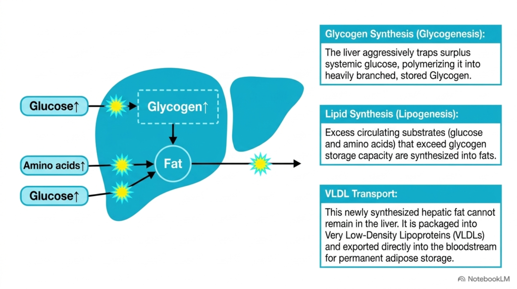
Serving as the central metabolic processing hub, the liver executes pivotal biochemical pathways during the absorptive state. When glucose levels surge in the portal circulation, hepatocytes rapidly trap incoming glucose through high-capacity glucokinase phosphorylation. The liver’s buffering role directs this hepatic glucose surplus into glycogenesis, polymerizing glucose units into highly branched glycogen for rapid future mobilization.
However, hepatic glycogen storage capacity is inherently finite and fills quickly during large meals. Once glycogen stores reach saturation, excess circulating glucose and amino acids are funneled into lipogenesis. Through de novo lipogenesis, hepatocytes convert excess carbon skeletons into fatty acids and triacylglycerols. This pathway, within the buffer function in organ metabolism, ensures that surplus dietary energy is preserved rather than excreted.
Newly synthesized hepatic triacylglycerols cannot remain stored within liver tissue without causing pathological steatosis. To overcome this limitation, hepatocytes package synthesized lipids into Very Low-Density Lipoproteins (VLDLs). In addition, hepatocyte metabolic enzymes are tightly regulated by allosteric mechanisms during feeding. Fatty acid synthase and acetyl-CoA carboxylase activity increases significantly under direct insulin stimulation. Allosteric activation of glycogen synthase ensures rapid conversion of phosphorylated glucose into liver glycogen.
The liver then secretes VLDLs directly into the bloodstream for transport to peripheral adipose tissue for long-term storage. Through this lipid exportation process, the buffer function in organ metabolism coordinates essential inter-organ lipid trafficking, preventing fatty liver accumulation and maintaining systemic metabolic balance across non-hepatic tissues while preserving liver cellular health. Hepatic lipid exportation prevents lipotoxicity and maintains structural integrity within hepatocyte cytoplasm.
Slide 6: Peripheral Tissue Anabolism and the Buffer Function in Organ Metabolism
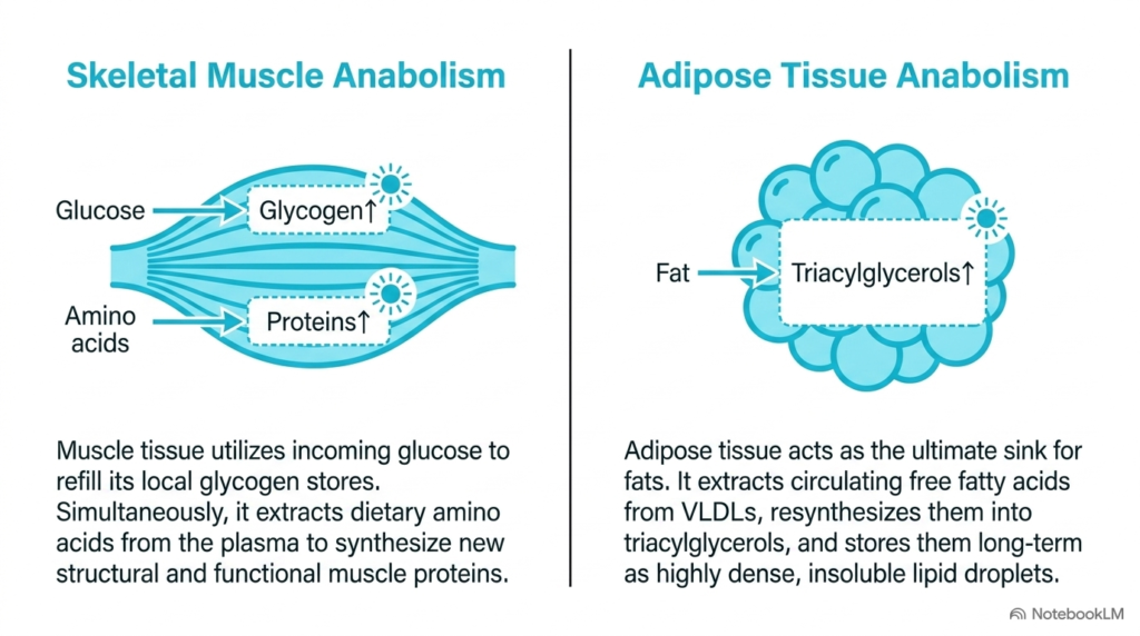
During the absorptive state, peripheral tissues cooperate closely with the liver to process circulating nutrients. Skeletal muscle tissue absorbs plasma glucose under insulin stimulation to replenish its internal glycogen stores. Simultaneously, muscle cells take up circulating amino acids from blood plasma to synthesize new structural and contractile proteins, exemplifying the buffer function in organ metabolism in peripheral muscle tissue.
Adipose tissue functions as the ultimate long-term energy sink during times of nutrient abundance. Adipocytes express lipoprotein lipase on their capillary walls, allowing them to hydrolyze circulating VLDLs and chylomicrons into free fatty acids. Inside the adipocyte, these fatty acids are resynthesized into triacylglycerols, illustrating how the buffer function in organ metabolism stores energy within dense, insoluble lipid droplets.
By sequestering surplus lipids within specialized adipose tissue, the body protects non-adipose organs from ectopic fat accumulation and lipotoxicity. Muscle protein accretion and adipose lipid storage work synergistically to clear excess nutrients from systemic circulation. Furthermore, skeletal muscle is a major site of insulin-stimulated post-meal glucose clearance, ensuring rapid glycemic normalization. Adipose tissue responsiveness to insulin prevents systemic free fatty acid elevation during feeding intervals.
This coordinated nutrient removal prevents toxic substrate overload in non-storage tissues. Thus, peripheral organ cooperation remains a critical element of the buffer function in organ metabolism, maintaining metabolic harmony and structural stability throughout the well-fed state. Adipocyte intracellular lipid droplets store energy efficiently without occupying excessive aqueous volume. Coordinated peripheral storage mechanisms safeguard metabolic flexibility across all non-hepatic organ systems and maintain cellular energy balance.
Slide 7: Transition to Catabolism and the Buffer Function in Organ Metabolism
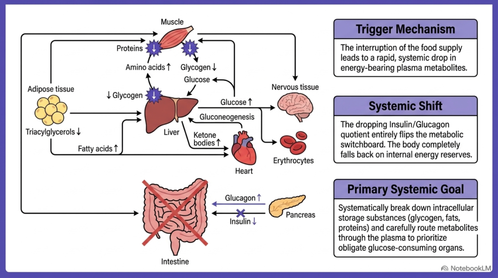
When gastrointestinal digestion and nutrient absorption cease, the body enters the postabsorptive state. The interruption of external food supply leads to a rapid, systemic decline in energy-bearing plasma metabolites. This decrease triggers a profound shift in metabolic buffering, prompting the organism to transition from storing dietary nutrients to mobilizing internal fuel reserves.
Falling blood glucose concentrations reduce pancreatic insulin secretion while elevating glucagon output. This dropping insulin-to-glucagon ratio acts as a metabolic switchboard, reversing enzyme phosphorylation states across major biochemical pathways. Under this catabolic hormonal signaling, the buffer function in organ metabolism activates intracellular degradation pathways to supply the circulatory system with essential energy substrates.
The overarching systemic objective during the postabsorptive state is to systematically break down stored glycogen, lipids, and proteins. Metabolites liberated into blood plasma are carefully routed to prioritize obligate glucose consumers, such as the brain and erythrocytes. In addition, the drop in circulating insulin disinhibits key catabolic enzymes in metabolic tissues, promoting efficient fuel mobilization. Glucagon binds G-protein-coupled receptors, activating adenylate cyclase and raising intracellular cyclic AMP levels in target hepatocytes.
These specialized tissues require constant nutrient delivery to maintain baseline cellular functions. Through precise inter-organ substrate routing, the buffer function of organ metabolism prevents hypoglycemic crises during fasting, protecting vital neural and vascular structures from irreversible energetic damage. Phosphorylase kinase and glycogen phosphorylase are activated to initiate rapid substrate breakdown. This signaling cascade activates protein kinase A, triggering coordinated glycogenolysis and lipolysis to safeguard systemic fuel supply. Regulating these enzymatic pathways is crucial for maintaining cellular bioenergetics during fasting.
Slide 8: Hepatic Altruism and the Buffer Function in Organ Metabolism
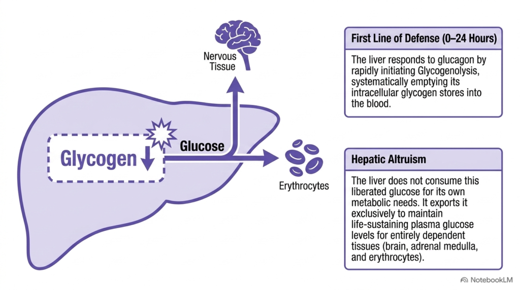
During the first 0 to 24 hours of fasting, the liver serves as the primary defense against falling blood glucose levels. In response to rising glucagon concentrations, hepatocytes rapidly initiate glycogenolysis. This biochemical pathway, within the buffer function in organ metabolism, breaks down stored hepatic glycogen into glucose-6-phosphate, which is then hydrolyzed into free glucose.
Unlike skeletal muscle cells, hepatocytes express the crucial enzyme glucose-6-phosphatase, allowing unphosphorylated glucose to exit into the bloodstream. Remarkably, the liver does not consume this liberated glucose for its own bioenergetic requirements. This physiological phenomenon, known as hepatic altruism, demonstrates how the buffer function in organ metabolism prioritizes systemic survival over local hepatic energy needs.
The glucose exported by altruistic hepatocytes is strictly reserved for tissues that depend entirely on continuous glucose availability, including the central nervous system, adrenal medulla, and erythrocytes. These specialized cells rely on steady glycemic levels to maintain active metabolic function. Furthermore, hepatic glucose production during early fasting matches systemic glucose consumption rates exactly. Hepatic glycogen phosphorylase activity ensures rapid release of glucose monomers during early fasting intervals.
Because these dependent tissues cannot utilize alternative fuels effectively during acute fasting, hepatic glucose release is vital. By supplying these organs with steady plasma glucose, the liver underscores the indispensable role of this buffer function in organ metabolism during short-term fasting. The absence of glucose-6-phosphatase in muscle prevents muscle glycogen from directly contributing to blood sugar. By prioritizing systemic glycemic maintenance, the liver prevents central nervous system bioenergetic collapse during food deprivation.
Slide 9: De Novo Gluconeogenesis in the Buffer Function in Organ Metabolism
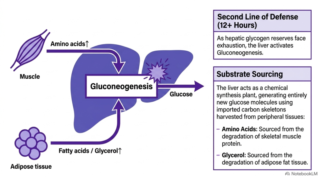
As fasting progresses past twelve hours, hepatic glycogen stores face exhaustion and cannot maintain blood sugar levels independently. To preserve systemic glycemic balance, the liver activates gluconeogenesis as its secondary line of defense. This biochemical transition expands the liver’s metabolic buffer from simple storage mobilization to active synthesis of new glucose molecules.
During gluconeogenesis, the liver functions as a synthetic chemical factory, generating new glucose molecules from non-carbohydrate carbon precursors. The liver harvests the required raw materials from peripheral tissues throughout the body. Glucogenic amino acids, such as alanine and glutamine, are liberated from skeletal muscle protein degradation, illustrating the buffer function in organ metabolism across tissues.
Concurrently, adipose tissue lipolysis releases glycerol into circulation, providing another key carbon substrate for hepatic glucose synthesis. Fatty acid oxidation within hepatocytes yields the ATP required to drive this endergonic synthetic process within hepatic cellular compartments. The coordination between muscle amino acid release and hepatic gluconeogenesis maintains stable plasma glucose. Bypassing irreversible glycolytic steps requires specialized gluconeogenic enzymes present in hepatocytes.
By coupling lipid catabolism to glucose synthesis, the liver maintains continuous metabolic output. Through de novo gluconeogenesis, this buffering function ensures an uninterrupted glucose supply to glucose-dependent tissues during extended starvation. Key gluconeogenic enzymes like pyruvate carboxylase and PEP carboxykinase are significantly upregulated under glucagon stimulation. Gluconeogenesis sustains glycemic stability during prolonged nutritional deprivation when glycogen reserves are empty, preserving systemic life across organs. This complex enzymatic machinery ensures blood glucose remains adequate to support vital organ functions throughout extended fasting.
Slide 10: Ketogenesis as an Alternative Fuel in the Buffer Function in Organ Metabolism
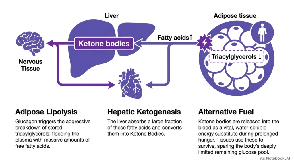
During extended fasting or starvation, elevated glucagon levels stimulate aggressive lipolysis in adipose tissue. Adipocytes break down stored triacylglycerols, flooding blood plasma with large quantities of free fatty acids. The liver absorbs a major fraction of these circulating lipids, triggering an adaptive pathway that supports the organ’s buffering role in metabolism. Ketogenesis represents a vital evolutionary adaptation.
Inside hepatic mitochondria, rapid fatty acid beta-oxidation produces an abundance of acetyl-CoA that exceeds the processing capacity of the citric acid cycle. The liver converts this excess acetyl-CoA into ketone bodies, generating water-soluble molecules like acetoacetate and beta-hydroxybutyrate. This metabolic pathway shows how the liver’s buffer function provides crucial alternative fuels.
Ketone bodies are released into circulation, where peripheral tissues, including cardiac muscle and the brain, utilize them as primary energy sources. By replacing glucose with ketone bodies, the organism dramatically decreases its overall glucose consumption rate across systemic tissues. Extrahepatic tissues oxidize beta-hydroxybutyrate and acetoacetate back to acetyl-CoA. Hepatic HMG-CoA synthase catalyzes the rate-limiting step in mitochondrial ketone body production during fasting.
This metabolic shift reduces the demand for gluconeogenic amino acids harvested from skeletal muscle. Consequently, this glucose-sparing adaptation, governed by the buffer function in organ metabolism, prevents excessive degradation of functional body proteins during prolonged nutritional deprivation. Ketone bodies serve as efficient water-soluble fuels that easily cross the blood-brain barrier. Ketone body oxidation supplies extrahepatic tissues with abundant acetyl-CoA for ATP generation during prolonged fasting phases, preserving structural tissues. Overall, hepatic ketogenesis provides a critical survival mechanism during severe caloric restriction, safeguarding systemic physiological homeostasis.
Slide 11: Muscle Adaptations in the Buffer Function in Organ Metabolism
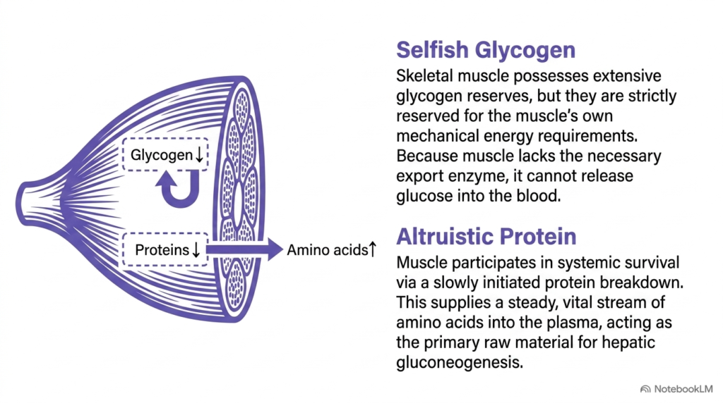
Skeletal muscle demonstrates a unique physiological role during starvation, balancing local energy needs against whole-body survival requirements. Although muscle contains substantial glycogen reserves, this glycogen is strictly reserved for the muscle’s own mechanical activity. Muscle cells lack glucose-6-phosphatase, preventing glucose release into plasma and showcasing a specialized limitation in the buffer function in organ metabolism.
Because muscle glycogen cannot be exported directly, muscle tissue contributes to systemic energy homeostasis through controlled proteolysis. During fasting, skeletal muscle slowly degrades structural and contractile proteins into free amino acids. This process releases a steady stream of amino acids into the bloodstream, highlighting another key aspect of the buffer function in organ metabolism.
These circulating amino acids travel directly to the liver, where they serve as the primary carbon skeletons for hepatic gluconeogenesis. While muscle glycogen remains selfishly sequestered for local muscle contraction, muscle protein acts altruistically to support whole-body glucose homeostasis. Alanine released from muscle travels to the liver via the glucose-alanine cycle for gluconeogenesis. The glucose-alanine cycle efficiently transfers muscle amino nitrogen to the liver for safe urea synthesis.
This tissue trade-off balances localized energy preservation with systemic survival requirements. It illustrates how the buffer function in organ metabolism mobilizes peripheral structural reserves to maintain glycemic stability during starvation while protecting essential cellular structures. Muscle proteolysis is regulated to prevent rapid wasting of vital functional proteins. Controlled proteolysis supplies gluconeogenic precursors without compromising essential skeletal muscle mechanical and respiratory functions during fasting. This careful conservation of muscle amino acids prevents premature structural wasting while supporting essential systemic glucose requirements.
Slide 12: Tissue Fuel Specificity in the Buffer Function in Organ Metabolism
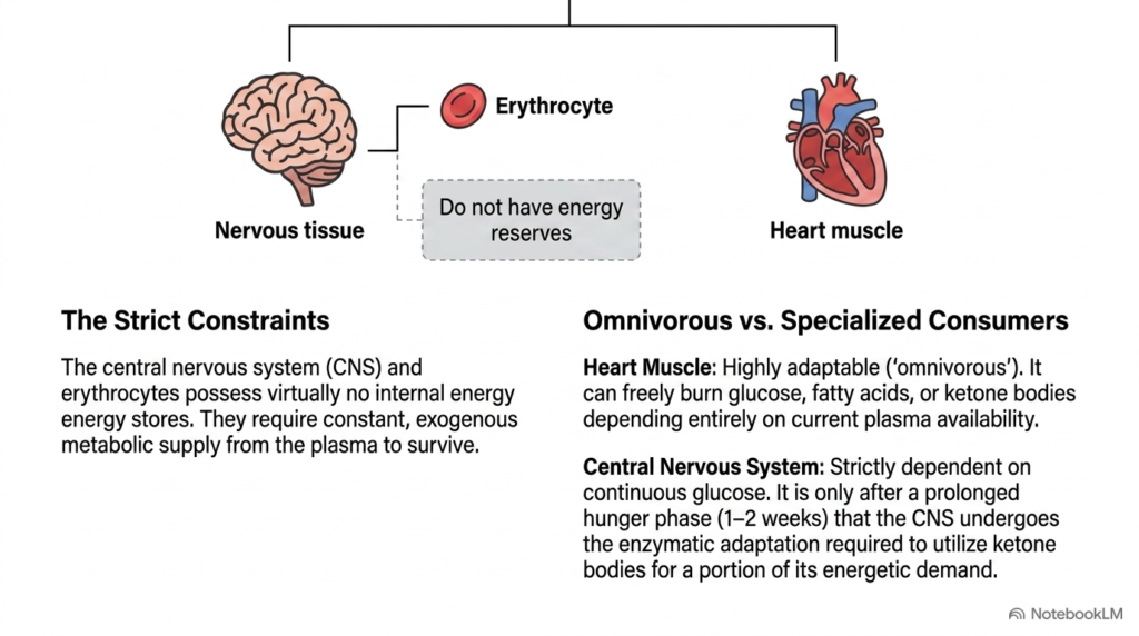
Metabolic coordination across organs depends heavily on the unique fuel requirements of specialized tissues. The central nervous system and erythrocytes face strict bioenergetic constraints because they lack significant internal energy reserves. Consequently, these organs depend entirely on continuous metabolic supply from plasma, regulated by the buffer function in organ metabolism.
Erythrocytes lack mitochondria entirely and rely exclusively on anaerobic glycolysis for ATP synthesis. The central nervous system relies almost exclusively on continuous glucose supply under non-starvation conditions. In contrast, cardiac muscle is omnivorous, flexibly oxidizing glucose, fatty acids, or ketone bodies based on plasma availability. The buffer function in organ metabolism caters to these distinct fuel preferences.
Only after several weeks of continuous starvation does the brain undergo enzymatic adaptations allowing it to derive two-thirds of its energy from ketone bodies. This metabolic shift reduces systemic glucose requirements, sparing structural proteins from degradation throughout the body. During chronic starvation, the brain gradually upregulates monocarboxylate transporters to import ketone bodies. Cardiac myocytes adjust substrate oxidation preferences dynamically based on circulating fatty acid and ketone levels.
By shifting fuel dependency toward ketone bodies, the brain adapts to severe fasting conditions. Through precise, tissue-specific fuel partitioning, the buffer function of organ metabolism preserves life during severe long-term nutrient deprivation. Erythrocytes continue relying on glycolysis throughout fasting, returning lactate to the liver via the Cori cycle. Substrate partitioning protects obligate glucose consumers while allowing flexible tissues to utilize alternative circulating fuels during starvation. This metabolic flexibility allows the central nervous system to survive prolonged food shortages without compromising baseline neurological function.
Slide 13: Comparative Overview of the Buffer Function in Organ Metabolism
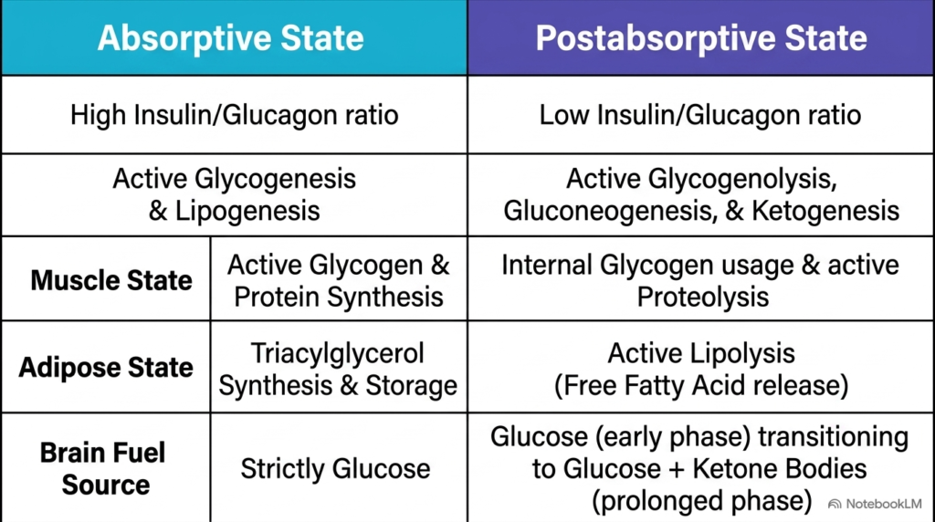
A direct comparison between the absorptive and postabsorptive states highlights the human body’s remarkable metabolic flexibility. In the absorptive state, a high insulin-to-glucagon ratio drives liver glycogenesis and lipogenesis, while peripheral tissues store glycogen and proteins. The buffer function in organ metabolism optimizes this well-fed phase to build robust energy stores.
Conversely, the postabsorptive state is directed by a low insulin-to-glucagon ratio, shifting organ metabolism toward glycogenolysis, gluconeogenesis, and ketogenesis. Adipose lipolysis liberates fatty acids, while muscle proteolysis provides amino acid precursors for hepatic glucose synthesis. This catabolic coordination illustrates how the buffer function in organ metabolism mobilizes internal fuels during starvation.
While peripheral tissues continuously adapt their fuel selection to current nutrient availability, obligate consumers like the brain and erythrocytes receive uninterrupted metabolic protection from plasma. Comparative analysis of metabolic states also clarifies how hormonal ratios direct substrate fluxes. Mastering these integrated pathways enables medical students to analyze complex metabolic disorders. Hormonal ratios act as master metabolic switches that align enzymatic activities across organ systems.
Through these highly coordinated inter-organ pathways, the body maintains bioenergetic stability. By balancing anabolic storage with catabolic mobilization, the buffer function in organ metabolism maintains systemic energy homeostasis across all physiological states, ensuring cellular survival. The reciprocal regulation of anabolic and catabolic pathways prevents futile biochemical cycles. Understanding this comprehensive inter-organ metabolic coordination provides a solid foundation for clinical biochemistry and human physiology. Ultimately, this inter-organ coordination highlights the elegance and efficiency of human metabolic adaptation across varying nutrient states.
Please read our Content Disclaimer Statement.
Check out our social media channels: