87. Protein Translation Initiation: A Molecular Overview
Imagine a bustling microscopic factory where genetic blueprints are transformed into physical reality. This cellular factory operates continuously, synthesizing the vital proteins required for life. The following slide deck explores this exact biochemical marvel. It provides a detailed breakdown of how ribosomes, messenger RNA, and specialized enzymes converge. By examining these fundamental mechanisms, medical and university students can build a robust understanding of cellular biology and structural biochemistry.
1. Protein Translation Initiation: An Introduction to Protein Biosynthesis
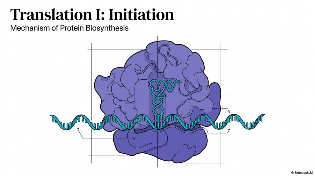
The process of building a protein from a genetic template stands as one of the most remarkable achievements of the biological world. At the heart of this process is Protein Translation Initiation, the critical first phase where cellular components gather to begin synthesis. The visual representation on this slide captures the elegance of the ribosome, a massive molecular machine, engaging with a strand of messenger RNA. This interaction represents the bridge between the information stored in DNA and the active proteins that carry out vital cellular functions. Understanding this initial step is fundamental for students exploring the depths of molecular biochemistry.
To appreciate the magnitude of this biological event, one must consider the precision required during Protein Translation Initiation. The ribosome does not simply attach anywhere on the genetic message; it must locate a specific starting point. If this complex engages the messenger RNA even one nucleotide out of place, the entire resulting protein will be structurally incorrect and nonfunctional. The coordination of the large and small ribosomal subunits, along with various nucleic acids, ensures this precise alignment. The visualization shows this macromolecular assembly clamping down on the genetic code, ready to begin its crucial task of biological manufacturing.
Furthermore, the rigorous study of Protein Translation Initiation holds profound implications in medicine and modern pharmacology. Many antibiotics and therapeutics function by disrupting this exact stage of the biological sequence in bacterial cells. By mapping out the structural and functional nuances of this starting phase, scientists can design targeted drugs that halt bacterial growth without harming human cells. When the ribosome recognizes the start signal, it locks into place, transitioning the cellular environment from preparation to active synthesis. This slide introduces that pivotal moment of molecular commitment, setting the stage for the entire biosynthetic pathway.
2. Protein Translation Initiation: Architecture of the 80S Eukaryotic Ribosome
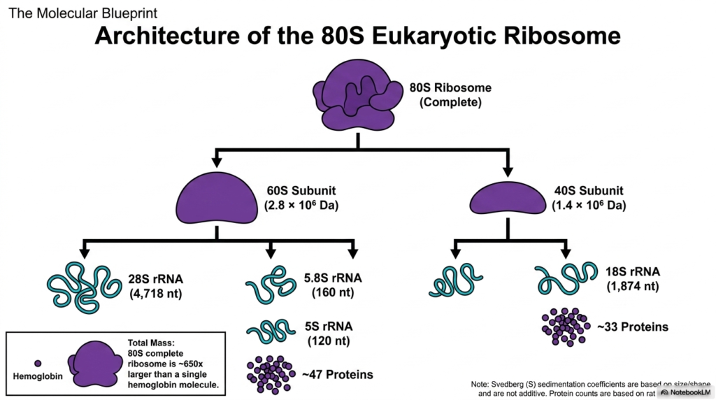
Before diving into the dynamic sequence of Protein Translation Initiation, it is necessary to examine the physical machinery responsible for the process. This slide details the architecture of the complete eukaryotic 80S ribosome, a massive ribonucleoprotein complex. The fully assembled 80S ribosome is exceptionally large, measuring approximately 650 times larger than a single hemoglobin molecule. This incredible mass underscores the structural complexity required to synthesize living tissue. It consists of two distinct halves: the large 60S subunit and the smaller 40S subunit. These molecular weights and sedimentation coefficients reflect the complex shape of these critical cellular structures.
Breaking down the individual components reveals an organized mixture of ribosomal RNA and structural proteins. The larger 60S subunit contains three distinct RNA molecules—the 28S, 5.8S, and 5S rRNAs—complexed with about 47 different proteins. Conversely, the smaller 40S subunit holds a single 18S rRNA chain bundled with approximately 33 proteins. These RNA strands do not merely serve as structural scaffolding; they possess catalytic properties essential for the chemical reactions that follow. For Protein Translation Initiation to succeed, these individual pieces must be fully formed and readily available in the surrounding cellular environment to guide the assembly process.
A critical feature of these subunits is their modular nature. In the cytoplasm, the large and small subunits exist independently of one another when inactive. They only clamp together to form the complete 80S structure when a specific messenger RNA transcript requires decoding. This structural separation is a vital regulatory mechanism, ensuring that cellular energy is not wasted on idle ribosomes. The accurate assembly of this massive 80S architecture represents a cornerstone of Protein Translation Initiation. Understanding this enormous molecular blueprint allows researchers to grasp how genetic mutations affecting ribosomal proteins can lead to severe developmental disorders and complex diseases.
3. Protein Translation Initiation: The Cellular Environment for Protein Biosynthesis
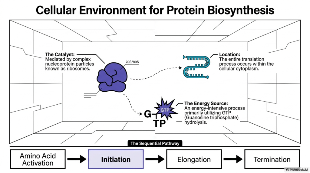
To fully grasp how cells manufacture their components, one must contextualize where and how these complex biochemical reactions occur. The cellular environment for protein biosynthesis is exclusively located within the aqueous cytoplasm. Here, the massive nucleoprotein particles known as ribosomes act as the primary catalysts for the entire operation. This slide illustrates the broad sequential pathway of building a polypeptide, which progresses from the preliminary amino acid activation directly into Protein Translation Initiation, followed by elongation, and finally termination. Placing this specific initiation phase within the broader biological context helps clarify its role as a regulated gateway for cellular production.
Progress through this sequential pathway requires a tremendous amount of cellular resources, making metabolic energy management a critical focus for the organism. The entire sequence, particularly the intricate molecular steps involving Protein Translation Initiation, is energy-intensive and tightly controlled. Instead of relying solely on standard ATP molecules, the ribosomal machinery primarily utilizes the hydrolysis of Guanosine triphosphate, commonly known as GTP. When the high-energy phosphate bonds of this GTP are broken down, the resulting energy release powers the mechanical movements, conformational shifts, and secure locking of the massive ribosomal subunits onto the genetic template.
Furthermore, defining the assembled ribosome as a biological catalyst emphasizes its vital role in lowering the activation energy required to subsequently form strong peptide bonds. Whether operating as a large 80S eukaryotic complex or a slightly smaller 70S prokaryotic complex, the ribosome provides a stable, protected micro-environment separated from the chaotic chemical fluctuations of the cytoplasm. By acting as a molecular sanctuary, it aligns the messenger RNA and transfer RNA molecules with spatial precision. Ultimately, a successful, well-coordinated event of Protein Translation Initiation sets off a continuous chain reaction that consumes GTP while meticulously assembling the foundational building blocks of biological life.
4. Protein Translation Initiation: Spatial Arrangement and Subunit Assembly
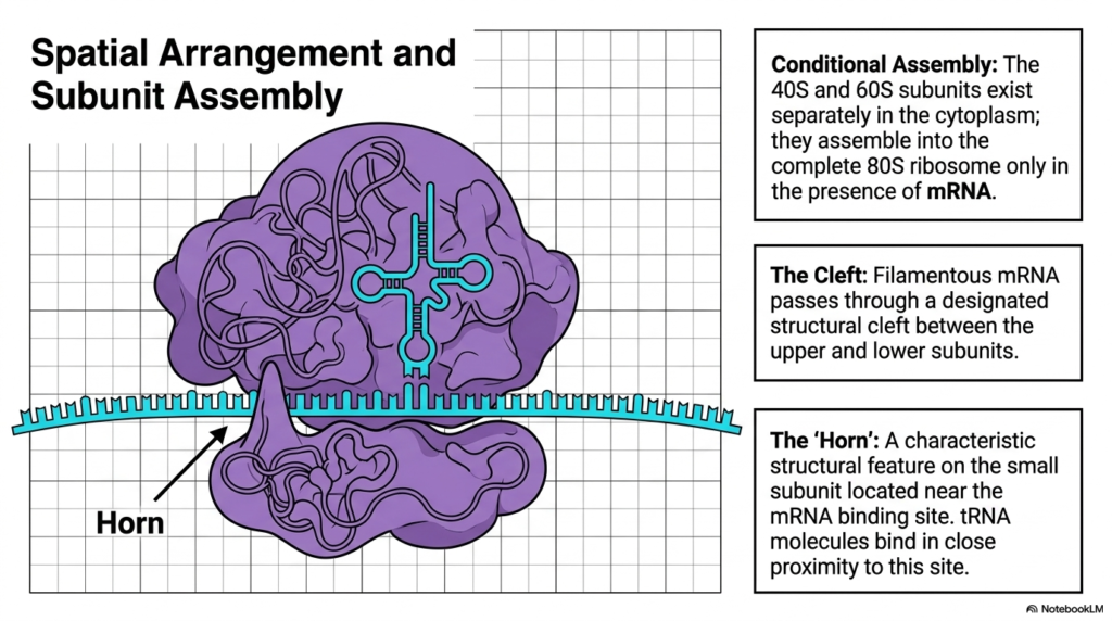
The intricate architecture of the ribosome dictates exactly how it functions during the earliest stages of protein synthesis. This slide focuses on the specific spatial arrangement and conditional subunit assembly required for successful Protein Translation Initiation. In the cellular cytoplasm, the small 40S subunit and the large 60S subunit exist independently of one another. They do not spontaneously combine; rather, their assembly is conditional and strictly regulated. The two massive halves only join together to form the complete, functional 80S ribosome in the direct presence of a mature messenger RNA transcript.
This conditional joining acts as a biological fail-safe mechanism, preventing empty ribosomes from wasting cellular energy or synthesizing erratic amino acid chains. A critical structural feature facilitating this process is the designated ‘cleft’ located exactly between the upper and lower subunits. During the precise steps of Protein Translation Initiation, the filamentous messenger RNA passes directly through this horizontal cleft. This physical arrangement allows the genetic template to be securely sandwiched and protected by the heavy ribosomal machinery. This ensures the fragile RNA code is read accurately and shielded from degradative enzymes present within the surrounding cellular fluid.
Additionally, this slide highlights a unique structural landmark on the small subunit referred to as the ‘horn’. Located near the designated messenger RNA binding site, this characteristic protuberance helps guide incoming molecules into their proper functional positions. Transfer RNA molecules must bind in close proximity to this horn to ensure spatial accuracy. The exact alignment of the cleft, the horn, and the joining subunits creates the perfect biochemical landscape. Without these specific topographical features, the flawless execution of Protein Translation Initiation would be structurally impossible, leading to a halt in biological protein production and ultimately cellular death.
5. Protein Translation Initiation: Comparing Eukaryotic and Prokaryotic Machinery
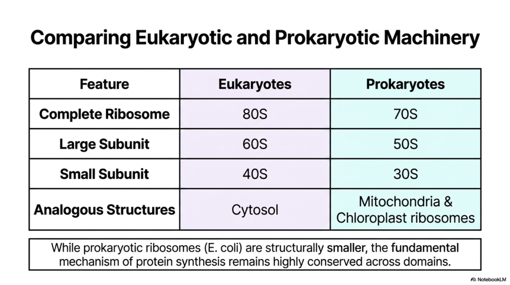
While the universal goal of building polypeptides remains identical across all domains of life, the precise machinery utilized exhibits notable differences. This slide provides a direct comparison between the ribosomal complexes of eukaryotes and prokaryotes, providing vital context for understanding Protein Translation Initiation across species. Eukaryotic organisms utilize the significantly larger 80S complete ribosome, which is subdivided into a massive 60S large subunit and a 40S small subunit. In contrast, prokaryotic organisms, such as Escherichia coli bacteria, rely on a more compact 70S complete ribosome consisting of a 50S large subunit and a 30S small subunit.
Beyond their overall size and density differences, the specific cellular locations of these analogous structures present an interesting evolutionary tale. In eukaryotic cells, the massive 80S complexes are predominantly located within the general cytosol or attached to the endoplasmic reticulum. However, organelles like mitochondria and chloroplasts possess their own distinct ribosomes that closely resemble the prokaryotic 70S variety. This structural similarity provides strong evidence for the endosymbiotic theory. Furthermore, analyzing these physical variations is vital because they dictate the subtle mechanistic differences observed during Protein Translation Initiation across different species and distinct intracellular compartments.
Despite these measurable physical differences in mass and subunit composition, the fundamental biochemical mechanisms driving the process remain conserved throughout evolutionary history. The core objectives of locking onto a messenger RNA template, identifying a starting codon, and establishing a reading frame are universal. Therefore, while prokaryotic ribosomes are structurally smaller and less complex, studying their version of Protein Translation Initiation serves as an exceptional and simplified model for understanding the more elaborate eukaryotic pathways found in human cells. This fundamental evolutionary conservation highlights the biological importance of preserving accurate protein manufacturing mechanisms across billions of years of existence.
6. Protein Translation Initiation: Macro-Translation via Polysomes
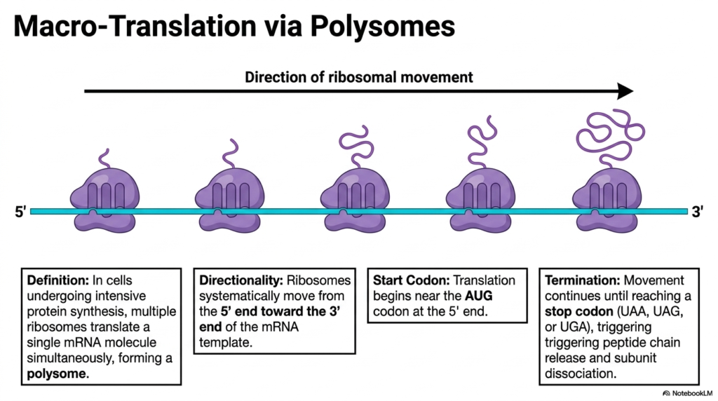
Cellular survival frequently demands the rapid, high-volume production of specific polypeptides in a short amount of time. This slide introduces the concept of macro-translation via polysomes, demonstrating how biological efficiency is maximized. In cells undergoing intensive protein synthesis, a single messenger RNA molecule does not simply host one lone ribosome at a time. Instead, immediately following successful protein translation initiation at the 5-prime end, the initial ribosome begins moving down the genetic template. This progressive movement quickly exposes the starting sequence again, allowing subsequent ribosomes to attach and begin reading simultaneously.
This remarkable cellular phenomenon forms a densely packed structural complex known as a polyribosome, or polysome. The directionality of this entire massive operation is strictly conserved, with every individual ribosome systematically tracking from the 5-prime end toward the 3-prime end of the messenger RNA transcript. Because Protein Translation Initiation always occurs near the specific AUG start codon located at the 5-prime region, a sequential train of molecular factories is established. Each ribosome on this transcript carries a growing polypeptide chain, with the chains becoming progressively longer as the complex nears the final 3-prime terminal end of the RNA sequence.
This synchronized mass-production continues flawlessly until each migrating ribosome physically encounters a designated stop codon, such as UAA, UAG, or UGA. Upon reaching these distinct chemical stop signals, a termination event triggers the release of the newly formed peptide chain and forces the large and small subunits to dissociate. The ability to form polysomes means that a single, fragile messenger RNA template can yield dozens or even hundreds of identical proteins rapidly. Without the rapid, repeated firing of Protein Translation Initiation at the genetic starting site, critical cellular adaptation and biological survival responses would be dangerously slow and ultimately inefficient.
7. Protein Translation Initiation: The Prokaryotic Model for Initiation
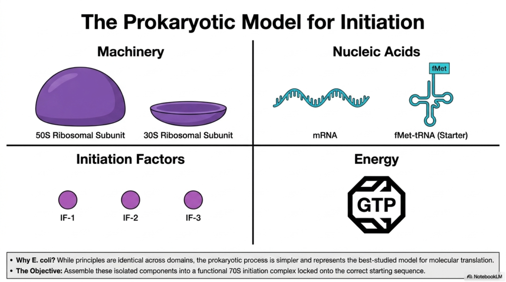
To fully understand the intricacies of protein assembly, scientists frequently turn to bacterial systems for clarity and experimental consistency. This slide introduces the isolated components necessary for Protein Translation Initiation within the heavily studied prokaryotic model. Researchers rely on Escherichia coli because its fundamental principles of molecular translation are identical to those of eukaryotes, yet the process is inherently simpler and easier to manipulate in a laboratory setting. The core objective of this complex biological phase is to seamlessly assemble a multitude of isolated cellular components into a functional 70S initiation complex locked onto a correct genetic starting sequence.
The essential machinery required for this assembly includes the 50S large ribosomal subunit and the 30S small ribosomal subunit, which initially float separately in the cytoplasm. Alongside these structural behemoths are the required nucleic acids: the filamentous messenger RNA template and the specialized starter molecule known as fMet-tRNA. Because Protein Translation Initiation cannot occur spontaneously or randomly, specialized helper proteins are absolutely mandatory for success. In the prokaryotic biological model, these helper proteins are identified as Initiation Factors 1, 2, and 3, or simply IF-1, IF-2, and IF-3. Each of these specific factors plays an indispensable, coordinated role.
Finally, assembling these independent structural components into a unified, reading-ready macromolecule requires a significant chemical energy investment from the cell. This energy is generously supplied by Guanosine triphosphate, prominently featured as the universal energy currency for this specific biological event. When examining the mechanics of Protein Translation Initiation, it becomes clear that this is an orchestrated convergence of specialized machinery, delicate nucleic acids, dedicated helper factors, and expendable molecular energy. The successful unification of these isolated elements ensures that the subsequent translation of the genetic code proceeds with flawless biochemical accuracy and efficiency.
8. Protein Translation Initiation: Phase 1 – Preparing the 30S Subunit
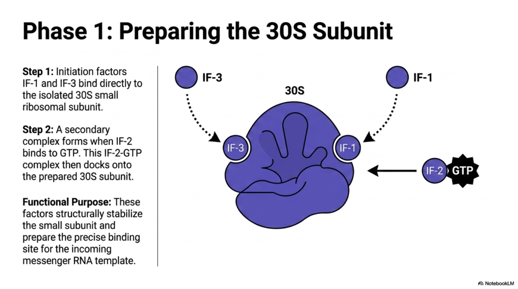
The complex assembly of the molecular reading machine occurs in regulated, sequential phases to guarantee absolute precision. This slide illustrates Phase 1 of Protein Translation Initiation, which specifically focuses on the careful preparation of the 30S small ribosomal subunit. Before any genetic templates or amino acids can be introduced to the system, the isolated 30S subunit must be physically modified and biochemically stabilized. This preparation begins when two specific helper proteins, known as initiation factors IF-1 and IF-3, physically bind directly to the irregular surface of the free-floating 30S small ribosomal structure in the bacterial cytoplasm.
Following this initial binding event, a crucial secondary complex must form to supply the necessary energy for the impending molecular shifts. The factor IF-2 tightly binds to a molecule of Guanosine triphosphate, creating a potent IF-2-GTP complex. This energetic complex then physically docks onto the already prepared 30S subunit. During Protein Translation Initiation, these three distinct initiation factors serve an essential functional purpose. They structurally stabilize the small subunit, preventing the premature and disastrous attachment of the massive 50S large subunit before the genetic code has been properly secured and accurately aligned within the internal reading cleft.
Furthermore, the strategic placement of IF-1, IF-2, and IF-3 physically alters the topographical conformation of the 30S subunit. This subtle shape change prepares the exact binding site for the incoming filamentous messenger RNA template. Without the successful completion of this initial preparatory phase, subsequent steps of Protein Translation Initiation would fail, leading to total biochemical chaos and cellular dysfunction. The delicate small subunit must be meticulously primed, structurally protected, and energetically charged by these specialized initiation factors before it can even attempt to correctly identify the chemical start signals embedded within the approaching genetic messenger RNA transcript.
9. Protein Translation Initiation: Phase 2 – Docking the Template and Starter tRNA
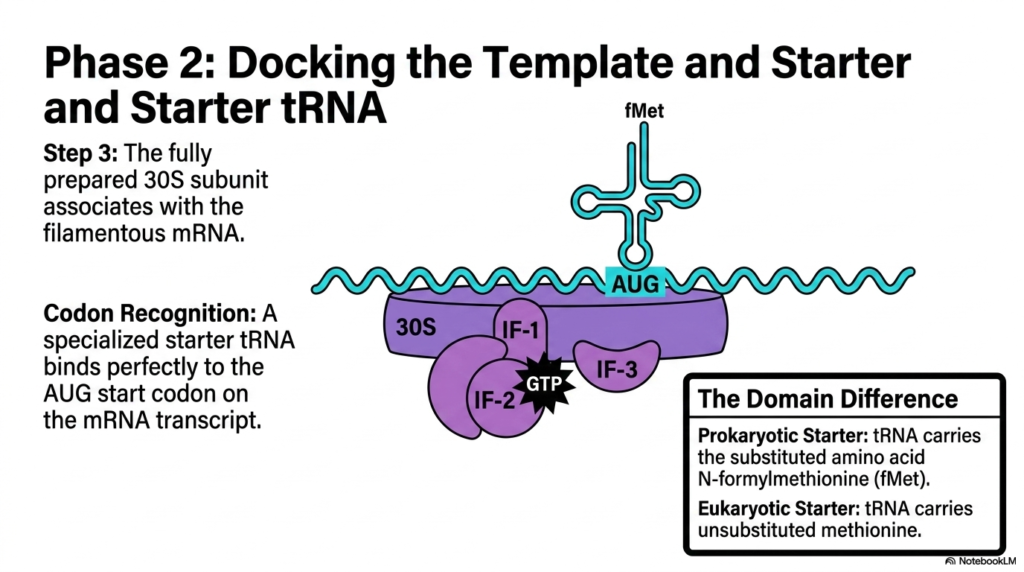
Once the small ribosomal subunit is fully prepared and structurally stabilized by the necessary helper factors, the biological machinery is ready to receive its instructions. This slide details Phase 2 of Protein Translation Initiation, focusing on the critical docking of both the genetic template and the specialized starter molecule. The fully prepared 30S subunit, still attached to its three initiation factors, physically associates with the filamentous messenger RNA transcript. This association is not random; the complex carefully scans the nucleotide sequence until it aligns with the designated chemical starting point, ensuring the correct reading frame is fundamentally established.
The precise establishment of this reading frame is entirely dependent on accurate codon recognition. A specialized starter transfer RNA molecule binds perfectly to the AUG start codon located on the messenger RNA transcript. During prokaryotic Protein Translation Initiation, this specific starter tRNA carries a chemically modified amino acid known as N-formylmethionine, abbreviated as fMet. This subtle chemical modification is a hallmark of bacterial organisms. The complementary base pairing between the AUG start codon on the template and the corresponding anticodon on the fMet-tRNA molecule securely locks the genetic message into the exact center of the small ribosomal subunit.
This slide also explicitly highlights a major domain difference regarding this starter molecule. While the prokaryotic system utilizes the uniquely substituted N-formylmethionine to begin its peptide chains, the eukaryotic version of Protein Translation Initiation relies on a standard, unsubstituted methionine molecule. Despite this distinct chemical variance in the starter amino acid, the overarching physical mechanics of the docking phase remain beautifully consistent across different life forms. The flawless docking of both the genetic template and the initial building block represents the ultimate point of no return for synthesizing a new biological protein within the active cellular environment.
10. Protein Translation Initiation: Phase 3 – Subunit Joining and Energy Hydrolysis
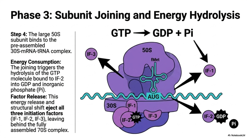
The final major sequence in this intricate molecular dance involves locking the massive structural components together to form a functional factory. This slide outlines Phase 3 of Protein Translation Initiation, characterized by large subunit joining and rapid energy hydrolysis. With the 30S small subunit securely bound to both the messenger RNA template and the starter fMet-tRNA, the heavy 50S large ribosomal subunit is finally permitted to bind to the pre-assembled complex. This massive physical union is the biological equivalent of closing a heavy lid onto a complex machine, sealing the genetic blueprint safely inside the ribosomal core.
However, this monumental structural joining cannot occur passively; it requires a rapid consumption of localized energy. The physical docking of the large 50S subunit immediately triggers the hydrolysis of the Guanosine triphosphate molecule that was previously bound to initiation factor IF-2. During this explosive phase of Protein Translation Initiation, the GTP is chemically cleaved into Guanosine diphosphate and a free inorganic phosphate ion. This sudden burst of localized energy consumption provides the mechanical force required to permanently lock the two massive ribosomal subunits together, finalizing the stable architecture needed for the subsequent steps of cellular protein assembly.
The chemical energy release and the resulting dramatic structural shift cause an immediate ejection event. The completed 70S ribosome forcibly ejects all three initiation factors—IF-1, IF-2, and IF-3—back into the surrounding cellular cytoplasm. Because their vital structural preparation and stabilization duties are now fully complete, these specialized factors are released to be efficiently recycled for another subsequent round of Protein Translation Initiation on a different messenger RNA transcript. The departure of these helper factors leaves behind an assembled, functional 70S initiation complex, completely primed and biochemically ready to begin eagerly linking amino acids together.
11. Protein Translation Initiation: Topology of the Complete 70S Initiation Complex
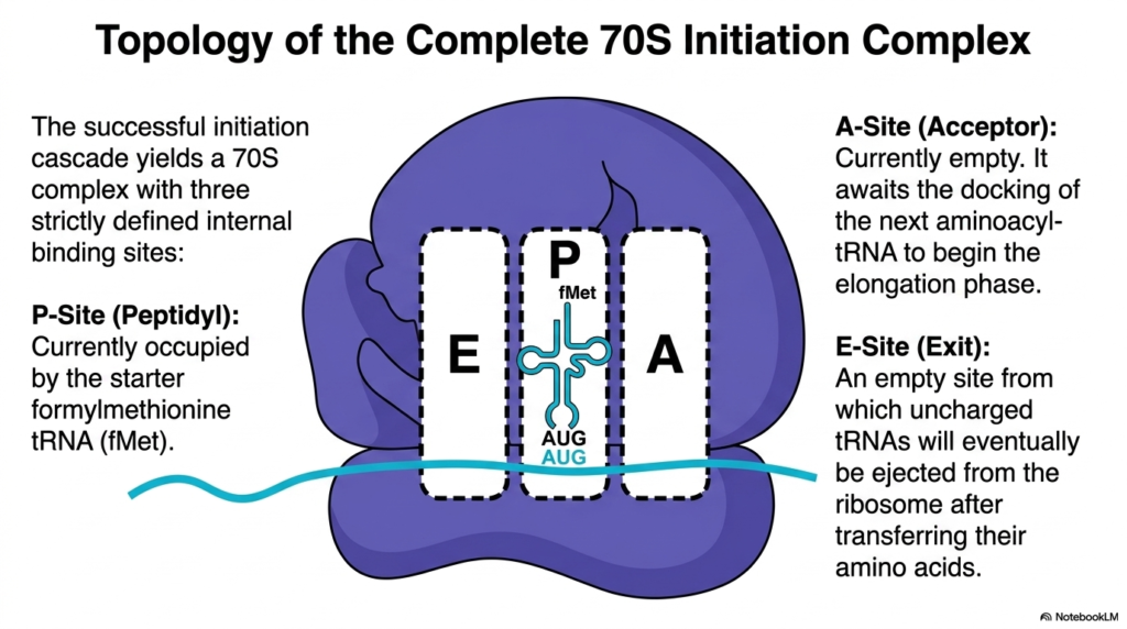
The successful culmination of the previous regulated phases results in a magnificent molecular structure primed for biological action. This final slide examines the internal topology of the completely assembled 70S initiation complex. Following a flawless sequence of Protein Translation Initiation, the resulting 70S ribosome possesses three strictly defined, distinct internal binding cavities: the Acceptor site, the Peptidyl site, and the Exit site. These three internal chambers, commonly abbreviated as the A-Site, P-Site, and E-Site, serve as a specialized mechanical conveyor belt that will systematically guide incoming transfer RNA molecules through the massive ribosomal core during elongation.
A defining characteristic of a newly formed initiation complex is the highly specific spatial positioning of the starter components. Because of the careful preparatory steps taken during Protein Translation Initiation, the starter formylmethionine transfer RNA molecule sits anchored within the central P-Site. This crucial peptidyl chamber is designed to hold the growing polypeptide chain, and placing the first amino acid directly into this middle slot is essential for proper biological function. The initial AUG start codon of the messenger RNA template is seamlessly aligned directly beneath this central chamber, dictating absolute molecular precision for the entire sequence.
Surrounding this occupied central chamber are two vacant, waiting cavities. The A-Site currently remains empty, patiently awaiting the docking of the next appropriate aminoacyl-tRNA molecule to officially begin the rapid elongation phase. Similarly, the E-Site is currently vacant, acting as a designated empty exit portal from which completely uncharged, depleted transfer RNAs will eventually be forcefully ejected from the massive ribosome after successfully transferring their molecular cargo. The architectural configuration of these three internal sites marks the official, highly successful conclusion of Protein Translation Initiation, definitively transforming the cellular machinery into an active protein-manufacturing powerhouse.
Please read our Content Disclaimer Statement.
Check out our social media channels: