85. RNA Maturation: Mechanisms of Eukaryotic RNA Processing
The intricate process of converting a raw genetic code into a readable set of instructions is a fascinating phenomenon in molecular biology. This slide deck explores the vital biochemical steps that prepare transcripts for protein synthesis. Designed for college and medical students, the content dissects complex molecular structures into clear, understandable mechanisms. By analyzing these visuals, learners will uncover the precise chemical modifications and structural changes required to protect and refine the genetic blueprint before it reaches the ribosome.
Slide 1: Introduction to RNA Maturation: Transforming the Genetic Blueprint
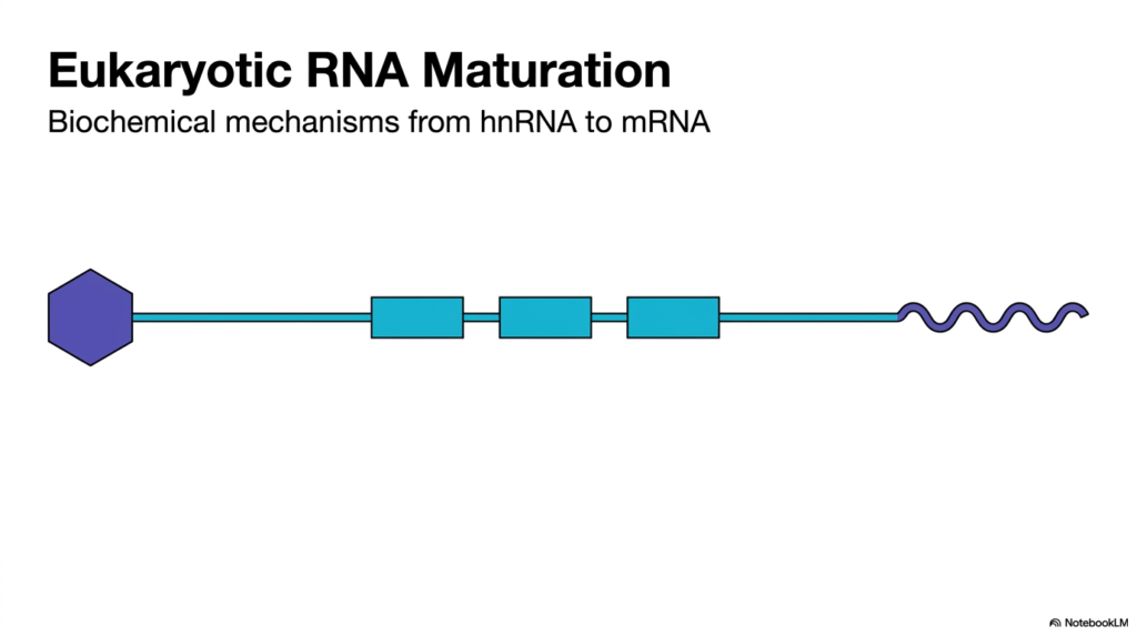
The journey of gene expression begins within the eukaryotic nucleus, but the initial genetic transcript is raw and unusable. This opening visual introduces the overarching concept of RNA Maturation, a mandatory biochemical overhaul that every transcript must undergo to become functional. Without the crucial steps of RNA Maturation, the newly synthesized blueprint—known as heterogeneous nuclear RNA or hnRNA—would remain unreadable and vulnerable to rapid degradation. The slide captures this transformation, illustrating how a basic linear sequence is chemically and structurally modified into a functional messenger RNA molecule ready for cellular action.
As the diagram demonstrates, the raw transcript undergoes massive structural changes at both ends and within its core coding region. A distinct chemical cap, represented by the purple hexagon, is attached to the starting end. Meanwhile, a wavy, repetitive tail is added to the trailing end. These physical modifications represent the defining features of RNA Maturation, serving as essential protective armor for the genetic code. The central blue boxes represent the vital coding regions that must be preserved. This visual simplification establishes the structural baseline for the complex molecular choreography that occurs before the transcript translates into proteins.
Understanding this transformation is critical for students studying molecular medicine, because errors in this specific pathway lead to severe genetic diseases. By mastering the sequential stages of RNA Maturation, learners gain profound insight into how eukaryotic cells regulate and protect their protein production machinery. The straightforward diagram shown here serves as an essential roadmap for the detailed biochemical mechanisms explored in the subsequent slides. This foundational view prepares the student to delve into the chemical nuances of transcript processing, from capping to complex splicing.
Slide 2: The Phases of RNA Maturation: Preparing Messages for Cytoplasmic Export
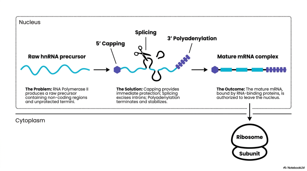
The transition from a newly transcribed sequence to a functional molecule requires distinct, sequential modifications. This slide breaks down the core phases of RNA Maturation occurring within the nucleus. RNA Polymerase II initially produces a raw precursor containing non-coding regions and unprotected termini. This presents a significant biochemical problem, as the transcript is vulnerable to nuclear exonucleases. The diagram outlines the solution through three primary events: 5′ capping, splicing, and 3′ polyadenylation. Each phase plays a distinct role in securing the transcript’s structural integrity.
The visual employs simple icons to represent the solutions provided by RNA Maturation. Capping provides immediate protection to the transcript’s front end. A pair of scissors symbolizes the splicing mechanism, which meticulously excises the intervening non-coding introns. Finally, polyadenylation terminates and stabilizes the sequence at the rear. These three concurrent steps ensure the raw transcript transforms into a secure, mature messenger RNA complex. The illustration links the problem of a raw precursor directly to its precise biochemical solutions.
The ultimate outcome of RNA Maturation is a transcript authorized to leave the nucleus. The diagram shows the mature mRNA complex, bound by specific RNA-binding proteins, migrating across the nuclear boundary into the cytoplasm. Here, it targets the ribosomal subunits for protein synthesis. RNA Maturation ensures that only fully processed and protected transcripts reach the cytoplasm, preventing the translation of faulty genetic codes. This spatial representation helps students visualize the crucial compartmentalization of eukaryotic gene expression.
Slide 3: The 5′ Cap in RNA Maturation: Securing the Terminus for Translation
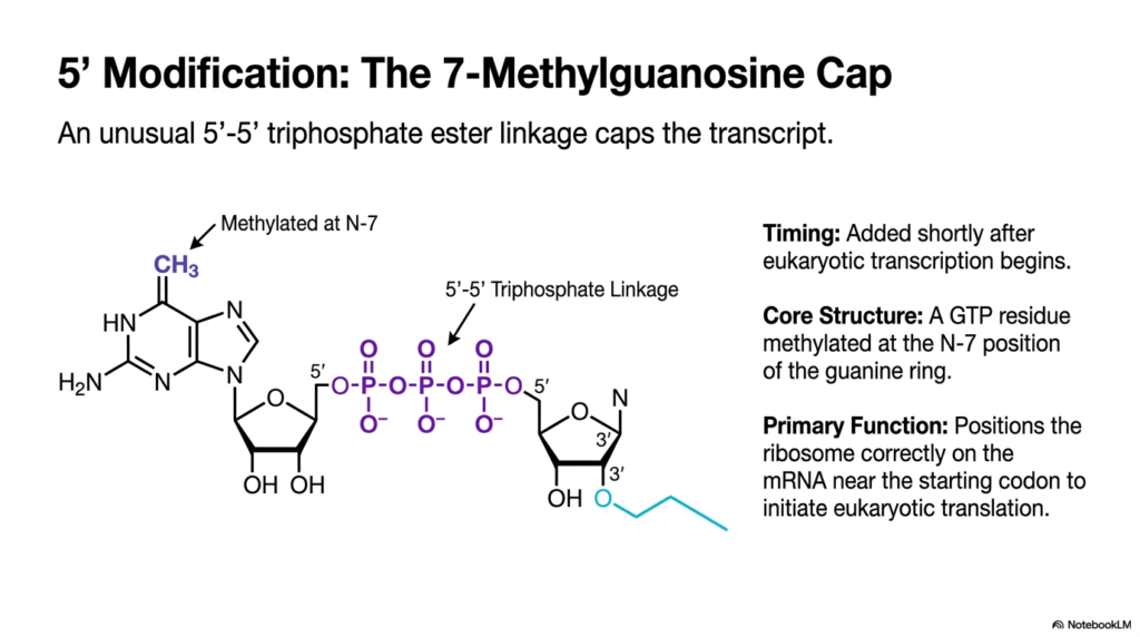
The first critical modification in transcript processing occurs almost immediately after transcription initiates. This slide delves into the biochemical structure of the 5′ cap, a fundamental component of RNA Maturation. A specialized enzyme complex adds a 7-methylguanosine cap to the free end of the nascent transcript. The visual displays the molecular structure of this cap, highlighting a guanine ring methylated at the N-7 position. This distinct chemical addition protects the transcript from rapid enzymatic degradation in the nuclear environment.
A defining biochemical feature shown in the diagram is the unusual 5′-5′ triphosphate ester linkage. Unlike the standard bonds that form the RNA backbone, this unique linkage in RNA Maturation connects the cap to the transcript in an inverted orientation. The three phosphate groups create a robust, enzyme-resistant bridge. This structural anomaly is a hallmark of eukaryotic transcripts, distinguishing them from other cellular nucleic acids. Students must recognize this specific chemical bond as it is a common target in pharmacological studies.
Beyond protection, the primary function of this cap is vital for later cellular processes. The diagram notes that the modification is critical for positioning the ribosome correctly on the mRNA. RNA Maturation ensures the translation machinery can locate the starting codon efficiently. Without this properly structured 5′ modification, translation initiation fails, rendering the entire transcript useless. Thus, the cap acts as both a protective shield and an indispensable molecular barcode for the translation apparatus.
Slide 4: The Polyadenylate Tail in RNA Maturation: Synthesizing a Protective Buffer
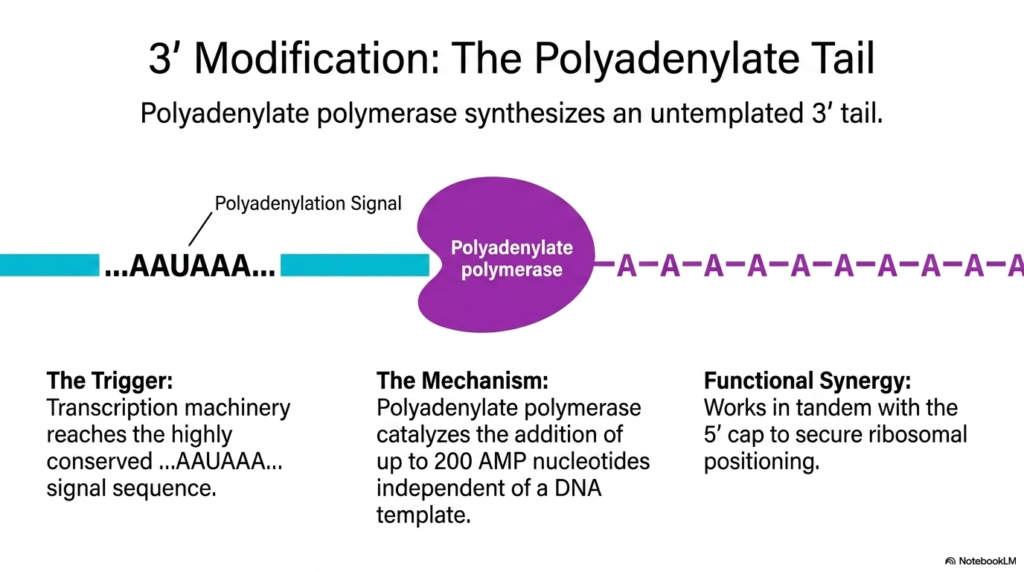
Following the protection of the front terminus, the transcript requires stabilization at its trailing end. This slide illustrates the 3′ modification process, a critical concluding phase of RNA Maturation. When the transcription machinery encounters a highly conserved AAUAAA signal sequence, it triggers the termination and modification cascade. The visual depicts a large purple enzyme complex, identified as polyadenylate polymerase, binding to the transcript. This enzyme drives the addition of the protective tail, securing the genetic message.
The biochemical mechanism of this enzyme is unique within RNA Maturation. Polyadenylate polymerase catalyzes the addition of up to 200 adenosine monophosphate nucleotides to the free 3′ end. Crucially, this synthesis is untemplated, meaning it occurs independent of a DNA template. The diagram shows the resulting chain of ‘A’ residues extending from the coding sequence. This creates a buffer zone that protects the critical upstream coding regions from gradual exonuclease degradation in the cytoplasm.
The functional synergy between the two transcript ends is a major theme in RNA Maturation. The slide text emphasizes that the poly-A tail works in tandem with the 5′ cap to secure ribosomal positioning. Together, these terminal modifications circularize the transcript during translation, vastly increasing the efficiency of protein synthesis. By understanding the creation of this untemplated tail, students can appreciate the dynamic, non-templated ways in which cells modify and protect their genetic investments.
Slide 5: Modification Matrix in RNA Maturation: Comparing Transcript Termini
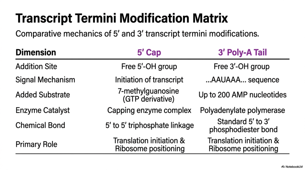
To consolidate understanding of transcript protection, this slide presents a comparative matrix of the terminal modifications. It places the 5′ cap and the 3′ poly-A tail side-by-side, summarizing a significant portion of RNA Maturation. The table contrasts the specific dimensions of each process, beginning with their addition sites on the free 5′-OH and 3′-OH groups. It also highlights the different signal mechanisms, contrasting the early initiation trigger of the cap with the specific AAUAAA sequence trigger for the tail.
The matrix further details the distinct chemical substrates and enzymes utilized in RNA Maturation. The 5′ end receives a 7-methylguanosine derivative catalyzed by a capping enzyme complex, forming an unusual 5′ to 5′ triphosphate linkage. Conversely, the 3′ end receives numerous AMP nucleotides via polyadenylate polymerase, connected through standard 5′ to 3′ phosphodiester bonds. This clear side-by-side comparison helps students memorize the precise biochemical differences between the two ends, which is frequently tested in biochemistry curricula.
Despite their structural and chemical differences, the matrix underscores a shared biological purpose. Both terminal modifications in RNA Maturation share the primary role of translation initiation and ribosome positioning. They function collaboratively to signal that the transcript is intact and ready for protein synthesis. This slide serves as a high-yield study tool, stripping away the complex visuals to deliver the core testable facts regarding the protective bookends of eukaryotic messenger transcripts.
Slide 6: The Intervening Sequence Problem: Ensuring Precision in RNA Maturation
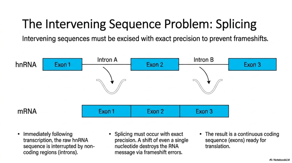
Eukaryotic genes are inherently fragmented, presenting a major obstacle for protein synthesis. This slide illustrates the intervening sequence problem, a massive challenge solved by RNA Maturation. Following transcription, the raw hnRNA sequence is interrupted by non-coding regions known as introns. The diagram depicts Exon 1, Exon 2, and Exon 3 separated by Intron A and Intron B. These intervening non-coding segments must be removed to create a functional genetic blueprint.
The visual highlights the strict requirement for exact precision during RNA Maturation. The excision of introns is represented by curved arrows pulling the sequences away from the transcript. Splicing must occur with exact accuracy. The text notes that a shift of even a single nucleotide destroys the RNA message via frameshift errors. If the splicing machinery cuts one base too early or too late, every subsequent amino acid in the resulting protein will be incorrect, leading to nonfunctional proteins.
The final result of this meticulous excision is a continuous coding sequence. The bottom of the diagram shows the mRNA with all three exons perfectly joined together. RNA Maturation ensures the transcript is ready for translation. This slide emphasizes the high stakes of the splicing process, reminding students that the genetic code relies entirely on the structural fidelity maintained during this exact phase of transcript processing.
Slide 7: The Spliceosome in RNA Maturation: Nature’s Complex Macromolecular Machine
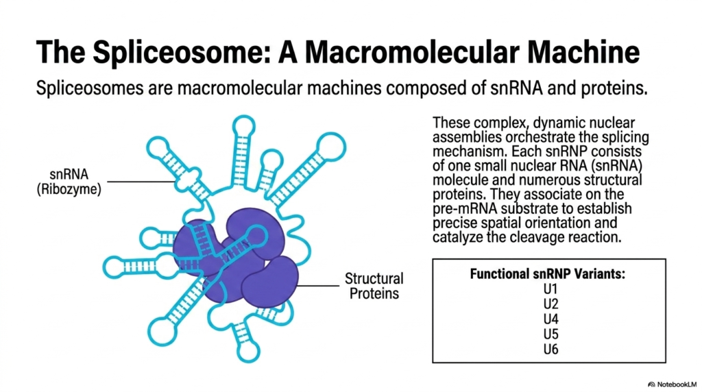
The removal of introns is not a simple chemical reaction; it requires a massive, dynamic apparatus. This slide introduces the Spliceosome, the primary engine of splicing during RNA Maturation. The spliceosome is described as a macromolecular machine composed of both RNA and proteins. The detailed illustration shows a central cluster of structural proteins surrounded by intricate, folded strands of snRNA acting as ribozymes. This assembly is responsible for orchestrating the precise cleavage and ligation of transcripts.
The composition of the spliceosome is vital for students to understand. Each subunit, termed a small nuclear ribonucleoprotein (snRNP), contains one small nuclear RNA molecule and numerous structural proteins. The table lists the functional snRNP variants: U1, U2, U4, U5, and U6. Throughout RNA Maturation, these specific variants dynamically associate and dissociate on the pre-mRNA substrate. Their coordinated interactions establish the precise spatial orientation required to catalyze the complex cleavage reactions.
This dynamic nuclear assembly highlights the complexity of eukaryotic gene expression. The spliceosome meticulously scans and binds to the transcript to perform RNA Maturation. The snRNAs provide the catalytic power, while the proteins build the scaffold. Understanding the sheer scale and coordination of this macromolecular machine allows students to appreciate the sophisticated evolutionary solutions cells have developed to manage fragmented genetic sequences.
Slide 8: Intron Anatomy in RNA Maturation: Recognizing Crucial Splicing Sites
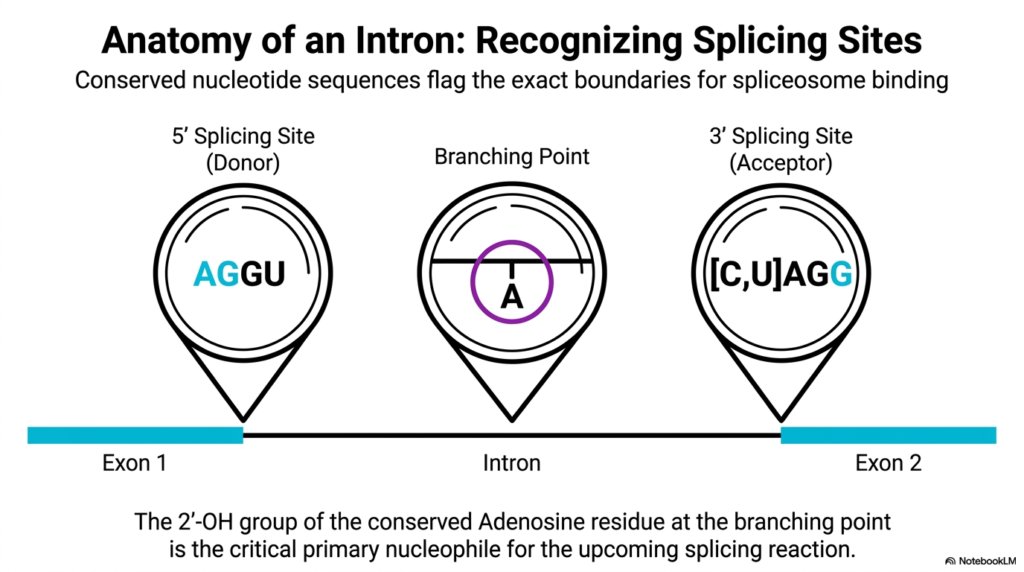
For the spliceosome to function accurately, it must know exactly where to cut. This slide maps out the anatomy of an intron, detailing the signals that guide RNA Maturation. Conserved nucleotide sequences flag the exact boundaries for spliceosome binding. The visual uses magnification circles to highlight three highly conserved regions: the 5′ splicing site, the branching point, and the 3′ splicing site. These sequence markers are non-negotiable requirements for the precise removal of non-coding regions.
The diagram explicitly defines the boundary markers that the machinery reads. The 5′ splicing site, or donor site, features the conserved sequence AGGU. At the opposite end, the 3′ splicing site, or acceptor site, is marked by [C,U]AGG. During RNA Maturation, the snRNPs scan the raw transcript to identify these exact nucleotide arrangements. Recognizing these boundaries ensures that the spliceosome does not accidentally cleave the critical coding sequences located in the adjacent exons.
Centrally located within the intron is the branching point, a feature of paramount chemical importance. The magnification highlights a specific Adenosine residue. The text points out that the 2′-OH group of this conserved Adenosine is the critical primary nucleophile for the upcoming reactions. RNA Maturation hinges on the reactivity of this specific hydroxyl group to initiate the complex chemical rearrangement of the transcript.
Slide 9: Intron Cleavage in RNA Maturation: Initiating the Transesterification Reaction
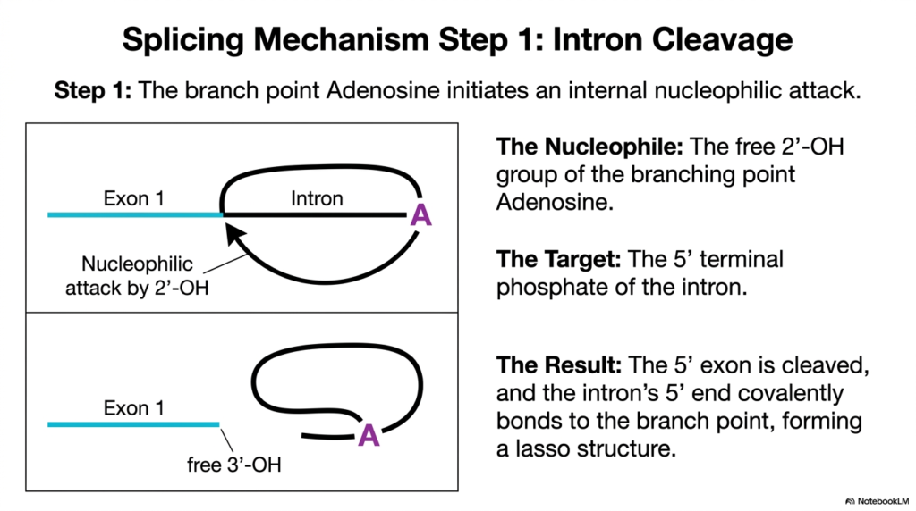
With the splicing sites recognized, the chemical transformation begins. This slide details Step 1 of the splicing mechanism in RNA Maturation, focusing on the initial intron cleavage. The reaction is driven by an internal nucleophilic attack initiated by the branch-point adenosine. The diagram illustrates a curved arrow demonstrating the 2′-OH group attacking the 5′ splice site. This chemical move breaks the continuous RNA backbone at a targeted location.
The text clarifies the specific chemical players involved in this step of RNA Maturation. The nucleophile is the free 2′-OH group of the branching point Adenosine. Its precise target is the 5′ terminal phosphate of the intron. The spliceosome carefully positions these two functional groups in close proximity, lowering the activation energy required for the attack. This transesterification reaction swaps one phosphodiester bond for another, altering the topology of the transcript.
The result of this first catalytic step is a dramatic structural rearrangement. The 5′ exon is cleaved and temporarily freed. Simultaneously, the 5′ end of the intron covalently bonds to the branch point Adenosine. As shown in the lower diagram, RNA Maturation at this stage creates a distinct, looped “lasso” structure. This physical tethering is a mandatory intermediate state before the final coding sequences can be joined.
Slide 10: The Lasso Structure in RNA Maturation: Exploring a Biochemical Anomaly
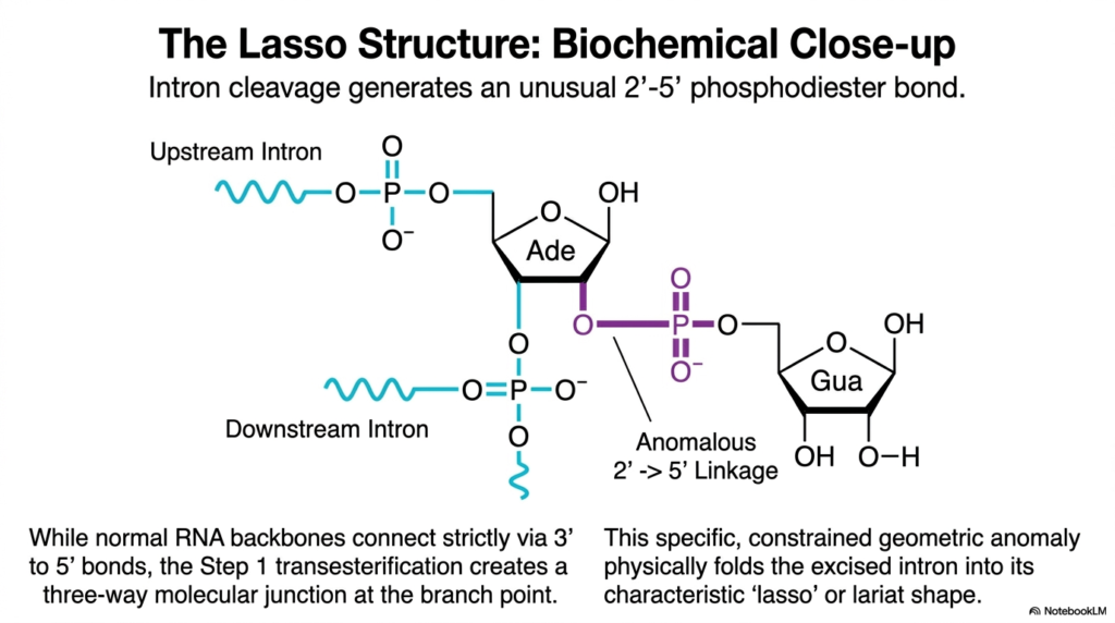
The intermediate stage of splicing produces a rare chemical linkage. This slide provides a biochemical close-up of the lasso structure formed during RNA Maturation. When the intron is cleaved, it generates an unusual 2′-5′ phosphodiester bond. The detailed molecular diagram shows the Adenine base forming three separate connections: one to the upstream intron, one to the downstream intron, and the anomalous linkage directly to the Guanine at the intron’s 5′ end.
This structural deviation is a fascinating aspect of RNA Maturation. While normal RNA backbones connect strictly via 3′ to 5′ bonds, this specific transesterification creates a three-way molecular junction at the branch point. The visual meticulously highlights the purple anomalous 2′ to 5′ linkage, emphasizing how the central Adenine residue acts as a chemical hub. This temporary architectural anomaly is a defining signature of eukaryotic splicing mechanisms.
The physical consequence of this bond is the characteristic shape of the excised sequence. This specific geometric anomaly physically folds the intron into its lasso or lariat shape. RNA Maturation relies on this tight folding to perfectly position the remaining free exon for the second step of the splicing reaction. By understanding this rare bond, students gain deeper insight into the flexible chemical properties of ribonucleic acids.
Slide 11: Exon Ligation in RNA Maturation: Unifying the Coding Sequences
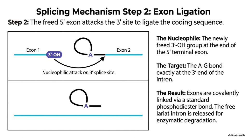
After the first cleavage, the severed coding regions must be permanently attached. This slide illustrates Step 2 of the splicing mechanism in RNA Maturation, which involves exon ligation. The previously freed 5′ exon now takes an active chemical role. The diagram shows a nucleophilic attack originating from the 3′-OH of Exon 1, targeting the 3′ splice site at the very end of the intron. This final chemical move seals the fate of the transcript.
The players in this second transesterification are clearly defined. The nucleophile is the newly freed 3′-OH group at the end of the 5′ terminal exon. Its target is the specific A-G bond exactly at the 3′ end of the intron. RNA Maturation requires the spliceosome to hold these reacting groups in perfect alignment. The attack breaks the final bond holding the intron to the transcript and connects the two exons.
The result is a unified and continuous genetic message. The exons are covalently linked via a standard phosphodiester bond, seamlessly joining the coding sequence. With the exons ligated, RNA Maturation concludes this phase by releasing the free lariat intron. This excised lasso structure is no longer needed and is quickly targeted for enzymatic degradation, recycling the nucleotides for future transcription events.
Slide 12: The Ribozyme Concept in RNA Maturation: RNA Driving Biochemical Catalysis
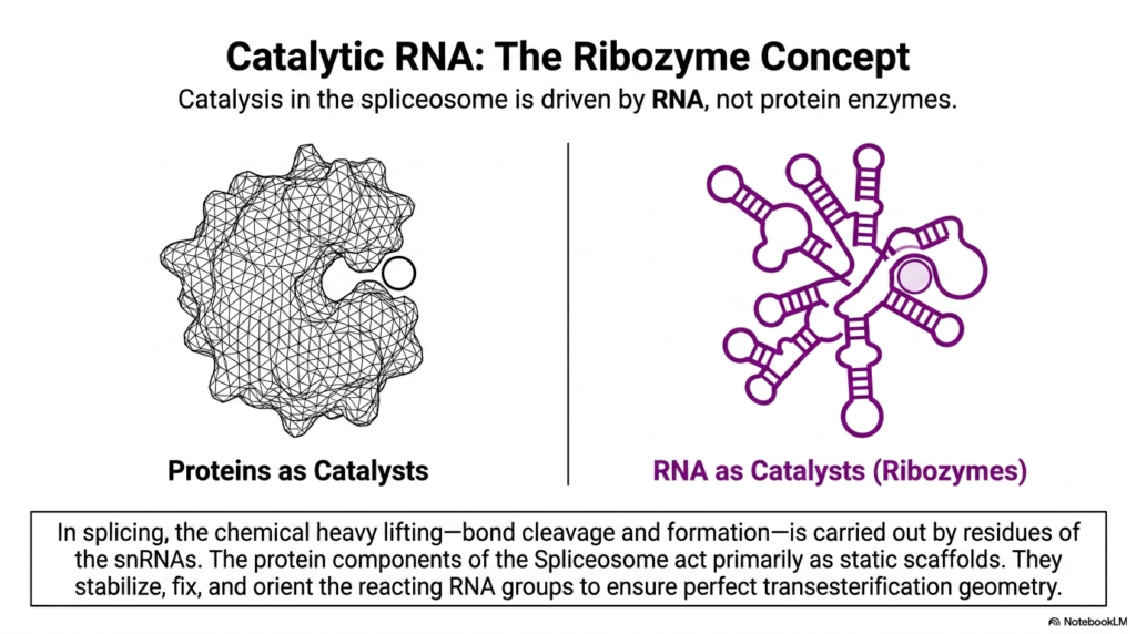
A profound paradigm shift in enzymology is showcased within the splicing machinery. This slide explains the Ribozyme concept, a defining feature of RNA Maturation. It illustrates a clear contrast between traditional protein catalysts, shown as a folded mesh surface, and RNA catalysts, depicted as complex folded loop structures. The core message is that catalysis within the spliceosome is driven by the RNA itself, rather than by standard protein enzymes.
In the context of RNA Maturation, the chemical heavy lifting is performed by the nucleic acids. The bond cleavage and formation observed in the transesterification steps are carried out by specific residues of the snRNAs. These snRNAs act as true ribozymes, possessing the chemical ability to lower activation energy and drive reactions. This highlights the evolutionary antiquity of RNA, demonstrating its capacity to perform complex biochemistry.
The structural proteins in the spliceosome still play an indispensable role. The slide notes that these protein components act primarily as static scaffolds. During RNA Maturation, they stabilize, fix, and orient the reacting RNA groups. By building a rigid architectural framework, the proteins ensure the perfect transesterification geometry required for the ribozymes to execute their precise catalytic cuts without error.
Slide 13: Spatial Orientation in RNA Maturation: Achieving Precision via Base Pairing
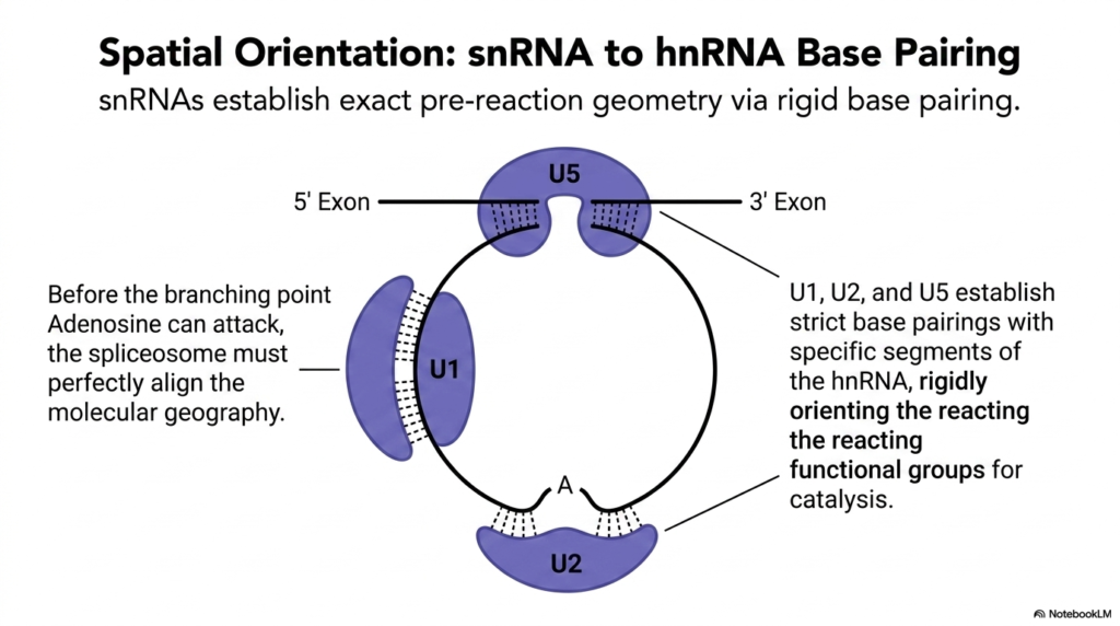
The catalytic power of ribozymes is useless without exact structural alignment. This slide explores the spatial orientation required during RNA Maturation. Before the branching point, Adenosine can initiate its nucleophilic attack; the spliceosome must align the molecular geography. The diagram visualizes how the snRNP complexes, specifically U1, U2, and U5, bridge the gaps between the exons and the intron, manipulating the transcript into a highly specific conformation.
This precise pre-reaction geometry is established via rigid base pairing. The snRNAs contain sequences that are perfectly complementary to the conserved boundary markers on the hnRNA. The visual shows dotted lines representing these hydrogen bonds, binding U1 to the 5′ site, U2 to the branch point, and U5 holding the two exons in close proximity. RNA Maturation depends on this extensive network of base pairing to stabilize the massive complex.
By establishing these strict base pairings, the snRNAs rigidly orient the reacting functional groups for catalysis. U1, U2, and U5 physically force the branching point into the correct spatial position to attack the 5′ splice site. RNA Maturation relies on molecular precision; without this three-dimensional scaffolding provided by complementary base pairing, the intricate cleavage and ligation steps would be biochemically impossible.
Slide 14: The Final Product of RNA Maturation: The Mature Eukaryotic mRNA
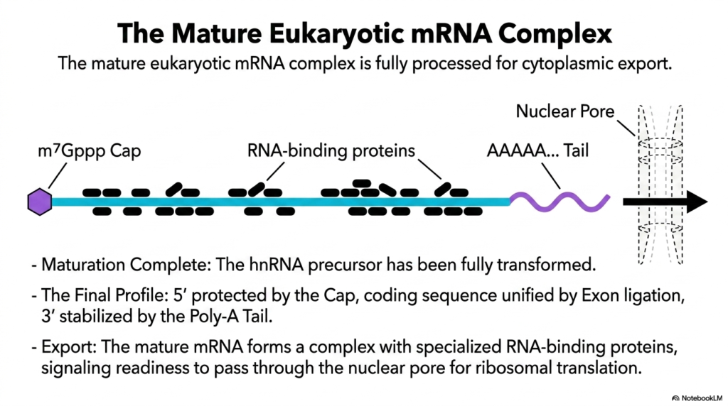
The culmination of all processing steps results in a transcript prepared for cellular action. This final slide showcases the mature eukaryotic mRNA complex, marking the completion of RNA Maturation. The diagram illustrates a polished transcript, featuring the m7Gppp Cap, a continuous coding sequence unified by exon ligation, and a stabilizing Poly-A Tail. Furthermore, the sequence is coated in specialized RNA-binding proteins, indicating it is fully processed.
The transcript has undergone a massive physical and chemical transformation. The initial hnRNA precursor has been overhauled. RNA Maturation has ensured the 5′ end is protected, the disruptive introns are removed, and the 3′ end is buffered. This final profile is vital for the molecule’s survival. The presence of the RNA-binding proteins acts as a molecular passport, signaling to the cell that the maturation process was successful and accurate.
With maturation complete, the transcript is authorized for cellular export. The visual depicts the mature mRNA complex moving toward a nuclear pore, symbolized by the dashed cylinder. It is now ready to pass through the nuclear envelope and enter the cytoplasm. The ultimate goal of RNA Maturation is achieved: delivering a pristine, secure, and readable genetic blueprint to the ribosome for the synthesis of vital cellular proteins.
Please read our Content Disclaimer Statement.
Check out our social media channels: