114. Monoclonal Antibodies in Clinical Diagnostics: Harnessing Molecular Specificity for Medicine
Modern medicine relies heavily on finding invisible signals within the human body. From detecting minor hormone fluctuations to identifying disease markers, diagnostic testing requires extreme accuracy. This slide deck breaks down how scientists engineer custom biological tools to detect trace molecules with unmatched precision.
Slide 1: Monoclonal Antibodies and Immunoassay Foundations

Targeted diagnostics transformed medicine by allowing clinical laboratories to measure molecules present at minute levels in blood serum. This visual overview establishes the foundational link between bioengineered protein tools and analytical detection techniques. At the core of this system are Monoclonal Antibodies, uniform proteins designed to bind specific target shapes with extreme precision.
The image depicts a characteristic Y-shaped immunoglobulin structure interacting directly with a target antigen molecule represented by a circle. In a natural biological system, immunoglobulins recognize specific foreign invaders. In diagnostic testing, however, Monoclonal Antibodies serve as custom probes that selectively attach to target analytes without binding to unrelated background molecules. This selective binding capability forms the physical basis for modern immunoassay systems used across research and medicine.
Understanding this initial concept is essential for mastering advanced biochemical assays. When an engineered protein binds its target, it forms a stable complex that can be measured using various detection methods. These detection methods convert tiny molecular interactions into readable signals, such as color changes or light emission. Consequently, Monoclonal Antibodies act as the critical recognition element that makes quantitative diagnostic testing possible.
By combining molecular biotechnology with clinical laboratory techniques, researchers can measure hormones, drugs, and disease markers at remarkably low concentrations. As students progress through this presentation, they will explore how cell fusion techniques produce these uniform proteins and how their unique binding properties drive competitive enzyme-linked assays. Mastering these biochemical principles provides a clear window into how laboratory findings guide clinical decisions every day.
Slide 2: Monoclonal Antibodies and the Specificity Advantage
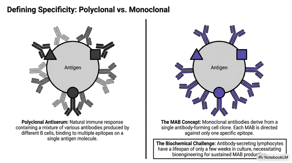
Clinical testing demands extreme specificity to distinguish target molecules from thousands of background proteins in serum samples. This comparison highlights the structural difference between natural polyclonal responses and targeted bioengineered solutions. Polyclonal antiserum consists of a mixed population of proteins derived from multiple B cell lineages. These diverse proteins bind to various distinct structural features, known as epitopes, across a single target antigen molecule.
While polyclonal responses provide broad protection in natural immunity, they introduce variability and cross-reactivity in diagnostic settings. In contrast, Monoclonal Antibodies originate from a single immortalized cell clone. This single-clone origin ensures that every produced protein possesses an identical molecular structure and binds exclusively to one specific epitope. Because every single protein molecule targets the exact same structural site, Monoclonal Antibodies offer reproducible specificity that eliminates unwanted background binding in laboratory assays.
However, harnessing this specificity presents a major biochemical challenge. Normal antibody-secreting B lymphocytes have a limited natural lifespan in laboratory culture dishes, typically dying after just a few weeks. This brief lifespan prevents scientists from maintaining a continuous, long-term supply of uniform recognition proteins. To overcome this biological expiration date, researchers must use advanced cellular engineering techniques to sustain monoclonal antibody production over time.
This cellular engineering approach preserves the desired single-epitope specificity while granting the host cells the capacity for endless replication. Fusing short-lived lymphocytes with immortal cancer cells ensures a permanent source of Monoclonal Antibodies for clinical applications. Students analyzing this process can observe how overcoming cellular aging paved the way for mass-producing identical molecular tools for high-precision diagnostic testing.
Slide 3: Monoclonal Antibodies and Hybridoma Generation
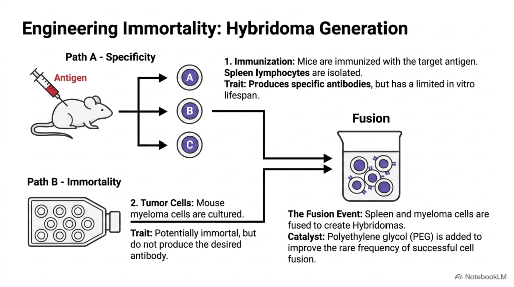
Generating a continuous supply of identical proteins requires combining two distinct cellular traits: specific target recognition and indefinite cell division. This diagram outlines the multi-step cellular fusion process that engineers immortalized hybrid cells, known as hybridomas. Path A begins by immunizing a mouse with a specific target antigen. The animal’s immune system responds by producing spleen lymphocytes that make specific target proteins, though these cells naturally have a brief lifespan in culture.
Path B focuses on cultivating mouse myeloma cells, cancerous plasma cells that can divide indefinitely. However, these specialized myeloma cells cannot synthesize the desired target protein. To combine the ideal traits of both cell lines, scientists mix isolated spleen lymphocytes with myeloma cells in the presence of polyethylene glycol. Polyethylene glycol acts as a chemical catalyst that alters membrane fluidity, dramatically increasing the low rate of successful cell membrane fusion.
The resulting cell fusion event joins the specific genetic machinery of the spleen lymphocyte with the endless proliferative capability of the myeloma cell. This engineered fusion cell is termed a hybridoma. Through this hybrid cell line, scientists establish a permanent biological factory capable of producing Monoclonal Antibodies continuously over time. Without this fusion technique, maintaining long-term monoclonal antibody production would remain biologically impossible.
By successfully merging these two pathways, biotechnology solves the fundamental problem of lymphocyte mortality in laboratory environments. These hybrid cell lines allow researchers to harvest Monoclonal Antibodies in virtually unlimited quantities. Students should note that cell fusion creates a mixed population containing fused hybridomas along with leftover unfused spleen cells and unfused myeloma cells that must be separated.
Slide 4: Monoclonal Antibodies and Biochemical Selection in HAT Medium
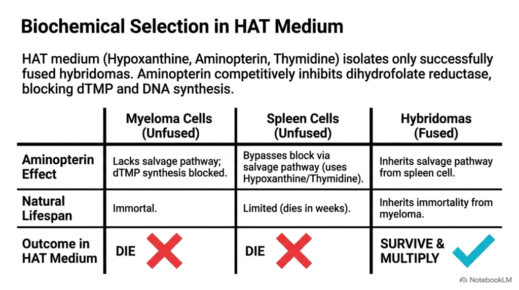
After cell fusion, laboratory cultures contain a mixture of unfused myeloma cells, unfused spleen cells, and successfully fused hybrid cells. This chart illustrates how HAT medium, containing hypoxanthine, aminopterin, and thymidine, selectively eliminates unwanted cells while isolating hybridomas that can produce monoclonal antibodies. Aminopterin acts as a powerful competitive inhibitor of dihydrofolate reductase, an enzyme required for de novo nucleotide synthesis. By blocking this primary pathway, aminopterin prevents cells from synthesizing thymidine monophosphate and DNA.
To survive this metabolic block, cells must rely on the salvage pathway, which requires specific salvage enzymes to utilize hypoxanthine and thymidine provided in the growth medium. Unfused myeloma cells lack these vital salvage pathway enzymes. Consequently, when aminopterin blocks de novo synthesis, unfused myeloma cells cannot manufacture DNA and quickly die out in culture.
Unfused spleen cells do possess the necessary salvage pathway enzymes and can survive the metabolic block temporarily. However, because normal lymphocytes have a brief natural lifespan, unfused spleen cells naturally die off within a few weeks. In contrast, successfully fused hybridomas inherit the functional salvage pathway from their spleen-cell parent and the infinite proliferative capacity from their myeloma-cell parent. As a result, hybridomas survive aminopterin inhibition and multiply rapidly in HAT medium.
This metabolic selection mechanism ensures that only true hybrid cells capable of producing monoclonal antibodies survive. By exploiting metabolic differences between cell lines, scientists isolate pure colonies of monoclonal antibody-producing cells without damaging them. Understanding metabolic pathways and enzymatic inhibition helps students see how basic biochemistry solves complex biotechnology problems in medical research.
Slide 5: Monoclonal Antibodies and Mass Production Methods
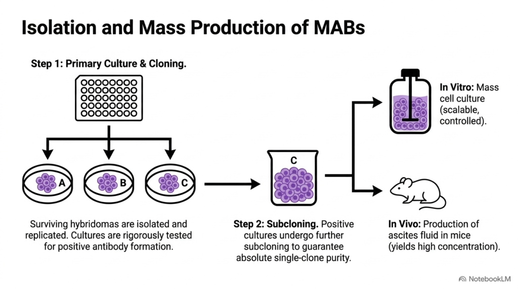
Isolating surviving hybridomas is only the first step in establishing a reliable manufacturing pipeline for diagnostic testing. This flowchart details how positive cell cultures undergo primary screening, subcloning, and large-scale expansion. After metabolic selection, surviving hybridoma colonies are placed in individual microtiter wells and rigorously tested to identify which cultures secrete functional Monoclonal Antibodies against the target antigen.
Once positive cultures are identified, they undergo a critical process called subcloning. Subcloning involves diluting cell suspensions until single cells are isolated into individual culture wells. This step ensures absolute clonal purity, so every cell in the resulting population arises from one original hybridoma cell. Absolute clonal purity is vital because even minor cell heterogeneity can compromise the consistency of manufactured Monoclonal Antibodies over long production runs.
After securing pure single-cell clones, production shifts to mass expansion through either in vitro or in vivo pathways. In vitro production uses large-scale bioreactors, offering a controlled, scalable environment that yields consistent protein batches without requiring animal hosts. Alternatively, in vivo production involves injecting hybridoma cells into the peritoneal cavity of mice, generating ascites fluid containing extraordinarily high concentrations of Monoclonal Antibodies.
Both production routes supply the high volumes required for commercial diagnostic kits and therapeutic applications worldwide. Through these systematic isolation and expansion steps, laboratory discoveries transform into reliable biological tools. Students should recognize that rigorous quality control and single-clone purification ensure monoclonal antibodies maintain identical binding characteristics across global diagnostic manufacturing facilities for clinical applications.
Slide 6: Monoclonal Antibodies and Immunoassay Principles
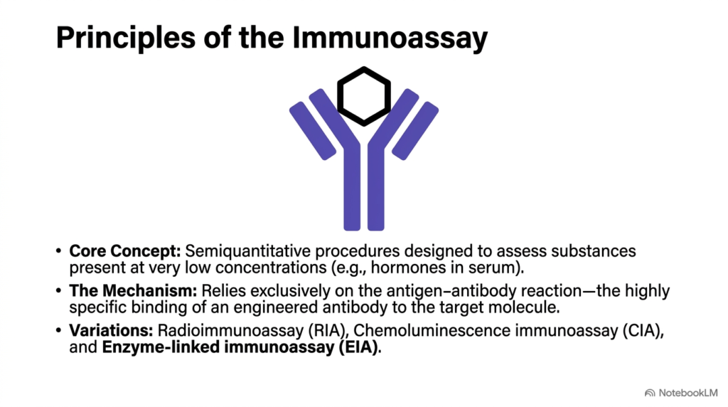
Now that we’ve established how uniform recognition proteins are bioengineered, we turn to how these biological tools function within diagnostic testing systems. This slide introduces the fundamental principles of the immunoassay, an analytical technique designed to measure trace biochemicals in complex clinical samples. Immunoassays quantify vital substances present at extremely low concentrations, such as thyroid hormones, steroid molecules, and cardiac markers in human blood serum.
The core mechanism of any immunoassay relies entirely on the antigen-antibody reaction. In these diagnostic setups, highly specific Monoclonal Antibodies act as molecular capture probes, seeking out and binding exclusively to the target molecule among millions of background serum components. Because this binding interaction displays high affinity and selectivity, Monoclonal Antibodies capture minute quantities of target molecules without interference from structurally similar compounds in the sample.
Diagnostic laboratories employ several analytical variations based on this core recognition principle. Radioimmunoassays use radioactive isotopes to trace binding events, offering high sensitivity but requiring specialized radiation-safety handling procedures. Chemiluminescence immunoassays measure light emitted during chemical reactions, providing an excellent dynamic range. Enzyme-linked immunoassays leverage enzymatic catalysts to generate visible color changes proportional to analyte concentration in test wells.
Across all these technical variations, Monoclonal Antibodies remain the essential molecular foundation that grants the assay its analytical specificity. Without highly specific binding from Monoclonal Antibodies, non-specific background interference would obscure minor biochemical signals in blood samples. Students examining these analytical systems will see how fundamental binding kinetics enable clinicians to detect silent biochemical imbalances early in disease progression.
Slide 7: Monoclonal Antibodies in Competitive Immunoassay Setups
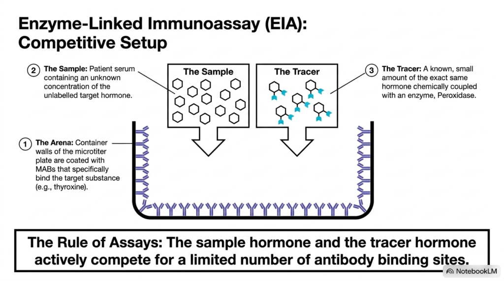
Understanding competitive analytical setups shows how laboratories quantify small molecules with a single binding epitope. This diagram details the structural layout of a competitive enzyme-linked immunoassay designed to measure serum hormones like thyroxine. The reaction container consists of a microtiter plate whose solid interior walls are coated with a uniform layer of Monoclonal Antibodies specific for the target hormone.
The assay setup brings two distinct fluid components into the antibody-coated reaction well. The first component is the patient sample, which contains an unknown concentration of unlabelled target hormone molecules. The second component is the tracer solution, which contains a known, fixed concentration of the exact same hormone chemically attached to a reporter enzyme, such as peroxidase.
When both solutions enter the reaction vessel, a direct molecular competition begins. Because the number of immobilized Monoclonal Antibodies on the vessel wall is strictly limited, the unlabelled patient hormone and the enzyme-labeled tracer hormone actively compete for the same binding sites. If the patient sample contains a high concentration of hormone, those unlabelled molecules occupy most available Monoclonal Antibodies, leaving very few sites open for tracer molecules.
Conversely, if the patient sample contains very little target hormone, the enzyme-labeled tracer molecules bind to most available antibody sites. This competitive binding rule forms the physical basis of quantitative measuring systems. Students must realize that Monoclonal Antibodies drive this competition by providing a fixed, highly uniform number of binding sites that strictly control molecule capture in clinical assays.
Slide 8: Monoclonal Antibodies and the Incubation and Wash Phase
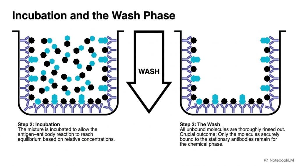
Accurate quantitative measurements require allowing competitive molecular binding to reach thermodynamic equilibrium, followed by removing non-bound background substances. This two-stage diagram illustrates the incubation period and subsequent washing step in a competitive assay setup. During the initial incubation phase, the patient sample and enzyme-labeled tracer remain in direct contact with the solid phase coated with Monoclonal Antibodies.
This incubation period provides sufficient time for the reversible antigen-antibody binding reaction to achieve thermodynamic equilibrium in solution. During this time, unlabelled patient hormones and enzyme-linked tracer molecules continuously collide with and bind to the immobilized Monoclonal Antibodies based on their relative concentrations. Higher concentrations of patient hormone successfully displace tracer molecules from available monoclonal antibody binding sites during this equilibrium phase.
Once equilibrium is reached, the reaction well undergoes a rigorous wash phase. A buffer solution rinses out the fluid contents, flushing away all unbound patient hormones, non-bound tracer molecules, and interfering serum proteins. The crucial outcome of this wash phase is that only molecules securely anchored to the stationary Monoclonal Antibodies remain attached to the microtiter well wall for subsequent diagnostic steps.
Removing non-bound enzyme-labeled tracer is vital for assay accuracy, as leftover free enzymes would generate false signal activity during subsequent chemical steps. By isolating only antibody-bound complexes, the wash phase prepares the assay vessel for precise signal generation. Students observing this step can appreciate how physical washing allows Monoclonal Antibodies to retain only target molecules, ensuring clean analytical measurements for laboratory diagnostics and patient evaluation.
Slide 9: Monoclonal Antibodies and Triggering the Indicator Reaction
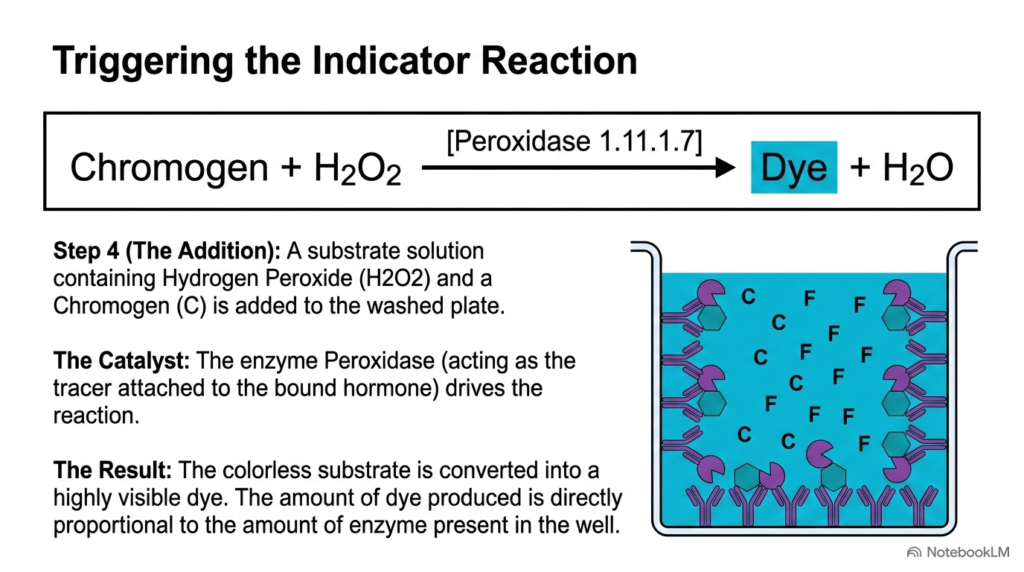
After removing unbound molecules during the wash phase, the remaining immobilized enzyme activity must be converted into a measurable visual signal. This diagram details the chemical indicator reaction that visualizes bound tracer molecules attached to immobilized Monoclonal Antibodies. A clear substrate solution containing hydrogen peroxide and a colorless chromogen compound is introduced into the washed reaction well.
The attached enzyme, peroxidase, acts as a biological catalyst that speeds up the chemical transformation. Peroxidase breaks down hydrogen peroxide while transferring electrons to the colorless chromogen molecule. This enzymatic reaction converts the colorless chromogen into a highly visible, intensely colored dye product, releasing water as a neutral byproduct in the well environment during incubation.
Because every bound tracer molecule carries a functional peroxidase enzyme, the rate of dye formation depends directly on the amount of tracer captured by the stationary Monoclonal Antibodies. The enzyme acts as a signal multiplier, converting single binding events into thousands of colored product molecules over time. This catalytic amplification allows clinical detection systems using monoclonal antibodies to detect extremely small amounts of target analyte in solution.
If many tracer molecules remain bound to the vessel wall, the enzyme reaction produces an intense color signal. If few tracer molecules bind due to high patient hormone competition, the color output remains faint. Monoclonal Antibodies ensure that enzyme attachment reflects true competitive binding rather than non-specific adhesion. Students can clearly see how enzyme catalysis translates sub-microscopic binding events into vivid visual results for accurate medical diagnosis and patient management.
Slide 10: Monoclonal Antibodies and Photometric Quantitative Analysis
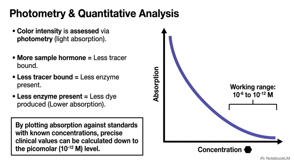
The final analytical stage converts visual color intensity into precise numeric analyte concentrations suitable for patient care. This slide illustrates photometric measurement and the resulting inverse calibration curve used in competitive assays. Clinical instruments pass specific wavelengths of light through the colored reaction well to measure light absorption, which reflects total dye production in the sample vessel during analysis.
In a competitive immunoassay setup, the relationship between patient analyte concentration and light absorption is strictly inverse. When a patient sample contains high hormone concentrations, those molecules occupy most Monoclonal Antibodies, leaving few binding sites for enzyme-linked tracer molecules. Consequently, less enzyme remains anchored to the well wall after washing, leading to lower dye production and reduced light absorption in the photometer.
Conversely, a sample with a low hormone concentration allows many tracer molecules to bind to available Monoclonal Antibodies, resulting in strong enzyme activity, high dye accumulation, and elevated absorption readings. Laboratory technicians plot these photometric absorption values against a standard curve generated from samples of known analyte concentrations. This standard curve allows precise calculation of patient hormone levels down to the picomolar range.
The wide working range spanning six orders of magnitude demonstrates the extraordinary sensitivity achieved by combining enzymatic amplification with single-clone binding specificity. Monoclonal Antibodies provide the constant binding affinity needed to maintain consistent calibration-curve geometry across different testing runs. Medical students analyzing this curve will understand how Monoclonal Antibodies enable clinical laboratories to transform raw light absorption values into actionable medical data for patient monitoring.
Slide 11: Monoclonal Antibodies: Synthesis from Clone to Clinical Result
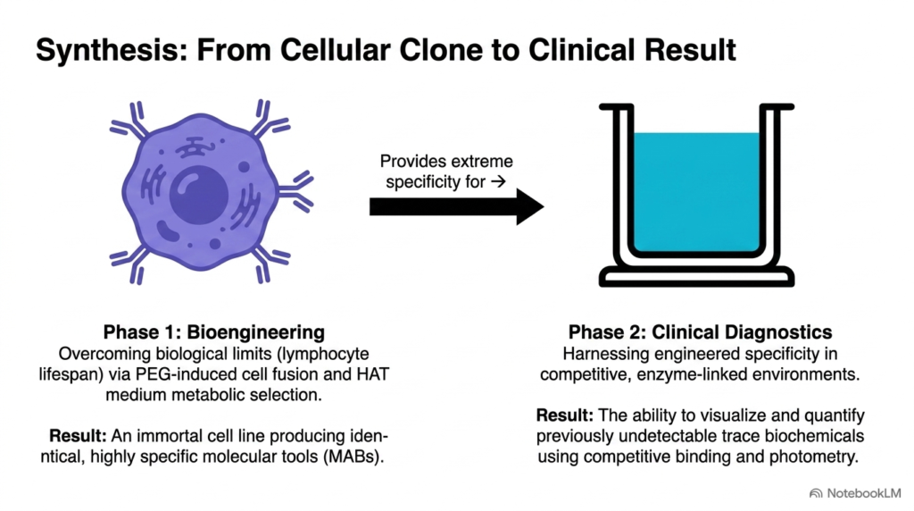
This summary slide integrates the entire journey of diagnostic biotechnology, linking cellular engineering directly to clinical laboratory outcomes. The left panel illustrates Phase 1: Bioengineering, where cell fusion and metabolic selection overcome basic biological limits. By combining short-lived antibody-producing lymphocytes with immortal myeloma cells, scientists create hybridomas that can manufacture unlimited quantities of identical Monoclonal Antibodies for medical use.
The right panel represents Phase 2: Clinical Diagnostics, where these engineered protein tools are deployed within competitive assay setups. Immobilized Monoclonal Antibodies act as high-precision biological filters, capturing target serum analytes with absolute specificity. Combined with competitive enzyme-linked tracers and photometric light detection, these assays allow clinical laboratories to detect trace hormones and disease markers that were previously impossible to measure accurately in routine practice.
This integrated process yields a powerful diagnostic platform that effectively bridges cellular biology and patient management. By moving from a single cell fusion event to quantitative light absorption readings, modern medicine can identify subtle disease states early and accurately. Understanding this full trajectory helps students see how monoclonal antibodies turn fundamental research into lifesaving clinical tools for global healthcare systems.
Throughout this series, Monoclonal Antibodies stand out as the central technology enabling reproducible, high-sensitivity diagnostic testing in modern medicine. By mastering both the cellular synthesis and the analytical chemistry behind these systems, future healthcare professionals can better interpret diagnostic data and appreciate the biotechnology powering modern patient care across clinical settings.
Please read our Content Disclaimer Statement.
Check out our social media channels: