112. Immunoglobulin Structure and Dynamics: How Antibodies Safeguard the Human Body
Have you ever wondered how the human body neutralizes millions of foreign invaders every single day without destroying its own tissues? The secret lies within a remarkably specialized family of protective proteins that serve as precision-guided molecular defense systems. This comprehensive slide deck breaks down the structural biochemistry, modular domain architecture, and physiological mechanisms that allow these protective proteins to recognize, bind, and eliminate diverse microbial threats. By systematically exploring their quaternary arrangements, chemical modifications, and functional classes, students will gain a clear foundation in how humoral immunity operates at a precise molecular level.
Slide 1: Molecular Architecture and Biological Function of Antibodies
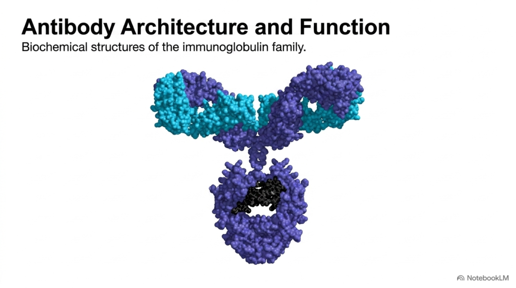
The human immune system relies on specialized defensive proteins to identify, neutralize, and eliminate foreign pathogens circulating throughout bodily fluids. These protective molecules, broadly known as antibodies, form the core structural framework of adaptive humoral immunity. This opening slide shows the three-dimensional molecular architecture of these Y-shaped protein complexes. Their elegant spatial arrangement allows them to circulate through blood and extracellular fluids, continuously scanning for matching pathogen surfaces while maintaining essential structural integrity under demanding physiological conditions within host tissues.
At a detailed biochemical level, antibodies consist of symmetrical polypeptide chains arranged to perform two distinct, highly coordinated biological functions. The upper branching arms bind specific target sites with extraordinary affinity, while the lower stem communicates directly with host cellular defense networks. Rather than destroying foreign invaders directly through intrinsic catalytic activity, antibodies function as molecular tags that mark targets for destruction by other immune components. This dual functionality demonstrates how structural modularity enables broad protective coverage across diverse host defense mechanisms without causing non-specific tissue damage.
Understanding this three-dimensional spatial conformation is foundational for mastering advanced immunology and biomedical engineering. Modern therapeutic development heavily utilizes engineered antibodies to combat autoimmune disorders, infectious diseases, and various malignancies. By studying their fundamental architecture, college and medical students can appreciate how subtle amino acid variations yield incredible functional diversity across biological systems. Ultimately, antibodies serve as indispensable tools in both natural immunity and modern clinical medicine, highlighting the remarkable elegance of biological macromolecular design across human health and disease.
Slide 2: Mechanisms of Humoral Defense Mediated by Antibodies
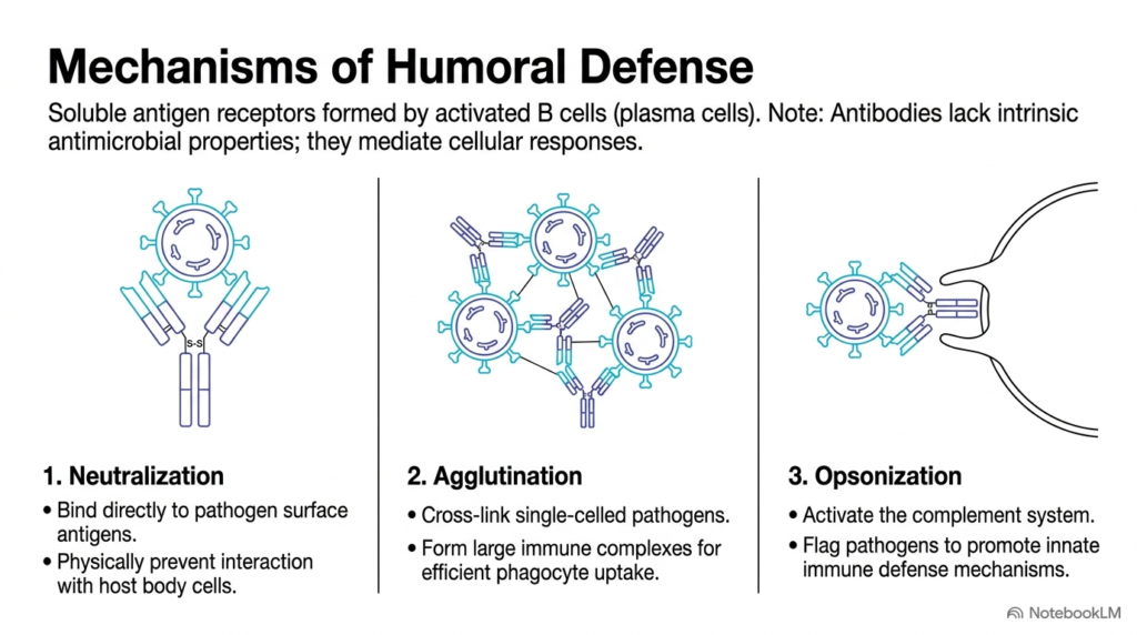
Humoral defense mechanisms depend heavily on how specialized soluble receptors produced by activated B cells interact with invading pathogens. Antibodies lack intrinsic antimicrobial or enzymatic properties, so they cannot directly degrade or kill foreign microbes on their own. Instead, antibodies function as critical molecular adapters that recruit and activate host cellular immune responses through three major pathways: neutralization, agglutination, and opsonization. These cooperative mechanisms ensure that foreign threats are rapidly contained, neutralized, and eliminated from the systemic circulation before causing extensive tissue damage.
Neutralization occurs when antibodies bind directly to pathogen surface antigens, physically blocking viruses or bacterial toxins from interacting with host cell receptors. In agglutination, multivalent antibodies cross-link single-celled pathogens into large, insoluble immune complexes, significantly enhancing phagocytic uptake by immune cells. Opsonization involves antibodies coating foreign surfaces to activate the complement system and flag pathogens, thereby promoting rapid recognition and engulfment by innate defense cells like macrophages and neutrophils. Together, these processes prevent widespread tissue invasion and systemic infection across vulnerable body organs.
Through these three coordinated mechanisms, antibodies effectively coordinate systemic cellular responses across the bloodstream and peripheral tissues. Neutralization prevents viral attachment, agglutination aggregates swimming bacterial populations, and opsonization dramatically boosts phagocyte engulfment. Recognizing how antibodies mediate these distinct pathways explains how humoral immunity efficiently neutralizes microbial threats without requiring direct catalytic destruction. This underscores the collaborative relationship between humoral recognition and innate cellular destruction in maintaining human health, highlighting why functional antibodies are essential for survival against pathogenic micro-organisms.
Slide 3: Immunoglobulin G as the Structural Prototype for Antibodies
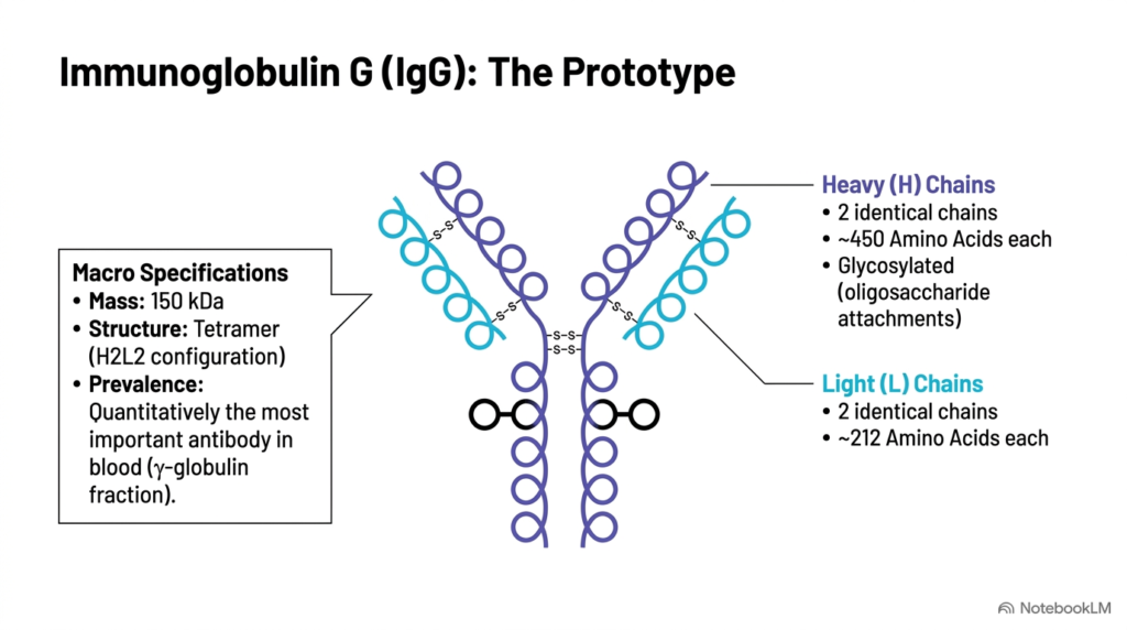
Immunoglobulin G, commonly abbreviated as IgG, serves as the classic structural prototype for the entire family of circulating antibodies. Possessing a total molecular mass of approximately 150 kilodaltons, IgG exists as a standard tetrameric unit arranged in an H2L2 polypeptide configuration. Quantitatively, IgG is the most abundant antibody class in human blood serum and interstitial fluids, constituting the vast majority of the gamma-globulin fraction during routine protein electrophoresis. Its high stability and long half-life make it a primary defender against systemic infections throughout the human body over extended periods.
The quaternary architecture of IgG consists of two identical Heavy chains and two identical Light chains held together by covalent interactions. Each Heavy chain contains roughly 450 amino acids and features attached oligosaccharide chains, classifying it as a glycosylated protein. The smaller Light chains each consist of approximately 212 amino acids and pair symmetrically with the Heavy chains. These structural features ensure that intact antibodies possess two functional antigen-binding sites per molecule, allowing them to bind bivalently to matching microbial epitopes with high avidity, structural stability, and molecular specificity.
The carbohydrate moieties attached to the Heavy chains play critical roles in maintaining overall structural stability and mediating interactions with cellular receptors. Because IgG serves as the structural baseline, understanding its H2L2 tetrameric blueprint is essential for evaluating other immunoglobulins. By mastering this foundational monomeric unit, students can better understand how more complex antibodies assemble into higher-order polymeric complexes to perform specialized protective duties across various anatomical compartments during acute and chronic immune responses.
Slide 4: Modular Domain Architecture of Functional Antibodies
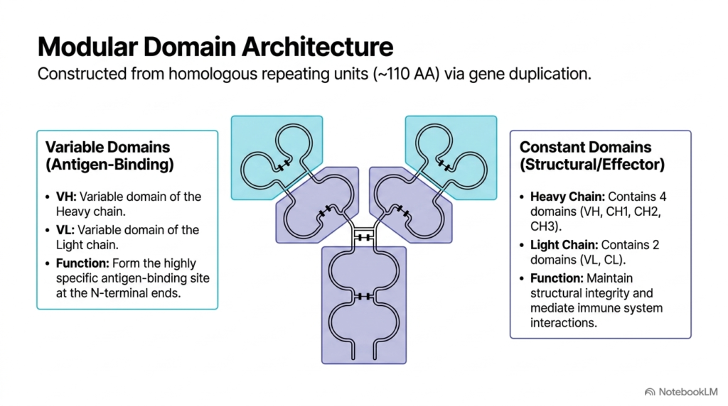
The overall structural framework of an antibody is constructed from homologous repeating units of approximately 110 amino acids, which arose historically through gene duplication events. These modular domains are functionally divided into Variable domains and Constant domains. Together, these repeating structural units allow antibodies to combine extreme antigen-binding specificity with uniform effector signaling, ensuring that recognition and immune activation remain tightly linked yet structurally independent. This modular organization provides the flexibility needed to adapt to the infinite variety of foreign antigens encountered in diverse environmental microenvironments and clinical settings.
Variable domains are located at the N-terminal ends of both Light and Heavy chains, designated as VL and VH, respectively. The precise spatial pairing of VH and VL constructs the highly specific antigen-binding pocket, enabling individual antibodies to discriminate between subtle chemical variations on target molecules. In contrast, Constant domains maintain structural framework integrity while mediating interactions with complement proteins and specialized Fc receptors expressed on host immune cells. This structural segregation prevents binding-site modifications from disrupting essential downstream effector functions during active immune responses in host tissues.
Regarding domain distribution, each Light chain consists of one Variable domain and one Constant domain (VL and CL). Each Heavy chain contains one Variable domain and three Constant domains (VH, CH1, CH2, and CH3). This modular domain architecture shows how functional antibodies separate variable target recognition from constant effector functions within a single, highly organized protein complex. Understanding this spatial distribution helps students appreciate how genetic recombination generates vast structural antibody diversity within the adaptive immune repertoire.
Slide 5: Structural Stabilization and Flexibility in Antibodies
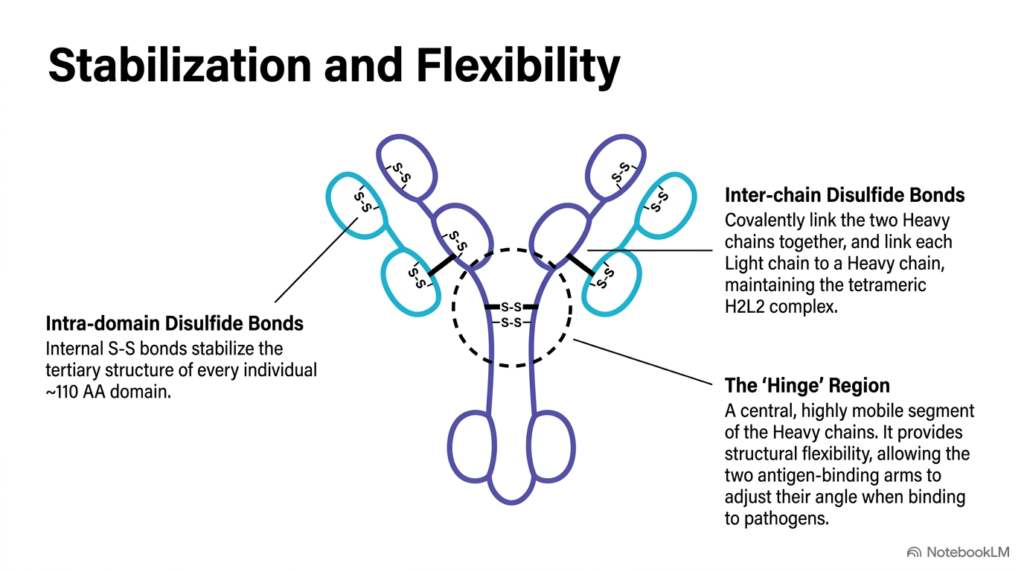
Maintaining structural durability while allowing dynamic spatial flexibility is essential for fully functional immunoglobulins. To achieve both mechanical stability and conformational adaptability, antibodies rely on strategic internal chemical bonds combined with a central mobile hinge region. These structural features ensure that antibodies can bind effectively to surface antigens with variable spatial arrangements on invading microbial targets, accommodating diverse surface geometries found on viruses, bacteria, and extracellular toxins circulating within host tissues during active immune challenges across human organs and physiological compartments.
Intra-domain disulfide bonds located inside each 110-amino-acid domain stabilize the tertiary immunoglobulin fold across all polypeptide chains. Simultaneously, inter-chain disulfide bonds covalently link the two Heavy chains at the central region and connect each Light chain to its neighboring Heavy chain. These covalent disulfide linkages are indispensable for maintaining the tetrameric H2L2 complex under physiological mechanical stress in circulating body fluids, preventing premature dissociation during turbulent blood flow, tissue migration, and cellular engagement during systemic immune responses throughout the human circulatory system.
The central hinge region consists of an extended, highly flexible segment on the Heavy chains between constant domains. This hinge lets the two antigen-binding arms pivot and rotate freely, allowing antibodies to adjust their angle as they encounter varying distances between target epitopes on surface membranes. Thanks to this inherent structural flexibility, antibodies efficiently cross-link pathogens and form dense immune complexes across diverse biological surfaces, maximizing their protective capacity in dynamic physiological environments throughout the human vascular, lymphatic, and extracellular tissue systems.
Slide 6: Enzymatic Cleavage into Fab and Fc Fragments of Antibodies
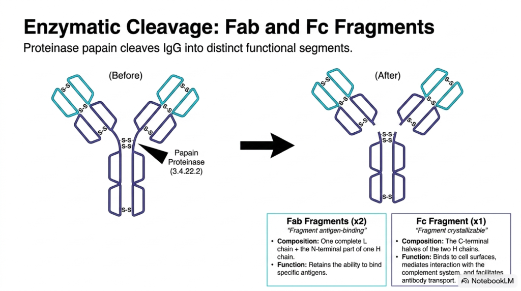
Biochemists historically mapped the functional anatomy of immunoglobulins by subjecting them to targeted enzymatic digestion using the cysteine proteinase papain. When papain cleaves the Heavy chains slightly above the central hinge region, it splits intact antibodies into distinct functional segments. This classic biochemical experiment proved that antibodies possess separate structural units dedicated exclusively to antigen binding and downstream physiological signaling, revolutionizing our early understanding of protein structure, immunology, molecular recognition mechanisms, and effector function regulation across biomedical sciences and modern biotechnology applications.
Papain digestion yields two identical Fab fragments and a single Fc fragment from each antibody molecule. The Fab segments, standing for “Fragment antigen-binding,” each consist of one complete Light chain paired with the N-terminal portion of a Heavy chain. These Fab fragments retain the ability of intact antibodies to recognize and bind specific target antigens, but they can no longer cross-link pathogens or trigger cellular destruction because enzymatic cleavage during experimental and clinical analysis removes their structural multivalency and effector stems.
The remaining product is the Fc fragment, or “Fragment crystallizable,” composed of the interacting C-terminal halves of both Heavy chains. The Fc region binds to cell surface receptors, interacts with the complement system, and facilitates antibody transport across cellular membranes. Enzymatic digestion shows how individual antibody domains divide essential immunological duties, allowing scientists to isolate antigen-binding domains for research, diagnostic assays, structural biology studies, and targeted biotherapeutic applications in modern pharmaceutical design and clinical medicine.
Slide 7: Five Structural Classes of Human Antibodies
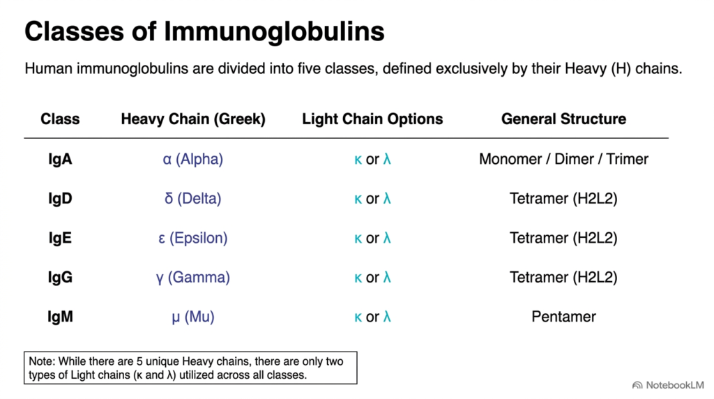
Human immunoglobulins are categorized into five distinct biological classes, or isotypes: IgA, IgD, IgE, IgG, and IgM. These five classes are defined exclusively by the chemical structure of their Heavy chains, designated by the Greek letters alpha, delta, epsilon, gamma, and mu. Across all classes, human antibodies use only two types of Light chains, kappa and lambda, which pair indiscriminately with any Heavy chain without altering class identity or broad physiological function during host immune defense across tissues.
Although Light chain selection remains restricted to kappa or lambda types, Heavy chain variations confer vastly different structural configurations and biological properties to each class. IgG, IgD, and IgE exist predominantly as basic four-chain tetramers in an H2L2 monomeric format. Conversely, IgA can assemble into monomers, dimers, or trimers, whereas IgM forms massive pentameric structures designed to maximize antigen-binding capacity during initial immune responses. These structural variations allow different antibody classes to specialize in distinct anatomical microenvironments across the human body during microbial infection, tissue repair, and environmental exposures.
This systematic classification highlights how nature modifies basic protein frameworks to generate functionally diverse antibodies. While all classes share identical antigen-binding principles, variations in heavy-chain constant regions determine where specific antibodies localize within tissues and how they interact with host effector pathways. Understanding these five classes provides students with a clear roadmap for studying humoral immunity, diagnostic assays, clinical pathology, immune deficiency disorders, and isotype-specific therapeutic interventions in modern medical science, pharmacology, and clinical immunology.
Slide 8: Polymeric Architectures of Specialized Antibodies
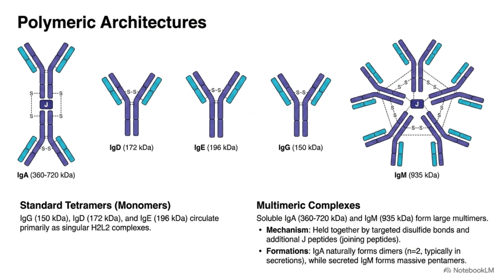
While several immunoglobulin classes circulate exclusively as monomeric four-chain units, other classes form high-molecular-weight polymeric complexes. Standard monomeric tetramers include IgG at 150 kilodaltons, IgD at 172 kilodaltons, and IgE at 196 kilodaltons, all circulating as single H2L2 structures. However, specialized mucosal and primary-response antibodies assemble into larger multimeric complexes that dramatically increase binding avidity when they encounter foreign invaders in secretions or blood during active infection phases throughout host tissues, mucous secretions, and epithelial surfaces across the body.
Soluble IgA and IgM form large multimeric assemblies held together by specialized inter-chain disulfide bonds and an additional polypeptide known as the J chain, or joining peptide. Soluble IgA ranges from 360 to 720 kilodaltons and naturally forms dimers in mucosal secretions like saliva, tears, and intestinal fluids. Secreted IgM forms massive pentameric structures reaching approximately 935 kilodaltons, creating ten potential antigen-binding sites per complex to rapidly trap circulating pathogens during early infection stages, before adaptive responses fully mature into memory populations across lymphoid organs and vascular channels throughout human physiology and immune networks.
The assembly of multimeric architectures confers significant operational advantages upon these specialized antibodies during pathogen defense. By clustering multiple binding sites, polymeric antibodies exhibit remarkably high avidity, enabling them to bind tightly to repetitive microbial surface structures even when individual epitope affinity is modest. This clustering allows multimeric antibodies to aggregate pathogens, prevent mucosal adherence, and clear foreign threats from extracellular fluids before systemic invasion can occur in host tissues, mucous membranes, and vital internal organs.
Slide 9: Functional Specialization and Distribution of Antibodies
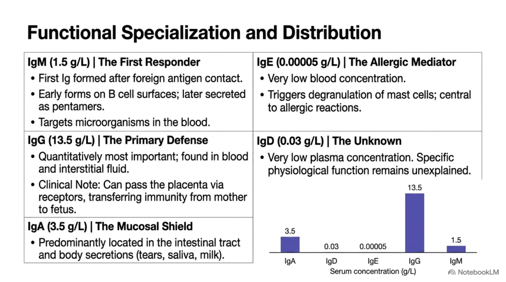
The five immunoglobulin classes display distinct physiological distributions and serum concentrations throughout the human body. IgG is the most abundant class, with a serum concentration of 13.5 grams per liter in blood and interstitial fluid. IgG provides primary systemic defense and uniquely crosses the placenta, conferring passive immunity to the developing fetus. IgA represents the second most abundant class at 3.5 grams per liter, acting as a mucosal shield in body secretions. Meanwhile, specialized antibodies perform crucial roles in targeted mucosal and tissue-specific defense networks across body barriers.
In contrast, IgM circulates at 1.5 grams per liter, operating as a pentameric first responder following initial antigen exposure in systemic circulation. IgD exists at a very low plasma concentration of 0.03 grams per liter, primarily serving as a B cell surface receptor with an enigmatic physiological role that remains under active scientific study in modern immunological laboratories. IgE occurs at trace levels of 0.00005 grams per liter, acting as a specialized mediator that triggers mast cell degranulation during allergic reactions and anti-parasitic defense mechanisms across vulnerable mucosal surfaces and peripheral tissues.
These dramatic concentration gradients demonstrate how human antibodies are strategically positioned across anatomical compartments to optimize host defense against environmental pathogens. High-concentration IgG protects the vascular system, mucosal IgA guards entry barriers, and trace IgE mediates immediate hypersensitivity responses. Examining these tissue distributions allows medical and college students to appreciate how specialized antibodies collaborate to provide complete, multi-layered systemic protection against environmental pathogens throughout human life, ensuring lifelong resilience against infectious microorganisms.
Please read our Content Disclaimer Statement.
Check out our social media channels: