113. Antibody Biosynthesis: How Molecular Genetics Generates Infinite Immune Diversity
The human body constantly defends itself against millions of unfamiliar pathogens without knowing what threat will arrive next. Rather than storing millions of separate genes, the immune system uses a clever genetic manufacturing process to craft specialized proteins on demand. This slide deck explores the genetic and cellular machinery of antibody biosynthesis. Students will discover how lymphocytes rearrange DNA segments and introduce targeted mutations to create vast protein diversity. By breaking down heavy and light chains, recombination steps, and RNA splicing, this visual guide illuminates how molecular biology protects human health.
Slide 01: Introduction to Antibody Biosynthesis and Immunoglobulin Architecture
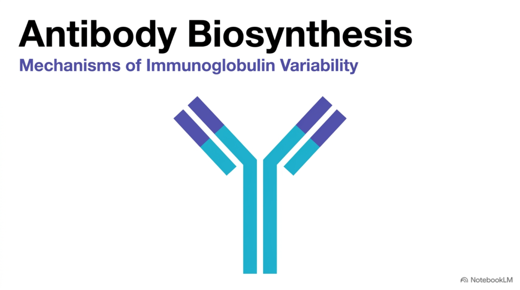
The adaptive immune system relies on specialized proteins known as immunoglobulins to recognize and neutralize foreign invaders. At the heart of this defensive system is antibody biosynthesis, the complex biological mechanism by which lymphocytes produce highly specific antigen receptors. This introductory slide establishes the fundamental structural blueprint of immunoglobulins, highlighting the characteristic Y-shaped architecture that defines these defensive molecules.
The classical Y-shaped antibody structure consists of two identical heavy chains and two identical light chains bound together by disulfide bonds. Understanding antibody biosynthesis requires examining how cells assemble these polypeptide chains into distinct functional domains. The stem of the Y-shape forms the constant region, which communicates with other immune system components. Meanwhile, the tips of the Y-shape form the variable regions, which physically attach to specific foreign antigens.
The primary objective of studying antibody biosynthesis is to uncover how the body generates an enormous variety of unique binding sites from a limited genome. B lymphocytes must balance structural stability in the constant regions with extreme structural variability in the binding tips. By establishing this structural baseline, the slide prepares students to explore the underlying molecular mechanisms that drive antibody production.
In medical and biological sciences, grasping antibody biosynthesis provides essential context for immunology, vaccinology, and therapeutic antibody engineering. Cellular machinery coordinates gene selection, transcript processing, and protein folding to yield functional immunoglobulins that safeguard the host. As students progress through this series, they will see how genetic rearrangement transforms simple germline sequences into a sophisticated defense network that can recognize virtually any foreign biochemical threat across a human lifetime.
Slide 02: The Adaptive Immune Repertoire and Antibody Biosynthesis
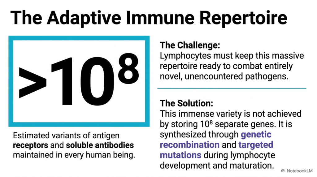
The human body maintains a staggering defense network that can recognize over one hundred million distinct molecular targets in the environment. This incredible diversity creates a fundamental biological dilemma that antibody biosynthesis resolves. If the human genome attempted to encode a separate, dedicated germline gene for every required receptor, the necessary genetic material would easily exceed the physical storage capacity of the human cell nucleus.
Lymphocytes overcome this severe storage barrier by utilizing dynamic genetic processes during early cellular development rather than relying on static inherited templates. Through antibody biosynthesis, immature B cells continuously synthesize fresh antigen receptors by rearranging modular gene fragments. This combinatorial strategy allows a modest pool of inherited genetic building blocks to produce an astronomical variety of functional receptor proteins ready to confront novel microbial pathogens.
The core challenge for the adaptive immune system is staying prepared for completely unfamiliar chemical threats that the host organism has never encountered before. The biological solution lies in the flexible execution of antibody biosynthesis within maturing lymphocytes. By combining physical DNA segment recombination with targeted point mutations, developing B cells construct custom molecular recognition sites without needing pre-existing genetic blueprints for every specific pathogen.
Understanding this combinatorial strategy shows why antibody biosynthesis is a cornerstone of modern immunology and clinical medicine. Rather than inheriting finished molecular weapons, human cells inherit a versatile genetic construction set. As B cells mature, they assemble these modular pieces into unique receptor configurations, ensuring that the host maintains a vast defense repertoire ready to neutralize emerging biological hazards throughout life.
Slide 03: Isotypes and Chains in Antibody Biosynthesis
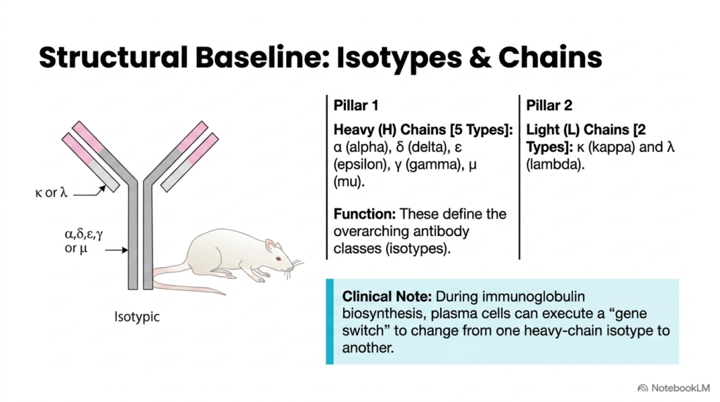
The structural framework of an immunoglobulin molecule relies on two distinct structural pillars that are assembled during antibody biosynthesis. The first pillar consists of the heavy chains, which occur in five primary functional types: alpha, delta, epsilon, gamma, and mu. These heavy chains define the antibody’s overarching functional classes, or isotypes, determining how the molecule interacts with effector cells and complement proteins.
The second structural pillar in antibody biosynthesis involves the light chains, which are classified into two types: kappa and lambda. Every individual antibody molecule contains two identical light chains paired with two identical heavy chains. These heavy- and light-chain combinations establish the structural scaffold on which antigen-specific binding regions are later constructed during cellular maturation.
A key clinical feature of antibody biosynthesis is the ability of plasma cells to undergo gene switching, or class switching. During this specialized genetic transition, a mature B cell alters the heavy-chain constant region it expresses while maintaining the exact same antigen-binding specificity. This mechanism lets the immune system shift from producing early IgM antibodies to secreting IgG, IgA, or IgE as an infection progresses.
Understanding how cells regulate heavy- and light-chain expression is fundamental to mastering antibody biosynthesis. By coordinating the production of specific chain combinations, lymphocytes tailor their immune responses to different tissue environments and anatomical locations. This structural versatility ensures that immunoglobulins can perform diverse protective roles, ranging from mucosal defense to systemic pathogen neutralization throughout the human body.
Slide 04: Three Tiers of Variation in Antibody Biosynthesis
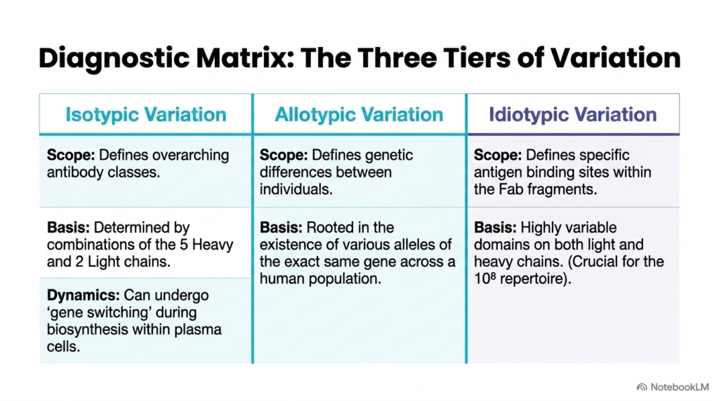
Immunoglobulin diversity can be categorized into three distinct hierarchical tiers that emerge during antibody biosynthesis. The first tier is isotypic variation, which defines the basic structural classes shared by all healthy individuals within a species. Isotypic differences arise from specific combinations of the five heavy chain classes and two light chain types, providing baseline functional templates for immune activity and cellular interaction.
The second tier, allotypic variation, reflects the genetic diversity present across different individuals within a population during antibody biosynthesis. Allotypes represent subtle amino acid differences in the constant regions of heavy or light chains that are inherited as allelic variants of the same gene. These inherited variations serve as useful genetic markers in population studies and forensic science, highlighting how normal genetic polymorphisms influence antibody structure.
The third and most functionally dynamic tier is idiotypic variation, which focuses on the unique antigen-binding sites generated during antibody biosynthesis within the Fab fragments. Idiotopes arise from highly customized amino acid sequences in the variable domains of both heavy and light chains. This tier of variation directly generates the vast repertoire of over one hundred million unique antigen receptors required for effective defense.
Analyzing these three tiers helps students understand how antibody biosynthesis balances species-wide consistency with individual diversity and receptor specificity. Isotypes provide shared effector functions, allotypes reflect population genetics, and idiotypes deliver precise target recognition. Mastering this diagnostic matrix enables future healthcare professionals to interpret immunological assays, diagnose immunodeficiencies, and comprehend therapeutic antibody design.
Slide 05: Idiotypic Variation and Hypervariable Regions in Antibody Biosynthesis
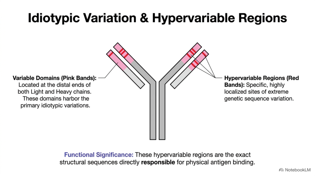
The molecular basis of precise antigen recognition lies in localized structural features formed during antibody biosynthesis. While the constant regions of immunoglobulins maintain structural stability, the distal ends of both heavy and light chains contain variable domains. Within these variable domains, specific clusters of amino acids show extraordinary sequence variability, forming the physical basis of idiotypic diversity.
These specialized sites are known as hypervariable regions, or complementarity-determining regions, and are formed during antibody biosynthesis. On each heavy- and light-chain variable domain, three distinct hypervariable loops project outward to form the antigen-binding surface. Because these loops vary dramatically in amino acid composition and length between different B cell clones, they allow antibodies to conform precisely to the shape of foreign molecules.
The functional significance of hypervariable regions in antibody biosynthesis cannot be overstated. These localized sequence hotspots create the exact three-dimensional contours needed to bind specific epitopes with high affinity. Surrounding these hypervariable loops are more conserved framework regions that maintain the essential structural fold of the protein domain, ensuring that extreme sequence variation does not compromise overall molecular stability.
By concentrating genetic mutation and physical recombination in these hypervariable zones, antibody biosynthesis achieves maximum antigen-binding diversity with minimum structural disruption to the protein backbone. Students can appreciate how evolutionary design preserves the core immunoglobulin fold while allowing infinite surface flexibility at the binding interface. This specialized molecular architecture forms the biological basis for how antibodies lock onto foreign viruses, bacteria, and toxins with exquisite chemical specificity.
Slide 06: Three Genetic Drivers of Diversity in Antibody Biosynthesis
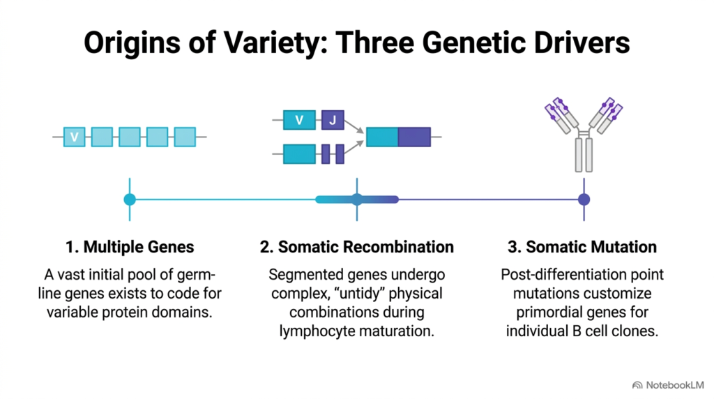
Generating an expansive immune repertoire requires a highly coordinated, multi-step molecular strategy during antibody biosynthesis. Rather than relying on a single genetic trick, developing lymphocytes use three distinct genetic mechanisms that operate at different stages of cellular maturation. Together, these three drivers transform a finite set of inherited germline genes into an almost endless array of unique antigen-binding proteins across the human body.
The first genetic driver of antibody biosynthesis is selection from multiple germline gene segments. The genome contains a large inherited library of distinct variable segments that provide the initial raw material for receptor diversity. Developing lymphocytes select specific segments from this germline pool, establishing the baseline genetic template for the variable domain before further modification.
The second driver in antibody biosynthesis is somatic recombination, which physically cuts and splices separate gene blocks together during lymphocyte maturation. This process creates untidy junctions between joining segments, generating novel mosaic genes that never existed in the germline DNA. Finally, the third driver is somatic mutation, which introduces targeted point mutations into the variable regions after B cells encounter antigen, fine-tuning binding affinity.
By combining germline selection, somatic recombination, and somatic point mutation, antibody biosynthesis achieves exponential combinatorial expansion. Each mechanism adds an additional layer of structural variability, ensuring that no two B cell lineages produce identical antigen receptors by chance. Understanding these three sequential genetic drivers gives students a clear roadmap for how molecular biology governs adaptive immune protection.
Slide 07: Multiple Gene Selection in Antibody Biosynthesis
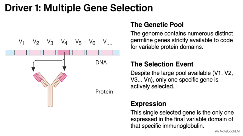
The initial phase of diversity generation relies on multiple gene selection, the foundational step in antibody biosynthesis. Each individual’s germline DNA contains a large library of distinct variable gene segments arranged in tandem along the chromosome. These inherited segments represent a diverse pool of potential coding sequences strictly dedicated to forming the variable domains of immunoglobulin chains.
During the early stages of antibody biosynthesis, a developing B lymphocyte undergoes stochastic selection. Out of the scores of available variable gene segments present in the germline DNA, the cellular machinery selects exactly one specific segment for expression. The unselected variable genes are either bypassed or rearranged, ensuring that only the chosen gene segment contributes to the final protein product.
This selection process guarantees that each individual B cell clone expresses a unique variable domain template as part of antibody biosynthesis. Because different developing lymphocytes select different variable segments from the germline pool, the overall B cell population instantly acquires a broad spectrum of baseline antigen receptors. This initial diversity forms the structural foundation upon which subsequent recombination and mutation events operate.
Understanding germline gene selection during antibody biosynthesis helps students grasp how cellular commitment strictly regulates gene expression during development. Once a specific variable gene is chosen and expressed in a developing B cell clone, that entire cell lineage remains committed to that baseline binding specificity. This slide highlights how selective gene expression converts an inherited genetic library into functional, individual cellular phenotypes necessary for effective systemic immune defense.
Slide 08: Somatic Recombination in Antibody Biosynthesis
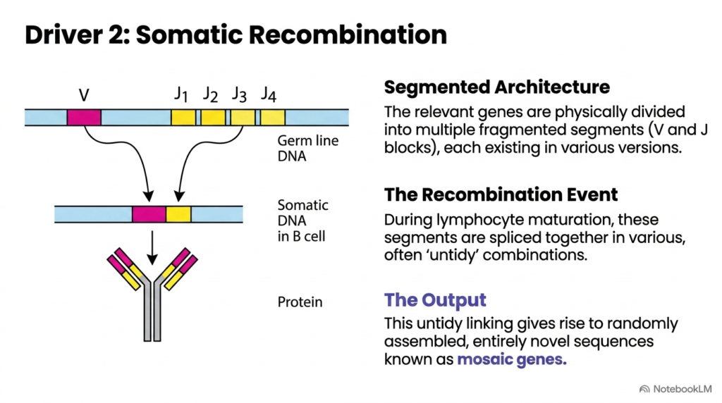
Somatic recombination is a dramatic genomic modification that occurs exclusively during antibody biosynthesis in developing B lymphocytes. Unlike standard non-immune somatic cells, which maintain static germline DNA throughout their lifespan, maturing B cells physically cut, rearrange, and rejoin their chromosomal DNA. This recombination process brings previously separated gene segments into direct physical proximity to form a contiguous coding sequence for immunoglobulins.
The genetic architecture responsible for this crucial driver of antibody biosynthesis consists of segmented DNA blocks, primarily variable and joining segments. During B cell maturation, specialized recombinase enzymes break the DNA double strand at specific recognition sequences bordering these segments. The cell then splices a chosen variable segment directly to a chosen joining segment, deleting the intervening genomic DNA loops.
A crucial feature of somatic recombination during antibody biosynthesis is its untidy or imprecise junctional joining mechanism. When the cut ends of the DNA segments are rejoined, specialized cellular enzymes randomly insert or delete nucleotides at the splice junction. This junctional flexibility creates novel, non-germline coding sequences known as mosaic genes, vastly increasing the potential structural variation at the site of physical antigen contact.
By physically altering the chromosomal sequence, somatic recombination permanently fixes the antigen receptor specificity for that individual lymphocyte and all of its cellular progeny. Students should note that this mechanism generates structural combinations that were never directly encoded in the inherited germline genome. This dynamic rearrangement demonstrates how genomic plasticity during antibody biosynthesis enables the adaptive immune system to construct custom molecular defenses on demand.
Slide 09: Somatic Mutation and Affinity Maturation in Antibody Biosynthesis
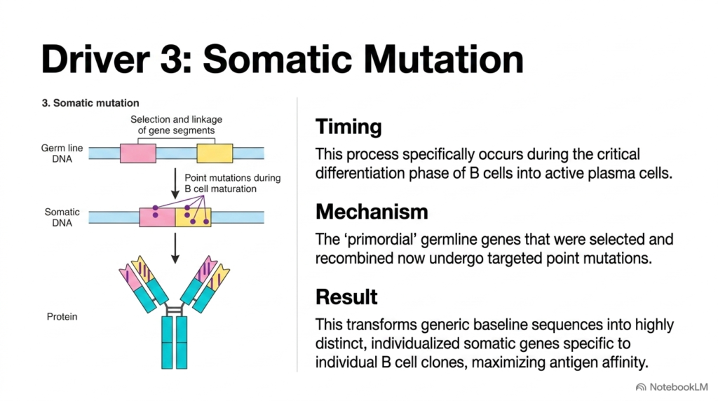
The final genetic mechanism for refining receptor specificity is somatic mutation, which operates during the later stages of antibody biosynthesis. While germline selection and somatic recombination occur during early B cell development in primary lymphoid organs, somatic mutation takes place in mature B cells after they encounter foreign antigens in secondary lymphoid tissues. This specialized process specifically fine-tunes the binding properties of the resulting immunoglobulin protein.
During this critical differentiation phase of antibody biosynthesis, activated B cells proliferate rapidly within germinal centers. As these cells divide, specialized enzymes introduce targeted point mutations into the rearranged variable domain genes. These single nucleotide changes alter individual amino acids within the hypervariable loops, subtly modifying the physical shape, surface contour, and electrostatic charge of the antigen-binding pocket.
The biological result of somatic mutation during antibody biosynthesis is the rapid transformation of baseline receptor sequences into highly customized, high-affinity antibodies. B cell clones carrying point mutations that increase antigen-binding affinity receive strong survival signals, whereas clones with neutral or deleterious mutations undergo programmed cell death. This evolutionary process, known as affinity maturation, ensures that the systemic immune response grows progressively more effective over time.
Mastering the concept of somatic mutation allows students to understand how antibody biosynthesis adapts dynamically during an active infection. By coupling targeted point mutations with selective cellular survival, the host organism continually optimizes its molecular defenses against mutating viral and bacterial pathogens. This fine-tuning mechanism is vital for establishing long-lasting immunological memory and forms the biological rationale behind booster vaccinations.
Slide 10: Germ-Line DNA Blueprint in Antibody Biosynthesis
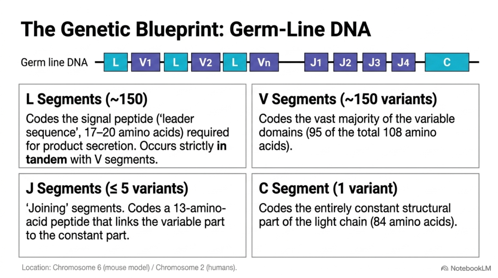
Examining the unarranged germline DNA layout reveals the raw genetic blueprint required for light chain antibody biosynthesis. Located on human chromosome 2 for kappa light chains, this genomic region contains a modular array of distinct segment types arranged in series. Understanding the organization and relative numbers of these germline segments is essential for calculating the theoretical diversity that a cell can generate.
The first component in this genetic blueprint for antibody biosynthesis consists of approximately 150 leader segments paired strictly in tandem with 150 variable segments. Each leader segment encodes a short 17- to 20-amino-acid signal peptide that directs the synthesized protein into the endoplasmic reticulum for secretion. The adjacent variable segment encodes the vast majority of the variable domain, accounting for roughly 95 of the 108 amino acids in the light chain variable region.
Downstream from the variable segments lie up to 5 joining segments, which play a dual role in antibody biosynthesis. Each joining segment encodes a short 13-amino-acid peptide that completes the variable domain while linking it directly to the constant region. Finally, the locus contains a single constant segment that encodes the 84-amino-acid structural framework shared by all kappa light chains within that class.
This organized germline structure highlights how efficient genetic storage underpins antibody biosynthesis across the genome. By grouping modular coding blocks along human chromosome 2, the genome maintains a highly compact arrangement that can be recombined into hundreds of functional gene variants. Students should recognize that this baseline germline organization provides the essential starting material for all subsequent DNA recombination, transcription, and translation events.
Slide 11: Biosynthesis Phase 1 – V/J Recombination in Antibody Biosynthesis
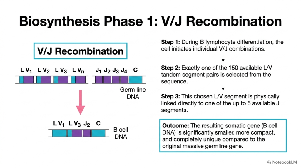
The first functional phase of light chain production centers on V/J recombination, a site-specific DNA rearrangement crucial for antibody biosynthesis. This nuclear process physically alters the chromosomal DNA of a developing B lymphocyte, converting a massive, unexpressed germline locus into a compact, functional somatic gene. This enzymatic cutting and rejoining step permanently sets the genetic identity of the individual cell line.
The step-by-step mechanism of V/J recombination during antibody biosynthesis begins when B lymphocyte differentiation signals activate specialized recombinase complexes. First, the cell initiates individual V/J segment combinations by selecting exactly one leader-variable tandem pair from the roughly 150 available options. Next, the selected segment pair is brought into proximity with one of the five available joining segments, and the intervening DNA is cleaved and excised.
Linking the chosen variable segment directly to the chosen joining segment completes Phase 1 of antibody biosynthesis. This crucial ligation event creates a shortened somatic gene in the B cell DNA, containing a unique leader-variable-joining sequence situated upstream of the single constant gene segment. The resulting somatic gene is significantly smaller, more compact, and completely unique compared to the original germline chromosome.
Understanding V/J recombination helps students see how targeted genomic editing initiates antibody biosynthesis at the nuclear DNA level. By permanently deleting unselected intervening germline segments, the developing B cell creates a streamlined transcriptional unit dedicated to producing a single receptor variant. This precise genomic modification guarantees that each maturing lymphocyte produces functional immunoglobulins with a defined, predictable antigen-binding specificity.
Slide 12: Biosynthesis Phase 2 – Transcription and Splicing in Antibody Biosynthesis
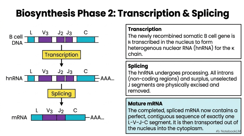
Once V/J recombination alters the chromosomal DNA, Phase 2 of antibody biosynthesis transfers the newly formed genetic instructions from nuclear DNA into messenger RNA. This phase involves nuclear gene transcription followed by post-transcriptional RNA splicing, transforming a recombined somatic gene into a mature messenger RNA transcript. These nuclear processing events ensure that only functional coding exons are retained for active protein synthesis in the cytoplasm.
During transcription, RNA polymerase II transcribes the newly recombined somatic B cell gene into primary pre-messenger RNA, also known as heterogeneous nuclear RNA. This large primary transcript contains the leader sequence, the selected variable segment, the chosen joining segment, any unselected surplus joining segments, non-coding intron regions, and the constant region segment followed by a protective poly-A tail.
The subsequent RNA splicing mechanism represents a vital molecular processing step in antibody biosynthesis. Specialized nuclear spliceosomes recognize specific consensus splice sites along the pre-mRNA strand, excising all non-coding introns and any remaining, unselected joining segments between the chosen joining block and the constant region. Splicing directly joins the leader, variable, joining, and constant exons into a single, contiguous coding sequence.
The resulting mature messenger RNA transcript is transported through nuclear pores into the cytoplasm to complete Phase 2 of antibody biosynthesis. This mature transcript now carries a perfect, contiguous reading frame consisting of exactly one leader, variable, joining, and constant segment. Students should appreciate how nuclear splicing removes non-coding genomic clutter, yielding a clean, continuous template ready for efficient ribosomal translation.
Slide 13: Biosynthesis Phase 3 – Translation and Protein Assembly in Antibody Biosynthesis
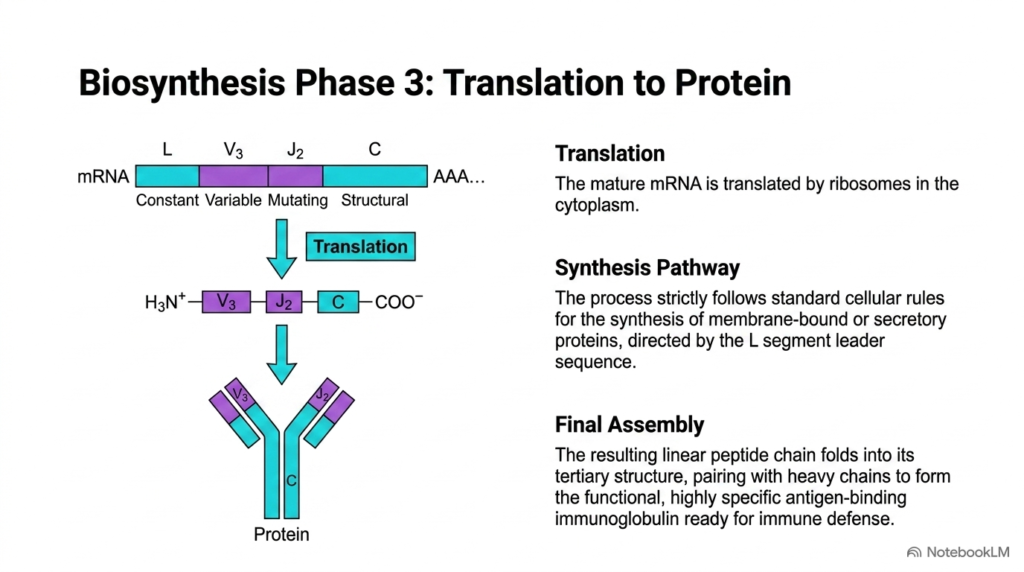
The third phase of immunoglobulin production converts mature messenger RNA into functional protein chains, completing the cytoplasmic pathway of antibody biosynthesis. Ribosomes binding to the mature messenger RNA in the cytoplasm initiate translation, synthesizing a linear polypeptide chain. This translational phase follows standard cellular rules for membrane-bound or secreted proteins, utilizing signal sequences to guide nascent protein transport directly into the endoplasmic reticulum.
The leader segment plays a critical role during this stage of antibody biosynthesis. As the nascent polypeptide emerges from the ribosome, the N-terminal leader sequence acts as a signal peptide, directing the ribosome-mRNA complex to the rough endoplasmic reticulum. Once the growing polypeptide translocates into the endoplasmic reticulum lumen, signal peptidase enzymes cleave off the leader sequence, leaving the mature variable domain at the amino terminus.
Following signal peptide cleavage, protein folding and quaternary assembly represent the final steps in antibody biosynthesis. The individual light chain folds into its distinct tertiary structure, forming intrachain disulfide bonds that stabilize the characteristic immunoglobulin domain fold. The folded light chain then pairs with a synthesized heavy chain, forming interchain disulfide bonds that yield a complete, divalent antibody monomer ready for immune defense.
Understanding Phase 3 highlights how post-translational processing fulfills the functional potential established during gene recombination and RNA splicing in antibody biosynthesis. Without proper endoplasmic reticulum targeting, enzymatic cleavage, disulfide bond formation, and heavy-light chain pairing, recombined genes could never produce functional surface receptors or secreted antibodies. Students can see how organelle-level coordination transforms genetic information into defensive immunological proteins.
Slide 14: Complete Pipeline Architecture of Antibody Biosynthesis
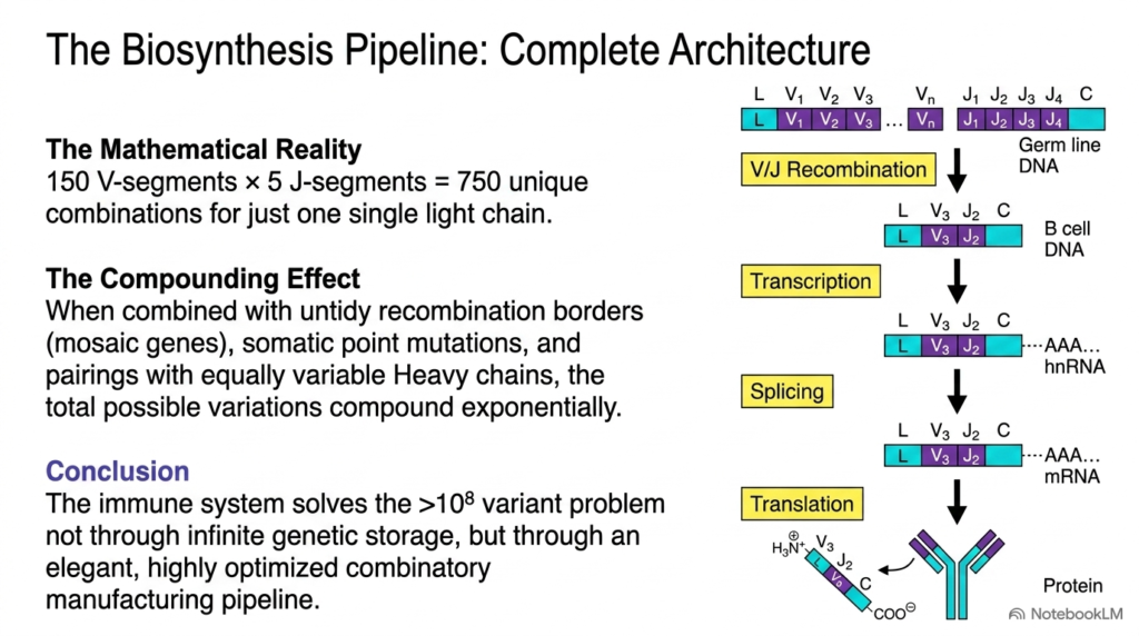
Synthesizing the entire biological pathway shows how the antibody biosynthesis pipeline solves the immune system’s immense repertoire challenge. By combining modular germline gene segments, junctional imprecision, RNA processing, and heavy-light chain pairing, the human body achieves exponential combinatorial expansion. This integrated system transforms a modest genomic footprint into a massive library of over one hundred million unique antigen receptors ready for defense.
Evaluating the mathematical reality of light chain production illustrates the power of antibody biosynthesis. Selecting from 150 variable segments and 5 joining segments yields 750 unique baseline combinations for a single kappa light chain. When this light chain diversity multiplies by the independent combinations possible for heavy chains, the total baseline variants easily reach hundreds of thousands before considering additional diversity mechanisms.
The compounding effect of untidy recombination borders and somatic point mutations further expands diversity during antibody biosynthesis. Junctional flexibility adds millions of potential sequence variations at the V-J interface, while somatic mutations continually fine-tune antigen affinity during active immune responses. This multi-tiered pipeline ensures that the host organism maintains an adaptable defense network ready to recognize virtually any pathogen structure.
In conclusion, antibody biosynthesis solves the receptor diversity problem not through infinite genetic storage, but through an elegant, highly optimized combinatorial manufacturing pipeline. Students should appreciate how DNA rearrangement, transcript splicing, translation, and somatic mutation operate as a unified biological assembly line. Mastering this pipeline gives future scientists and clinicians a foundational understanding of modern immunology, medicine, and biotechnology.
Please read our Content Disclaimer Statement.
Check out our social media channels: