90. DNA Mutation and Repair: Causes, Effects, and Mechanisms
Have you ever wondered how biological organisms survive sunlight, environmental toxins, and natural cellular errors without succumbing to genetic chaos? The answer lies in microscopic cellular mechanics. This presentation explores the core principles of mutagenesis, breaking down exactly how genetic information is threatened and protected. The following slide deck aims to provide medical and college students with a comprehensive understanding of these biochemical defense strategies that keep biological systems functioning.
Slide 1: DNA Mutation and Repair: The Foundational Blueprint of Life
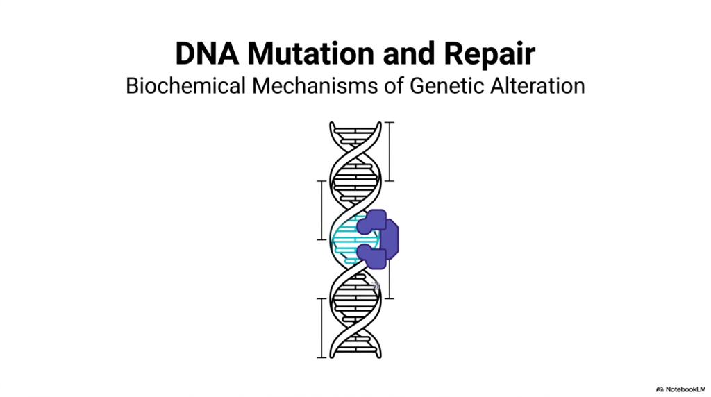
The journey into molecular biology begins with a broad overview of DNA Mutation and Repair. The first slide introduces the biochemical mechanisms of genetic alteration, presenting a clear visual of a double helix interacting with a specialized protein complex. This image perfectly encapsulates the continuous, dynamic relationship between genetic damage and cellular preservation. The foundational concept here is that the genetic code is not a static monolith. It is an active macromolecule constantly undergoing structural changes and immediate enzymatic corrections to ensure cellular viability.
Understanding the intricacies of DNA Mutation and Repair is absolutely essential for any student entering the medical or biochemical sciences. When the structural integrity of the double helix is compromised, the biological system must deploy specialized proteins to recognize and fix the structural anomaly. The illustration provided on this slide highlights this exact moment of physical interaction. A massive enzymatic complex binds directly to the localized site of the genetic defect, preparing to initiate a chemical fix. This visual sets the stage for a highly detailed exploration of how these molecular machines operate.
Ultimately, the overarching theme of DNA Mutation and Repair dictates the long-term survival of every living organism on the planet. Without these precise biochemical mechanisms operating continuously in the background, the fundamental blueprint of life would rapidly degrade into sequence gibberish. The genetic alterations and correction complexes introduced in this opening slide serve as the gateway to understanding complex biological pathologies. Students are now prepared for a deep dive into the fascinating world of genomic stability, physical damage, and cellular defense.
The profound importance of this topic cannot be overstated. A robust understanding of these foundational principles provides the necessary context for modern pharmacology, oncology, and genetic engineering. The visual introduction of the genetic helix being actively scanned and processed underscores the reality that molecular defense is an active, energy-consuming physiological priority for every living cell.
Slide 2: DNA Mutation and Repair: Balancing Evolution and Genomic Survival
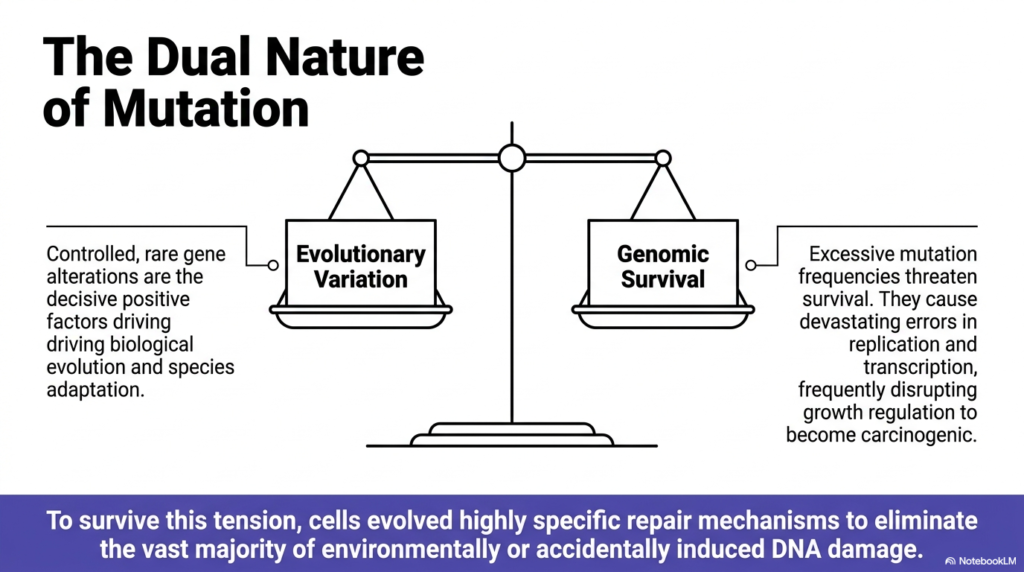
The concept of DNA Mutation and Repair involves a delicate biological balancing act. The second slide visually represents this tension using a classic scale, weighing evolutionary variation against genomic survival. On one side of the scale, controlled and rare gene alterations serve as the decisive positive factors driving biological evolution. Without these minor genetic shifts, species adaptation would be impossible, and life would remain stagnant in a changing environment. A minute level of genetic change is absolutely necessary for populations to develop new, advantageous traits over millennia.
Conversely, the opposite side of the scale illustrates the severe dangers of unmitigated genetic alteration. Excessive mutation frequencies directly threaten the survival of the organism. When the mechanisms of DNA Mutation and Repair fail to keep up with environmental damage, devastating errors occur during both replication and transcription. These unchecked errors frequently disrupt normal growth regulation pathways, ultimately causing cells to become malignant and carcinogenic. The biological system simply cannot tolerate a high volume of uncorrected genomic mistakes without facing catastrophic physiological consequences.
To survive this inherent tension, biological organisms rely on the intricate systems of DNA Mutation and Repair. Cells have evolved highly specific enzymatic defense mechanisms designed to eliminate the vast majority of environmentally or accidentally induced genomic damage. This allows the biological system to maintain genetic fidelity for individual survival while still permitting the extremely rare alterations required for long-term species adaptation. The scale analogy brilliantly highlights why these specific biochemical mechanisms are perfectly tuned to balance stability with adaptability.
For students in the medical field, understanding this delicate equilibrium is crucial for comprehending the origins of genetic diseases and cancer. When the balance tips toward excessive alterations, the clinical manifestations are often severe. Therefore, the robust systems dedicated to preserving the genomic code are not just abstract concepts; they are the primary biological barriers preventing rapid physiological deterioration and cellular death every day.
Slide 3: DNA Mutation and Repair: Categorizing Physical and Chemical Threats
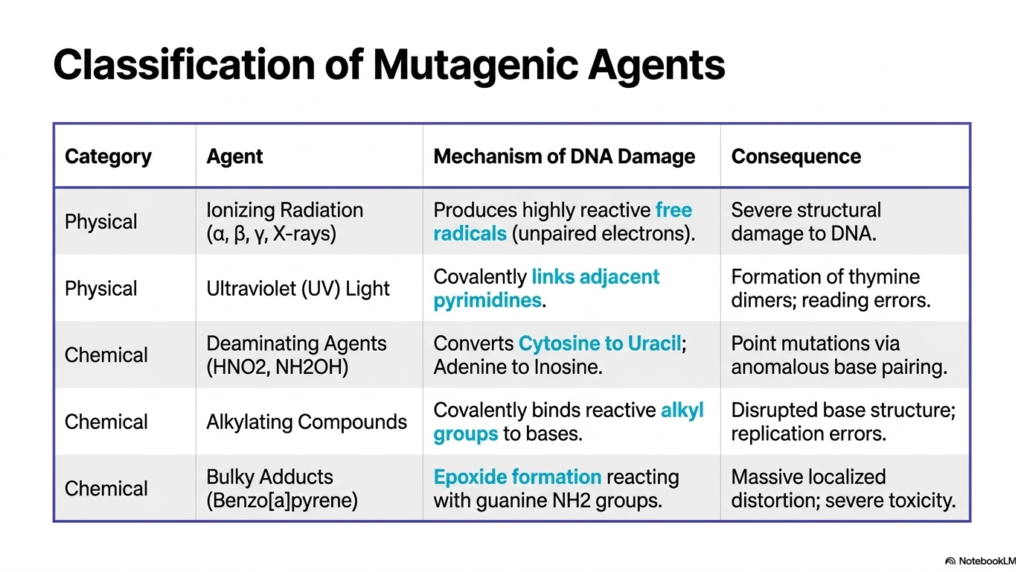
Any comprehensive study of DNA Mutation and Repair requires a thorough understanding of the specific agents that cause genetic damage. The third slide presents a detailed classification table that categorizes mutagenic agents into physical and chemical threats. Physical agents include ionizing radiation, such as X-rays and gamma rays, which produce highly reactive free radicals with unpaired electrons. These free radicals induce severe structural damage to the genetic backbone. Another major physical mutagen is ultraviolet light, which primarily targets pyrimidines to create dangerous covalent links.
The chemical agents categorized in this DNA Mutation and Repair overview are equally destructive but operate through different mechanisms. Deaminating agents, like nitrous acid, chemically strip functional groups from bases, converting cytosine to uracil and adenine to inosine. This causes point mutations through anomalous base pairing during replication. Alkylating compounds function by covalently binding reactive alkyl groups to specific bases, which disrupts the normal base structure and guarantees replication errors if left uncorrected by cellular defenses.
Finally, the table highlights bulky adducts, specifically polycyclic aromatic hydrocarbons like benzo[a]pyrene. These chemical mutagens undergo metabolic activation to form epoxides that react with guanine, causing massive localized distortion and extreme cellular toxicity. Understanding these specific categories is vital for mastering the topic of DNA Mutation and Repair. By identifying the exact mechanism of damage for each agent, medical students can better appreciate the specific enzymatic responses required to reverse these distinct structural threats and maintain cellular viability.
The diversity of these mutagenic agents highlights the relentless chemical assault that biological cells face continuously. Because the environment presents such a wide array of distinct physical and chemical threats, the cellular response must be equally varied and sophisticated. This classification table serves as a foundational roadmap, mapping out the precise molecular insults that will eventually require specialized enzymatic interventions to restore the pristine structural state of the double helix.
Slide 4: DNA Mutation and Repair: The Biochemistry of Pyrimidine Dimerization
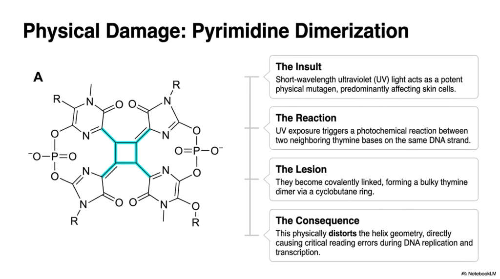
Diving deeper into specific physical threats, the fourth slide details the biochemical formation of pyrimidine dimers. This is a classic example of why DNA Mutation and Repair pathways are critical for organismal survival, particularly for skin cells exposed to the sun. The primary insult originates from short-wavelength ultraviolet light acting as a potent physical mutagen. When UV exposure occurs, it triggers a rapid photochemical reaction between two neighboring thymine bases located on the same nucleotide strand.
The resulting structural lesion is highly problematic for the cellular machinery. The two adjacent thymines become covalently linked together, forming a bulky thymine dimer connected via a specific cyclobutane ring. This molecular reaction is clearly visualized in the provided chemical diagram, which highlights the cyclobutane structure physically bridging the two pyrimidine rings. A dedicated system of DNA Mutation and Repair is absolutely necessary to identify and resolve this specific covalent linkage before the cell attempts to replicate its genetic material.
The ultimate consequence of this cyclobutane formation is a severe physical distortion of the normal helical geometry. This structural bending directly causes critical reading errors during vital cellular processes like replication and transcription. Without the intervention of DNA Mutation and Repair mechanisms, the polymerases would stall or insert incorrect bases opposite the dimer. This slide perfectly illustrates how a simple photochemical reaction can physically alter a macromolecule, disrupting its intended biological function and demanding immediate enzymatic attention to prevent permanent genetic damage.
For students analyzing this molecular event, the visual representation of the cyclobutane ring is essential. It demonstrates that genetic damage is not always a break in the backbone, but often an abnormal covalent bond that alters the three-dimensional shape of the molecule. The cell must sense this physical bulge in the double helix and recruit specific nucleases or photolyases to restore the original base-pairing structure.
Slide 5: DNA Mutation and Repair: Chemical Shifts via Base Deamination
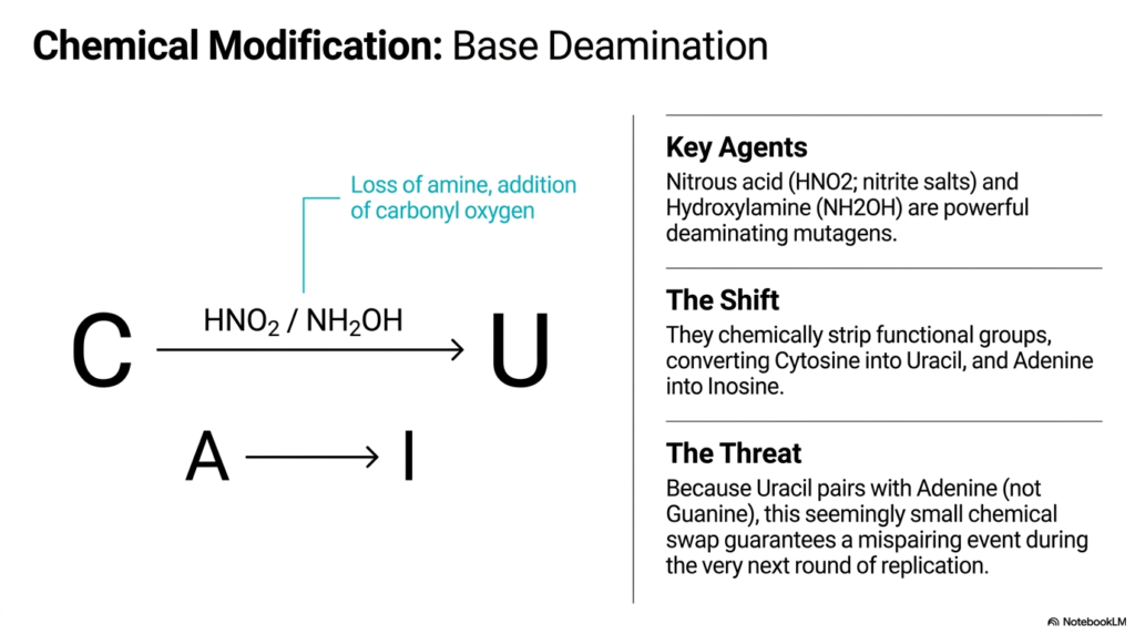
The fifth slide transitions to chemical modifications, specifically focusing on the insidious process of base deamination. In the context of DNA Mutation and Repair, deamination represents a subtle but highly dangerous chemical shift. Key agents driving this process include nitrous acid and hydroxylamine, which act as powerful deaminating mutagens. The slide illustrates the specific chemical reaction where a functional amine group is lost and a carbonyl oxygen is added, fundamentally altering the identity of the target nucleotide base.
This seemingly small chemical swap has profound biological implications. Through deamination, cytosine is directly converted into uracil, and adenine is converted into inosine. The primary threat is that uracil naturally pairs with adenine instead of guanine. If the cellular pathways for DNA Mutation and Repair do not quickly identify and remove the anomalous uracil base, a permanent mispairing event is guaranteed to occur during the very next round of genetic replication.
Understanding this mechanism is crucial for students studying DNA Mutation and Repair. Unlike the bulky physical distortions caused by UV light, deaminated bases look incredibly similar to normal bases and can slip past replication enzymes. This slide highlights the need for specialized surveillance proteins that constantly scan the genetic sequence for inappropriate bases like uracil, showing that the cellular defense system must be highly attuned to even minor chemical alterations to prevent permanent downstream errors.
The precise chemical alteration shown here underscores the fragility of nucleotide structures when exposed to reactive nitrogen compounds. Because these chemical agents can spontaneously strip away crucial amine groups, the biological system is in a constant state of chemical vulnerability. Therefore, the specialized enzymes tasked with recognizing these specific deaminated products operate continuously, performing microscopic surveillance to ensure that these stealthy chemical conversions do not permanently alter the genetic blueprint.
Slide 6: DNA Mutation and Repair: The Attack of Alkylating Agents
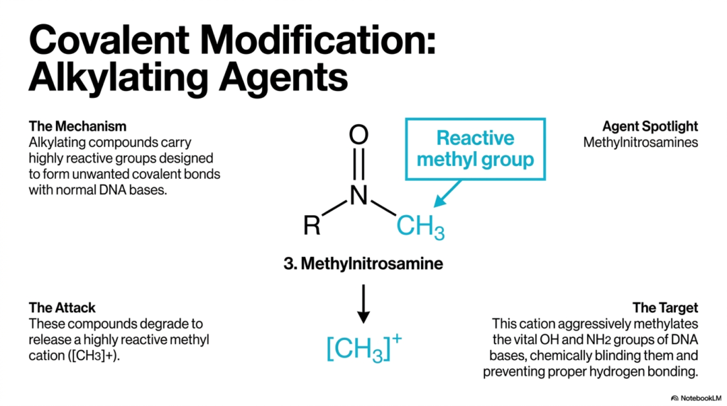
The sixth slide continues the exploration of chemical damage by detailing the covalent modification caused by alkylating agents. A thorough understanding of DNA Mutation and Repair requires students to recognize how compounds like methylnitrosamine attack normal genetic bases. These specific compounds carry highly reactive chemical groups that are naturally driven to form unwanted covalent bonds with the structural components of the genetic code. The slide focuses on the generation of a highly reactive methyl cation, which serves as the primary molecular weapon.
When these dangerous compounds degrade within the cellular environment, they release the methyl cation to seek out vulnerable biological targets. This cation aggressively methylates the vital hydroxyl and amine groups of the existing nucleotide bases. By attaching a reactive methyl group directly to these key positions, the alkylating agent chemically blinds the base. If the systems governing DNA Mutation and Repair fail to remove these additions, the affected bases are physically prevented from participating in proper hydrogen bonding.
The inability to form correct hydrogen bonds halts normal cellular replication and transcription. The pathways of DNA Mutation and Repair must deploy specific transferases or glycosylases to clear these alkyl groups from the affected nucleotides. This slide perfectly demonstrates how the targeted addition of a very small chemical group can massively disrupt the functional geometry of the double helix, demanding specialized enzymatic reversal strategies to preserve the structural integrity of the genome.
For medical and biochemistry students, the study of alkylating agents is particularly important because many chemotherapeutic drugs utilize these exact mechanisms to intentionally destroy the genetic material of rapidly dividing cancer cells. By understanding how the methyl cation attacks the specific amine groups, students gain insight into both environmental toxicity and targeted medical pharmacology. It is a perfect example of how chemical principles translate directly into profound biological consequences.
Slide 7: DNA Mutation and Repair: Carcinogenic Adducts and Epoxide Formation
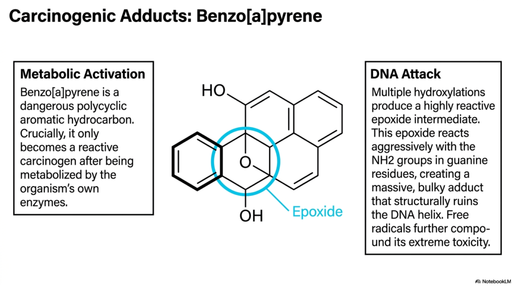
Slide seven tackles one of the most severe forms of chemical damage: carcinogenic adducts caused by massive molecules like benzo[a]pyrene. In the broad landscape of DNA Mutation and Repair, bulky adducts represent catastrophic structural roadblocks. Benzo[a]pyrene is a dangerous polycyclic aromatic hydrocarbon commonly found in environmental smoke. Crucially, the slide notes that it is not inherently reactive; it only becomes a highly dangerous carcinogen after being metabolically activated by the organism’s own internal enzymatic pathways.
This metabolic activation involves multiple hydroxylations that produce a highly reactive epoxide intermediate. The included chemical structure highlights this crucial epoxide ring. Once formed, this epoxide reacts aggressively with the amine groups found in guanine residues. For the pathways of DNA Mutation and Repair to manage this damage, they must contend with a massive, bulky adduct that physically ruins the delicate architecture of the genetic helix. Furthermore, the accompanying generation of free radicals severely compounds the event’s extreme cellular toxicity.
The presence of such a massive covalent addition completely stops replication enzymes in their tracks. A robust response from the DNA Mutation and Repair machinery is mandatory to cut out this immense structural distortion. Understanding the metabolic activation of benzo[a]pyrene is critical for medical students, as it perfectly illustrates how the biological system can accidentally weaponize environmental pollutants, turning inert molecules into direct threats that require massive enzymatic coordination to resolve.
The sheer size of the polycyclic ring structure chemically bonded to a single guanine base is visually striking. It helps conceptualize why standard replication mechanisms simply cannot bypass this type of lesion without catastrophic failure. Cellular defense systems are forced to use large-scale excision strategies to physically remove an entire segment of the affected strand, highlighting the immense energy and molecular coordination required to maintain genetic stability against severe environmental pollutants.
Slide 8: DNA Mutation and Repair: The Pathophysiological Fixation of Point Mutations
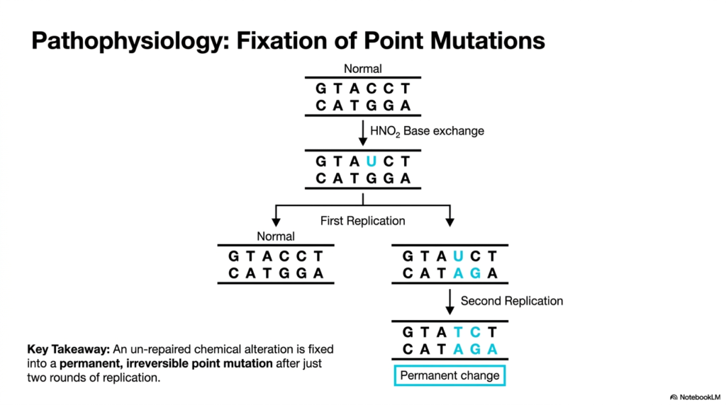
The eighth slide provides a crucial pathophysiological timeline showing exactly how chemical alterations become permanent genetic flaws. It demonstrates that the systems responsible for DNA Mutation and Repair are in a race against the biological clock of cellular division. The diagram illustrates an initial base exchange caused by nitrous acid, where a normal sequence is chemically altered. At this specific point, the structural change is still technically reversible if the cellular defense proteins intervene quickly.
However, the core lesson of this slide is the concept of permanent mutation fixation. If the mechanisms of DNA Mutation and Repair fail to correct the initial chemical alteration before the first replication cycle, the faulty strand serves as a template. By the time the second replication cycle completes, the unrepaired alteration is permanently fixed into the genetic code as an irreversible point mutation. The new base pairing is structurally sound, making it entirely invisible to standard biological surveillance systems.
This specific timing perfectly highlights why DNA Mutation and Repair pathways must be highly active and efficient. The key takeaway explicitly states that an unrepaired lesion becomes a permanent change after just two rounds of replication. For students studying genetics, this timeline emphasizes the urgency of cellular defense mechanisms. The biological machinery cannot wait to fix structural anomalies; it must operate continuously to catch chemical errors before the replication forks synthesize permanent mutations.
The visual flowchart in this slide is highly effective for tracking the progression from a temporary chemical error to a permanent genetic alteration. It forces the observer to realize that mutations are not instantaneous events, but rather multi-step processes driven by normal cellular mechanics acting upon faulty templates. This solidifies the understanding that the biological defense systems possess a very narrow, distinct window of opportunity to intervene and preserve the original genetic sequence.
Slide 9: DNA Mutation and Repair: The Catastrophic Consequences of Frameshifts
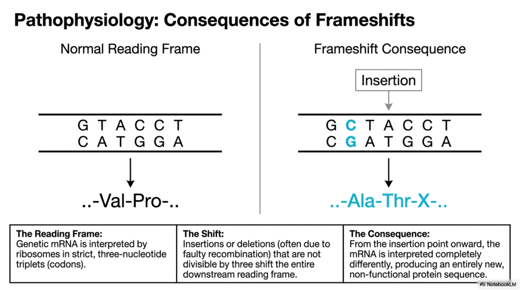
Slide nine shifts the focus to the devastating pathophysiological consequences of frameshift mutations. While point mutations alter a single amino acid, frameshifts represent a total collapse of the coding sequence, heavily emphasizing the need for robust DNA Mutation and Repair. The slide contrasts a normal reading frame with the consequence of a single nucleotide insertion. Genetic messenger RNA is strictly interpreted by biological ribosomes in three-nucleotide triplets known as codons, which makes the specific sequence order absolutely critical.
When insertions or deletions occur, often due to faulty recombination events that evade normal DNA Mutation and Repair systems, the entire downstream reading frame is shifted. Because the alteration is not divisible by three, every single triplet following the insertion point is completely scrambled. The visual provided clearly shows how a normal sequence encoding Valine and Proline is shifted to encode Alanine, Threonine, and unknown subsequent amino acids. The functional meaning of the original biological code is instantly destroyed.
The ultimate consequence of this shift is the production of an entirely new, completely non-functional protein sequence. The processes governing DNA Mutation and Repair are designed to guard against these specific insertions and deletions because their effect on cellular viability is so drastic. Understanding this specific reading frame vulnerability allows medical students to comprehend why certain genetic diseases are so phenotypically severe, as a single missing nucleotide can ruin an entire essential protein.
This concept is fundamental to molecular biology because it perfectly illustrates the fragile nature of the triplet codon system. A biological system operates much like a complex sentence; removing a single letter shifts all subsequent letters, rendering the entire message incomprehensible. The cellular machinery that guards against these specific structural shifts must be precise, constantly monitoring the replication forks to ensure that no extra bases are accidentally looped in or improperly deleted during synthesis.
Slide 10: DNA Mutation and Repair: Defense Mechanism 1 – Excision Repair
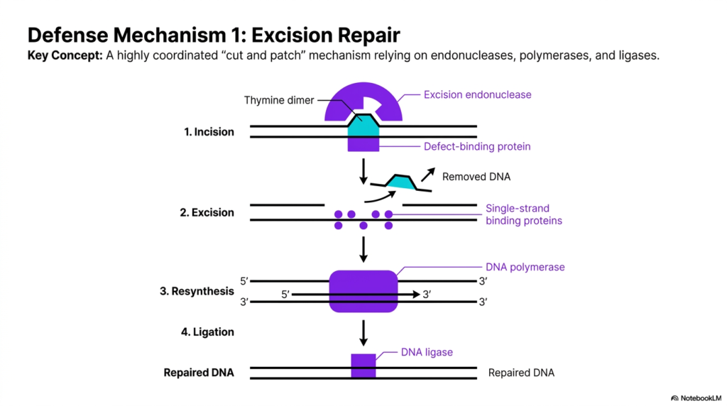
Moving from the causes of genetic damage to the solutions, the tenth slide details the first major defense strategy. Here, the true power of DNA Mutation and Repair is demonstrated through the highly coordinated mechanism of excision repair. This strategy is defined by a precise cut-and-patch mechanism that relies on the synchronized actions of endonucleases, polymerases, and ligases. The diagram visually breaks down this complex enzymatic ballet into four distinct, logical steps that restore the original genetic sequence.
The process begins with the identification of a thymine dimer by a specialized defect-binding protein. In the world of DNA Mutation and Repair, the first incision is critical; an excision endonuclease cuts the biological backbone near the bulky lesion. Following this initial cut, single-strand binding proteins help remove the damaged oligonucleotide segment completely. This physical removal leaves a noticeable structural gap in the double helix, clearing away the chemical problem but requiring immediate cellular resurfacing to prevent instability.
The final steps involve massive enzymatic resynthesis. A biological polymerase uses the undamaged opposite strand as a pristine template to fill the gap with correct nucleotides. Finally, a specialized ligase seals the remaining structural nick, fully restoring the biological molecule to its original state. For medical students, mastering this primary mechanism of DNA Mutation and Repair is essential, as it represents the most versatile and common cellular strategy for fixing bulky, environmentally induced structural anomalies.
The beauty of this specific biological mechanism lies in its elegant simplicity and reliance on the inherent redundancy of the double helix. Because genetic information is stored on two complementary strands, destroying the damaged section is safe as long as the opposing template remains intact. This intricate four-step enzymatic pathway is a marvel of evolutionary engineering, operating seamlessly in the background of every cell to ensure structural stability against environmental threats.
Slide 11: DNA Mutation and Repair: Defense Mechanism 2 – Photoreactivation
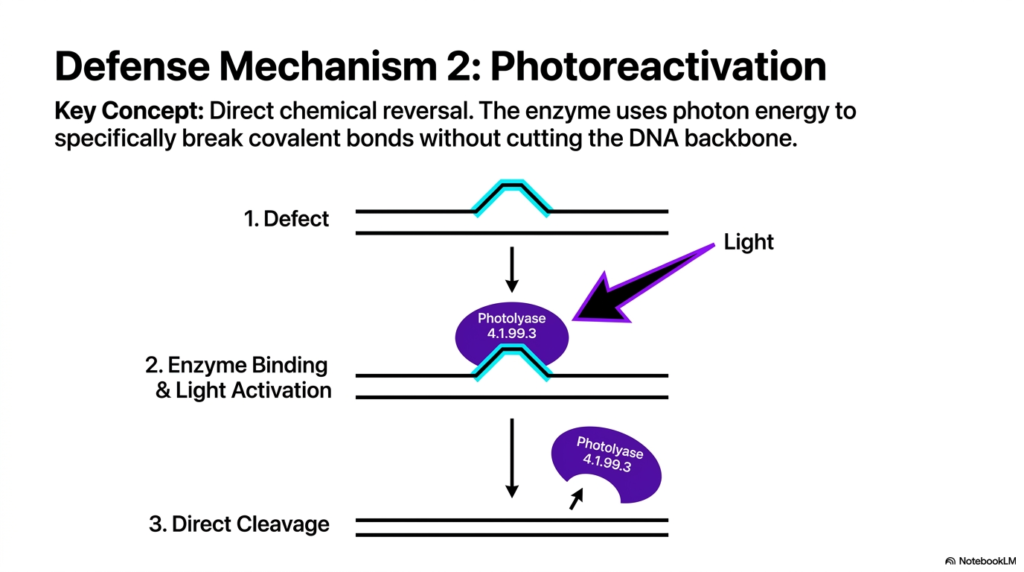
The eleventh slide introduces a highly specialized defense strategy known as photoreactivation, which provides a fascinating contrast to previous mechanisms. Unlike the complex cut-and-patch strategies common in DNA Mutation and Repair, photoreactivation is defined by the direct chemical reversal of a structural lesion. The visual clearly illustrates how this specific biological enzyme addresses the bulky defect caused by a pyrimidine dimer without ever breaking the underlying genetic backbone. It is a targeted, elegant, and efficient molecular solution.
The key enzymatic player in this specific process of DNA Mutation and Repair is a photolyase. The diagram demonstrates the enzyme binding directly to the structural defect on the double helix. However, the most critical aspect of this mechanism is its absolute requirement for external photon energy. The photolyase uses specific wavelengths of light to activate its internal chemical centers, providing the exact biological energy required to cleave the anomalous covalent bonds bridging the affected pyrimidine bases.
Once the light activation occurs and the direct cleavage is complete, the enzyme detaches, leaving the genetic sequence fully repaired. Understanding this form of DNA Mutation and Repair is crucial for biochemistry students because it highlights a unique evolutionary adaptation in which environmental light is used to cure the very damage that environmental radiation causes. It beautifully demonstrates the incredible specificity of biological enzymes designed for precise chemical intervention.
This mechanism represents a stunning biological efficiency that minimizes cellular energy expenditure. Because the backbone is never severed, the biological system does not need to recruit complex polymerases or ligases to finish the job. The direct reversal of the cyclobutane ring instantly restores the natural geometry of the macromolecule in a single enzymatic step. It is a specialized, rapid-response molecular tool dedicated exclusively to managing the continuous threat of ultraviolet radiation exposure in light-dependent biological organisms.
Slide 12: DNA Mutation and Repair: Defense Mechanism 3 – Recombinational Repair
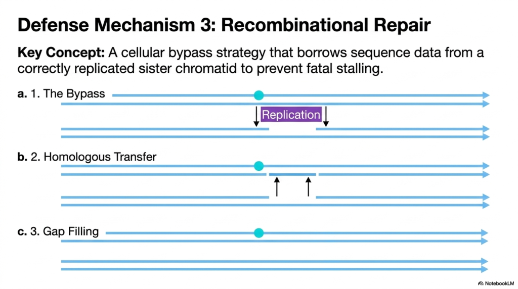
Slide twelve details the final major defense strategy: recombinational repair, a biological mechanism deployed when standard molecular fixes fail. In the broad scope of DNA Mutation and Repair, this represents a crucial cellular bypass strategy. When a rapidly moving replication fork encounters a massive, unrepaired lesion, it often stalls entirely. The diagram illustrates how the cellular machinery must intentionally bypass the structural defect to prevent a fatal halt in genetic synthesis, leaving a dangerous structural gap in the newly synthesized strand.
To resolve this massive physical gap, the systems governing DNA Mutation and Repair must borrow pristine sequence data from a correctly replicated sister chromatid. The second step of the diagram clearly shows a homologous transfer occurring between the parallel genetic strands. Biological recombinases facilitate the physical movement of a large sequence block from the undamaged sister strand into the missing gap of the new strand. This structural exchange ensures that the stalled replication process can eventually continue.
After the homologous transfer is complete, specialized polymerases are recruited to fill the newly created gap in the donor sister strand. This specific form of DNA Mutation and Repair is vital for medical students to understand because it heavily involves genetic recombination processes. It beautifully illustrates that biological cells will utilize incredibly complex structural swapping and sister-strand borrowing to survive massive physical lesions that would otherwise completely terminate the cellular life cycle during active division.
The sheer molecular coordination required for this bypass maneuver is truly astounding. The biological system recognizes that a complete halt in replication is far more dangerous than leaving a temporary structural gap. By utilizing the newly created sister chromatid as a pristine genetic backup, the cell demonstrates remarkable structural resilience. This specific pathway underscores the immense biological importance of sequence redundancy and the lengths a biological system will go to ensure total genomic replication.
Slide 13: DNA Mutation and Repair: The Diagnostic Synthesis of Genomic Defense
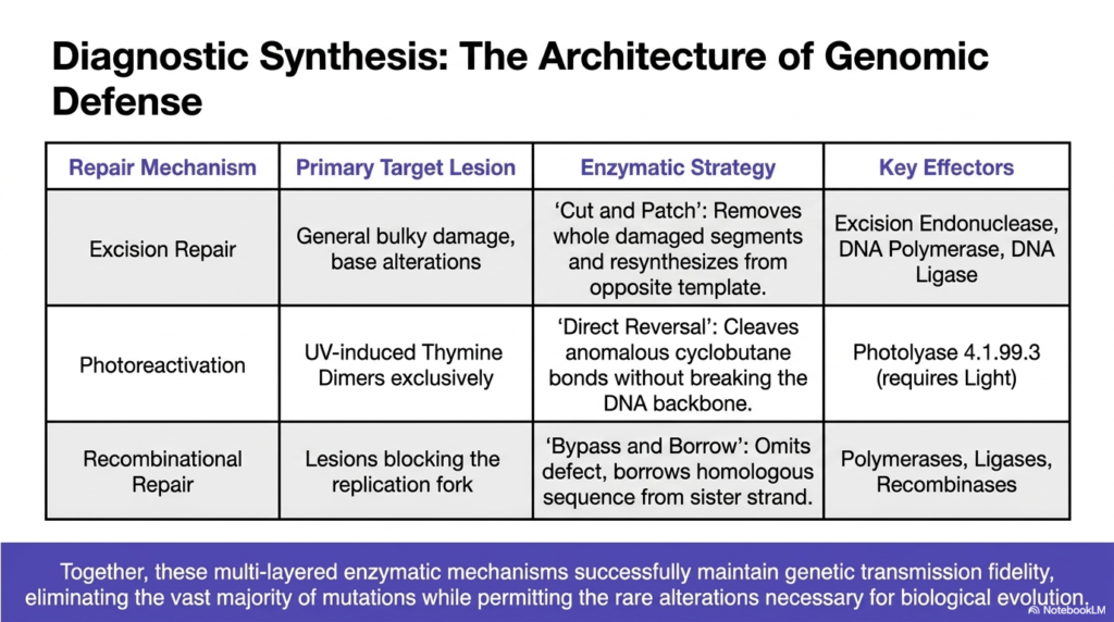
The final slide provides a comprehensive diagnostic synthesis, perfectly summarizing the entire architecture of genomic defense. This table serves as the ultimate study guide for mastering DNA Mutation and Repair, categorizing the three primary repair mechanisms against their specific structural targets and enzymatic strategies. Excision repair is highlighted for managing general bulky damage and base alterations through its highly coordinated cut-and-patch strategy, relying on the combined molecular efforts of endonucleases, polymerases, and ligases.
The summary table continues by contrasting this with the highly specialized mechanisms of DNA Mutation and Repair. Photoreactivation is explicitly listed as targeting UV-induced thymine dimers exclusively, utilizing direct chemical reversal via the light-activated photolyase enzyme. Finally, recombinational repair is defined as the biological solution for massive lesions that completely block the replication fork, using a bypass-and-borrow strategy heavily dependent on specialized polymerases and recombinases to transfer sequence data from a sister strand.
Together, these multi-layered enzymatic mechanisms of DNA Mutation and Repair successfully maintain genetic transmission fidelity across countless biological generations. The overarching conclusion is profound: these defense systems ruthlessly eliminate the vast majority of environmental and chemical mutations, ensuring cellular survival, while permitting the rare biological alterations strictly necessary for long-term species evolution. This presentation equips medical students with a highly detailed, functional understanding of how the fundamental blueprint of life is fiercely protected on a microscopic scale.
The synthesis presented in this final table perfectly encapsulates the sheer elegance of cellular biochemistry. Biological systems do not rely on a single, fragile method for structural preservation. Instead, they deploy a robust, multi-layered defensive network where diverse enzymatic strategies overlap to cover varying physical and chemical threats. A thorough comprehension of this architectural framework is the absolute key to understanding the profound resilience of biological organisms in an environment constantly trying to degrade their genetic information.
Please read our Content Disclaimer Statement.
Check out our social media channels: