93. Fundamentals of Polymerase Chain Reaction and Genetic Engineering
The modern medical landscape relies heavily on engineered therapeutics to treat complex human diseases. How do scientists actually construct these life-saving molecules in a laboratory? The answer begins with understanding a foundational technique known as the polymerase chain reaction. This slide deck explores the core biochemical principles underlying the polymerase chain reaction and the broader pipeline of genetic engineering. By mastering these concepts, future healthcare professionals and researchers can better grasp how targeted therapeutics are synthesized from the molecular level up.
Slide 1: Introduction to Polymerase Chain Reaction: Building Therapeutics
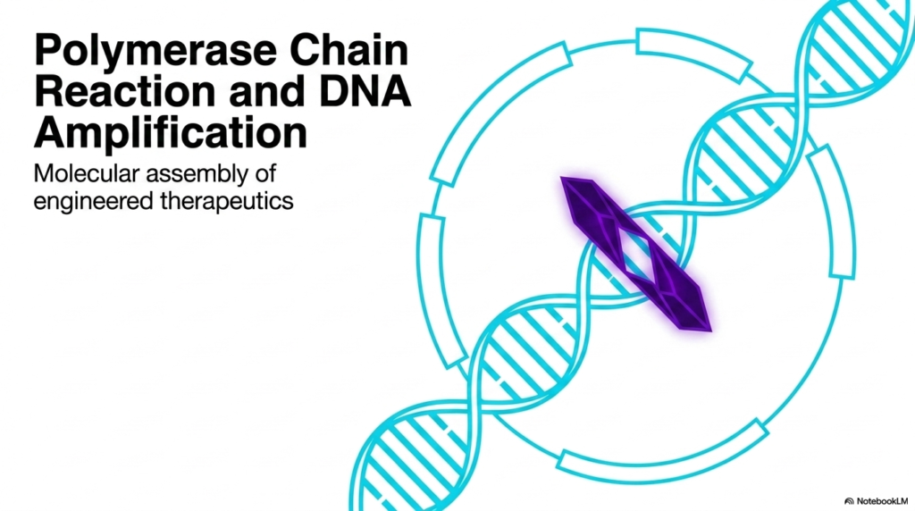
The first major concept introduced is the polymerase chain reaction, an ingenious method for in vitro DNA amplification. To engineer a therapeutic protein, scientists must first obtain massive amounts of the specific gene encoding that protein. The polymerase chain reaction solves this problem by utilizing thermal cycling and specialized enzymes to rapidly copy a target DNA sequence. This mechanism mimics natural cellular DNA replication but occurs outside a living host, offering a controlled environment for generating genetic material.
Furthermore, the slide emphasizes that the polymerase chain reaction is the critical first step in the molecular assembly of therapeutics. Without the exponential amplification of target genes provided by the polymerase chain reaction, downstream processes like protein overexpression would lack the necessary genetic blueprints. The image of the DNA double helix symbolizes the target molecule, while the central crystal-like structure represents the precise, targeted nature of this biotechnology.
This continuous cycle of DNA amplification through the polymerase chain reaction ensures that even the smallest trace of genetic material can be expanded into a workable sample. For medical students, recognizing the power of the polymerase chain reaction translates to a deeper appreciation of diagnostics, forensics, and recombinant drug manufacturing. The journey from a single DNA strand to a functional medicine is complex but highly logical.
Slide 2: The Genetic Engineering Pipeline: Where Polymerase Chain Reaction Fits
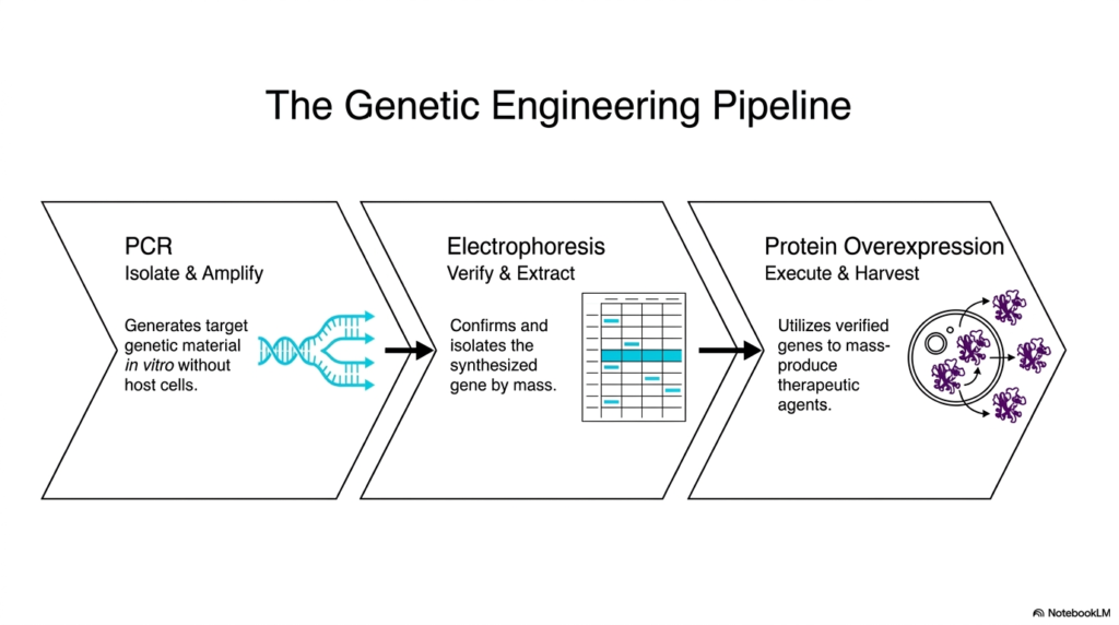
Understanding how a single gene becomes a mass-produced therapeutic requires visualizing the entire genetic engineering pipeline. It can seem daunting to map out the journey from microscopic DNA to a tangible medication. The core purpose of this slide deck is to demystify these steps, starting with the polymerase chain reaction. This slide lays out the three essential phases of the pipeline, providing students with a clear roadmap of the biochemical processes required to isolate, verify, and manufacture recombinant proteins.
The pipeline begins with the polymerase chain reaction, designated as the isolation and amplification phase. The polymerase chain reaction is deployed to generate massive quantities of a specific target gene. This process occurs in vitro, meaning it happens outside a living organism in a controlled laboratory setting. By using the polymerase chain reaction, scientists can bypass the slow and unpredictable nature of natural cellular replication when initially acquiring the genetic blueprint.
Following the polymerase chain reaction, the newly synthesized genetic material moves into the second phase: electrophoresis. This step is crucial for verifying and extracting the desired DNA fragment. Electrophoresis separates molecules by mass, ensuring that the product generated by the polymerase chain reaction is the correct length and free of significant errors or artifacts. It acts as a biological quality control checkpoint before the gene is introduced into a host cell.
The final phase of the pipeline relies on the verified DNA to execute protein overexpression. The target gene is inserted into host cells, which act as microscopic factories to mass-produce the therapeutic agents. Without the initial abundance of DNA provided by the polymerase chain reaction, this massive scale-up would be impossible. The pipeline demonstrates a beautiful synergy between synthetic chemistry, biophysics, and cellular biology, culminating in the harvest of functional proteins.
Slide 3: Amplification Anatomy: The Building Blocks of Polymerase Chain Reaction
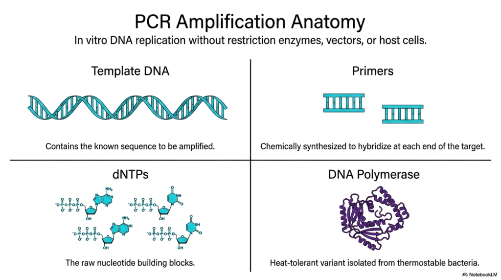
Before diving into the mechanics of DNA replication, one must understand the essential ingredients required for the process. Imagine trying to build a complex architectural model without knowing the materials; genetic engineering is no different. The core purpose of this slide deck is to detail these foundational elements, specifically focusing on the anatomy of the polymerase chain reaction. This slide breaks down the four vital components needed to execute in vitro DNA amplification successfully.
The first essential component for the polymerase chain reaction is the template DNA. This molecule contains the known genetic sequence that scientists intend to amplify. The template provides the exact blueprint necessary for the subsequent steps. Next, the reaction requires primers, which are short, chemically synthesized strands of nucleic acids. These primers are designed to perfectly match and hybridize at the specific flanking ends of the target sequence, acting as starting markers for the replication machinery.
To physically build the new DNA strands, the polymerase chain reaction requires raw materials known as deoxynucleotide triphosphates, or dNTPs. These are the individual nucleotide building blocks—adenine, thymine, cytosine, and guanine—that float freely in the reaction mixture. When the DNA is synthesized, these dNTPs are chemically linked together to form the new complementary strands. Without an ample supply of these precursors, the polymerase chain reaction would quickly stall.
The ultimate driver of the polymerase chain reaction is the DNA polymerase enzyme itself. In this context, scientists utilize a heat-tolerant variant isolated from thermostable bacteria. Because the polymerase chain reaction requires extreme temperature shifts to separate DNA strands, standard human enzymes would instantly denature and fail. This specialized, heat-resistant enzyme seamlessly incorporates the dNTPs onto the growing strand, reading the template DNA and constructing its mirror image with remarkable speed and precision.
Slide 4: The Thermal Cycle: Regulating the Polymerase Chain Reaction
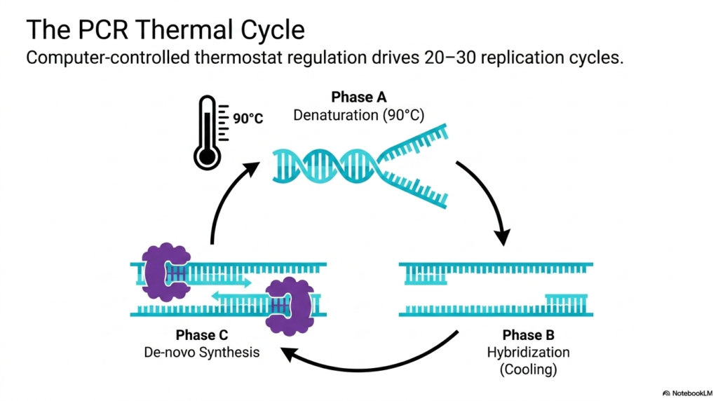
Heating and cooling are basic physical processes, but in the laboratory, they can be harnessed to manipulate the very code of life. How do temperature changes drive molecular synthesis? The core purpose of this slide deck is to explain this phenomenon through the lens of the polymerase chain reaction. This slide illustrates the automated thermal cycle, a computer-controlled thermostat regulation system that repeats 20 to 30 times to exponentially multiply target DNA.
The first phase of the polymerase chain reaction thermal cycle is denaturation. The reaction mixture is heated to an extreme temperature of 90 degrees Celsius. At this heat, the weak hydrogen bonds holding the double-stranded template DNA together break apart. This thermal energy forcefully separates the double helix into two distinct, single strands, exposing the genetic sequence so that the cellular machinery can access the crucial nucleotide bases required for the next phase of the polymerase chain reaction.
Phase B of the polymerase chain reaction involves a rapid cooling process known as hybridization, or annealing. As the temperature drops, the chemically synthesized primers introduced in the previous step seek out their complementary sequences on the single-stranded template DNA. The cooling allows hydrogen bonds to reform, securing the primers to the specific starting locations on the target gene. This precise binding is essential, as it dictates exactly where the amplification will initiate.
The final step in the thermal cycle is Phase C, de-novo synthesis. The temperature is adjusted to the optimal working condition for the heat-tolerant DNA polymerase. The enzyme binds to the primer-template junction and races along the single strand, pulling in free dNTPs to build the new complementary DNA sequence. Once this synthesis is complete, the cycle repeats. Each round of the polymerase chain reaction doubles the amount of target DNA, leading to massive amplification.
Slide 5: Amplification Kinetics: Growth Rates in the Polymerase Chain Reaction
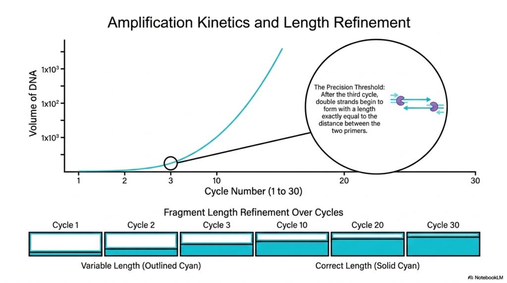
Exponential growth is a mathematical concept that can be difficult to visualize in a biological context. Seeing how a single invisible molecule becomes a workable laboratory sample is a fundamental lesson for medical students. The core purpose of this slide deck is to trace this exponential kinetic curve and show how the polymerase chain reaction refines molecular length. This slide demonstrates how consecutive replication cycles isolate the exact target sequence.
The graph illustrates the explosive volume of DNA generated by the polymerase chain reaction. In the earliest cycles, the amount of genetic material increases slowly. However, as each newly synthesized strand becomes a template for the next round, the amplification kinetics become purely exponential. By cycle 30, the polymerase chain reaction has created billions of copies from a single starting molecule. This immense scale is what makes clinical genetic analysis and therapeutic manufacturing possible today.
A critical milestone in the polymerase chain reaction is known as the precision threshold, which occurs during the third thermal cycle. In the first two cycles, the DNA polymerase synthesizes strands of variable lengths because there is no biological stop signal. However, after the third cycle of the polymerase chain reaction, double strands begin to form with a length exactly equal to the distance between the two specific primers.
This fragment length refinement is a hallmark of the polymerase chain reaction. The slide’s bottom graphic depicts how the variable-length fragments, outlined in cyan, are rapidly vastly outnumbered by the correct, defined-length fragments, shown in solid cyan. By cycle 20 or 30, nearly 100 percent of the genetic material in the tube is the exact desired gene sequence. This guarantees that only the precise therapeutic blueprint is carried forward into the electrophoresis and overexpression phases.
Slide 6: Biophysics of Nucleic Acids: Post-Polymerase Chain Reaction Analysis
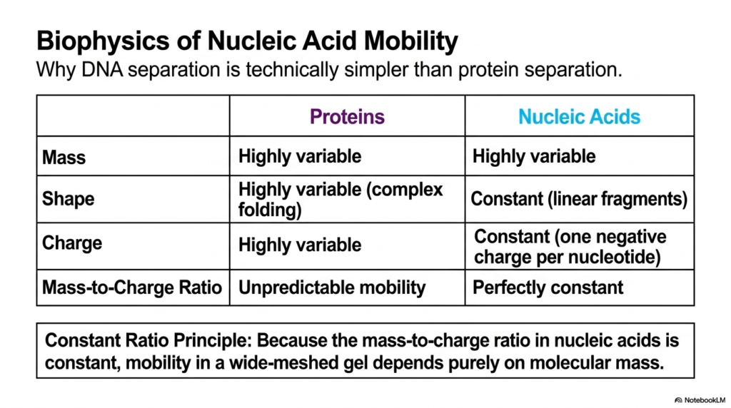
Separating complex biological molecules often requires immense technical effort and sophisticated machinery. Why is it that some molecules can be isolated much more easily than others? The core purpose of this slide deck is to explain the physical properties that enable researchers to verify the products of the polymerase chain reaction. This slide contrasts the biophysics of proteins and nucleic acids, explaining why DNA separation is technically simpler to achieve in the laboratory.
When analyzing the amplificates produced by the polymerase chain reaction, scientists rely on the unique physical characteristics of DNA. Proteins have highly variable mass, complex folded shapes, and highly variable charges based on their amino acid composition. This results in unpredictable mobility when subjected to electrical fields. In contrast, nucleic acids—like the DNA amplified by the polymerase chain reaction—have a much more uniform and predictable biophysical profile, making them ideal candidates for simple electrophoretic separation.
The slide highlights that nucleic acids maintain a constant, linear shape when fragmented, unlike folded proteins. More importantly, DNA holds a constant negative charge, with exactly one negative charge per nucleotide phosphate group. Because the mass increases at the exact same rate as the charge, nucleic acids possess a perfectly constant mass-to-charge ratio. This intrinsic physical property is a tremendous advantage for researchers working with the vast amounts of genetic material generated by the polymerase chain reaction.
This phenomenon leads to the Constant Ratio Principle. Because the mass-to-charge ratio in nucleic acids is unvarying, their mobility through a wide-meshed gel matrix depends entirely and purely on their molecular mass. Shorter fragments of DNA will travel faster and farther, while longer fragments will drag and move slowly. This elegant biophysical rule allows scientists to accurately measure the size of the genes amplified by the polymerase chain reaction, ensuring the correct therapeutic blueprint was assembled.
Slide 7: The Agarose Gel Apparatus: Processing Polymerase Chain Reaction Products
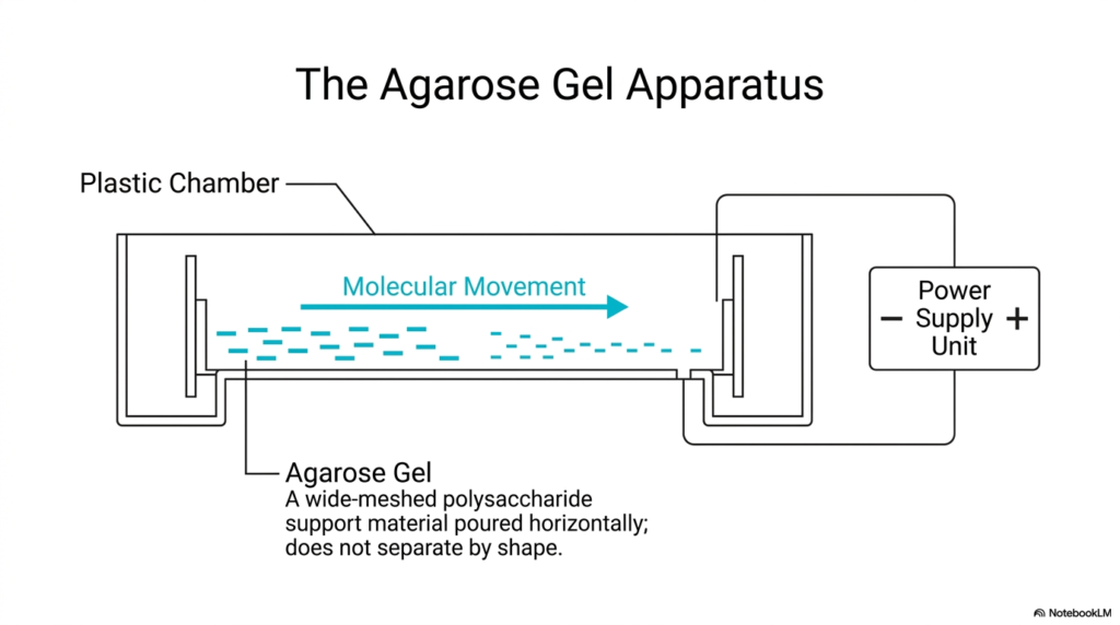
Visualizing an electrical circuit applied to biological material helps bridge the gap between physics and cellular biology. How do researchers actually harness the constant mass-to-charge ratio to separate molecules? The core purpose of this slide deck is to illustrate the tools used immediately after the polymerase chain reaction. This slide diagrams the agarose gel apparatus, the primary physical device used to sort and verify the DNA fragments created during amplification.
Once the polymerase chain reaction is complete, the resulting mixture is loaded into a specialized plastic chamber. This chamber contains a power supply unit that generates a horizontal electrical field across the entire device. Because the DNA fragments created by the polymerase chain reaction carry a uniform negative charge, activating the power supply causes molecular movement. The genetic material is forcefully pulled away from the negative terminal and drawn steadily toward the positive terminal.
The critical component inside this chamber is the agarose gel itself. Agarose is a wide-meshed polysaccharide support material that is poured horizontally to form a porous matrix. As the products of the polymerase chain reaction migrate toward the positive charge, they must navigate through this microscopic mesh. The gel acts like a biological sieve. It does not separate the molecules by shape, but strictly physically impedes them based on their overall length and mass.
In the context of the polymerase chain reaction, this simple apparatus is a powerful diagnostic tool. Smaller DNA fragments navigate the wide-meshed agarose gel easily and travel far, while massive fragments get tangled in the polysaccharide matrix and remain close to the starting point. By using this apparatus, scientists can sort the billions of identical molecules produced by the polymerase chain reaction into neat, highly organized physical bands based solely on their physical size.
Slide 8: Chemical Visualization: Seeing the Polymerase Chain Reaction Amplificates
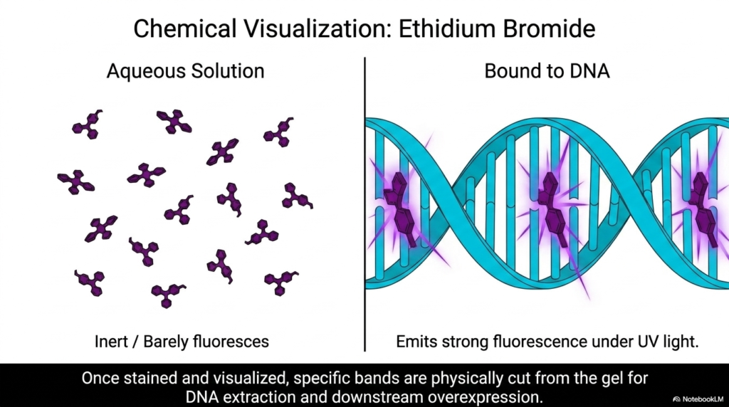
DNA is inherently microscopic and completely transparent, making it invisible to the naked eye even in large quantities. How can scientists possibly see the genetic material they have carefully sorted? The core purpose of this slide deck is to reveal the chemical tricks used to visualize the success of the polymerase chain reaction. This slide introduces ethidium bromide, a specialized chemical visualization agent that safely binds to nucleic acids for observation.
Before it interacts with the products of the polymerase chain reaction, ethidium bromide exists in an aqueous solution. In this free-floating state, the molecule is chemically inert and barely fluoresces under any lighting conditions. It is a completely dark dye. However, ethidium bromide has a highly specific affinity for the double helix structure of DNA. When introduced to the agarose gel containing the amplicons from the polymerase chain reaction, its physical properties fundamentally change.
The ethidium bromide molecules physically insert themselves between the base pairs of the DNA strands, a process known as intercalation. Once bound tightly to the DNA molecules generated by the polymerase chain reaction, the chemical undergoes a stark transformation. The previously inert molecules now emit a powerful, bright fluorescence when exposed to ultraviolet light. This chemical binding turns invisible genetic material into glowing, visible bands that scientists can photograph, measure, and analyze with complete clarity.
This visualization is the crucial final step of the electrophoresis process. Once the agarose gel is stained and visualized under UV light, researchers can physically pinpoint the exact location of the target gene amplified by the polymerase chain reaction. Specific fluorescent bands are literally cut directly from the gel with a scalpel. This allows for precise DNA extraction, providing the ultra-pure genetic sequence required for downstream cellular integration and therapeutic protein overexpression.
Slide 9: Gel Analysis: Verifying the Polymerase Chain Reaction Targets
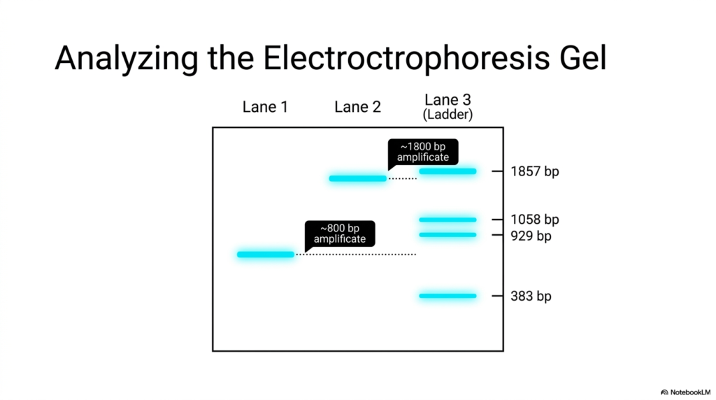
Interpreting laboratory data is a foundational skill for any modern biological researcher or medical student. What does the glowing biological data actually tell us about the synthesized genetic material? The core purpose of this slide deck is to teach students how to read the physical results of the polymerase chain reaction. This slide demonstrates how to analyze an electrophoresis gel by comparing sample lanes against a standardized molecular ladder.
The analysis begins by observing the physical lanes on the gel, which represent individual test samples. Lane 3 contains the ladder, a commercially prepared mixture of DNA fragments with known, perfectly calibrated molecular weights. This ladder serves as a standardized ruler for the entire gel. By looking at the known lengths in the ladder—such as the 383, 929, 1058, and 1857 base pair bands—researchers can estimate the size of the polymerase chain reaction products in other lanes.
In Lane 1, we observe a distinct glowing band produced by the polymerase chain reaction. By tracing a horizontal line from this band to the standardized ladder in Lane 3, the analyst can determine its mass. The band in Lane 1 sits slightly below the 929 base pair marker, indicating it is an amplificate of approximately 800 base pairs. This confirms that the polymerase chain reaction successfully synthesized a relatively short target gene sequence in that specific sample.
Conversely, Lane 2 displays a much heavier molecular product from a separate polymerase chain reaction experiment. This band has traveled a shorter distance through the agarose mesh. Comparing it horizontally to the ladder reveals that it aligns just below the highest marker. This signifies an amplification of roughly 1800 base pairs. Through this simple comparative analysis, scientists instantly verify whether the polymerase chain reaction accurately isolated and amplified the precise genetic blueprint required for therapeutic manufacturing.
Slide 10: Protein Overexpression: Beyond the Polymerase Chain Reaction
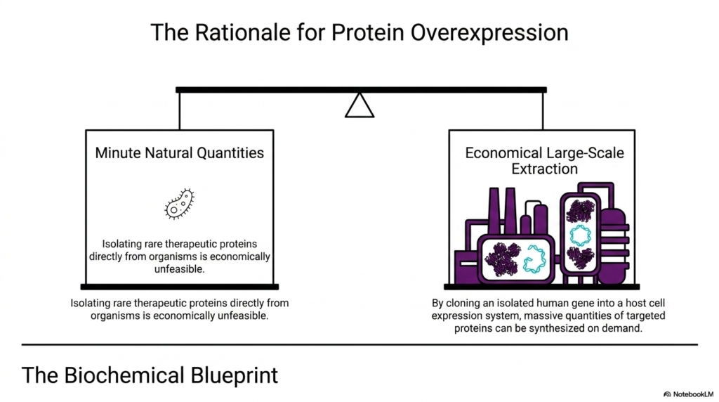
Isolating a gene is a monumental achievement, but a gene alone cannot cure a patient; it is merely an instruction manual. How do we translate this biological manual into massive quantities of functional medicine? The core purpose of this slide deck is to bridge the gap between DNA amplification via the polymerase chain reaction and physical drug manufacturing. This slide explains the economic and biological rationale for protein overexpression using host cell systems.
Historically, extracting rare therapeutic proteins directly from natural sources or systemic-therapeutic organisms yielded only minute natural quantities. It required harvesting massive amounts of biological tissue to produce a tiny fraction of usable medicine, a process that is economically unfeasible and highly inefficient. The advent of the polymerase chain reaction changed this paradigm entirely. By allowing scientists to isolate the exact human gene responsible for a therapeutic protein, the polymerase chain reaction opened the door to synthetic biology.
With the isolated human gene in hand—thanks to the polymerase chain reaction and gel extraction—scientists bypass natural limitations through economical large-scale extraction. The verified genetic blueprint is cloned into a specialized host cell expression system. These host cells act as microscopic bioreactors. Instead of relying on a human organ to slowly produce a specific hormone, researchers force thousands of liters of rapidly dividing bacterial or eukaryotic cells to manufacture the targeted protein simultaneously.
This biochemical blueprint strategy revolutionizes pharmacology. Massive quantities of targeted proteins can be synthesized on demand, without relying on unstable natural tissue supplies. However, this entire paradigm of large-scale manufacturing relies on the absolute precision of the upstream steps. If the initial polymerase chain reaction introduces errors, or if the wrong gene is amplified, the resulting overexpressed proteins will be useless or dangerous. Thus, flawless execution of the prior steps is clinically mandatory.
Slide 11: The Expression Plasmid: Delivering Polymerase Chain Reaction Products
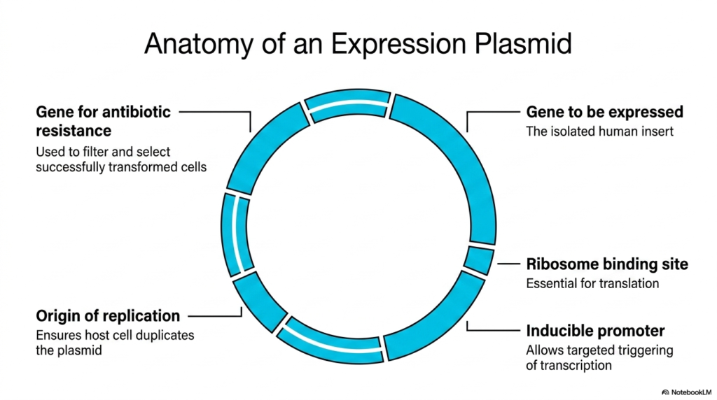
Host cells will not naturally accept or utilize foreign DNA floating in their environment; the genetic instructions must be smuggled inside. What vehicle is capable of safely delivering this targeted code into a microscopic factory? The core purpose of this slide deck is to explain how the genes isolated by the polymerase chain reaction are physically transported. This slide details the anatomy of an expression plasmid, the engineered circular vehicle used to deploy isolated therapeutic genes.
The most crucial component of the expression plasmid is the gene to be expressed. This section contains the isolated human insert, which exactly matches the biological sequence amplified by the polymerase chain reaction and verified during electrophoresis. To ensure the host cell can read this new manual, the plasmid also includes a ribosome-binding site. This site is essential for translation, signaling the cellular machinery to begin assembling amino acids based on the inserted code.
The plasmid also requires an inducible promoter, a specialized biological switch that allows researchers to trigger transcription. This means the gene amplified by the polymerase chain reaction remains dormant until the scientist introduces a specific chemical signal, causing the host cell to abruptly manufacture the protein on demand. Additionally, the plasmid features an origin of replication. This vital sequence guarantees that every time the host cell divides, it perfectly duplicates the plasmid along with its own natural genome.
Finally, the expression plasmid contains a gene for antibiotic resistance. When millions of host cells are exposed to the plasmids containing the polymerase chain reaction product, only a fraction will actually absorb the vehicle. By flooding the culture with a specific antibiotic, scientists rapidly eliminate any cell that failed to take up the plasmid. This clever filtering mechanism ensures that only successfully transformed cells survive to overexpress the crucial targeted therapeutic protein.
Slide 12: Host Cell Sequence: Expressing Polymerase Chain Reaction Genes
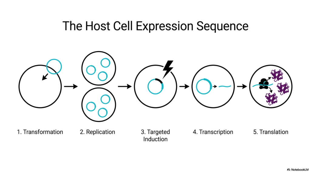
Inserting a microscopic vehicle into a living cell initiates a cascade of complex biological events. How does a single integrated plasmid reprogram an entire cellular organism to serve human medicine? The core purpose of this slide deck is to trace the biological timeline from DNA integration to final drug synthesis. This slide outlines the five-step host cell expression sequence, detailing how the blueprints generated by the polymerase chain reaction are executed to build tangible proteins.
The sequence begins with transformation, the physical process where the host cell membrane becomes permeable and absorbs the engineered expression plasmid containing the isolated therapeutic gene. Once inside, the second phase is replication. Driven by the plasmid’s origin sequence, the host cell blindly duplicates the foreign DNA alongside its own code. This internal biological copying multiplies the effects of the original polymerase chain reaction, ensuring every newly divided daughter cell carries the therapeutic instructions.
The third critical step is targeted induction. Researchers introduce a specific chemical trigger to the bioreactor, activating the inducible promoter on the plasmid. This signals the cellular machinery that it is time to read the gene originally isolated via the polymerase chain reaction. This leads immediately into the fourth phase, transcription. The host cell rapidly reads the targeted DNA sequence and converts it into transient messenger RNA molecules, preparing for physical protein construction.
The final phase of the sequence is translation. The host cell’s ribosomes bind to the messenger RNA and frantically assemble long chains of amino acids according to the exact human code. The host organism redirects its massive metabolic energy exclusively toward building the therapeutic agent. This beautiful sequence represents the culmination of the polymerase chain reaction pipeline: taking an isolated invisible gene and forcing a microscopic organism to manufacture massive, visible quantities of complex, life-saving medical proteins.
Slide 13: Protein Harvesting: Purifying Polymerase Chain Reaction End Products
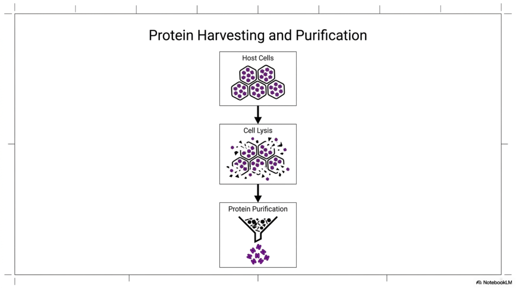
After microscopic cellular factories have finished manufacturing massive amounts of a drug, scientists face a massive logistical hurdle. How does one extract a pure therapeutic molecule from a chaotic soup of living organisms and cellular debris? The core purpose of this slide deck is to conclude the biological manufacturing journey that began with DNA amplification. This slide illustrates the final steps of protein harvesting and purification, ensuring the molecules derived from the polymerase chain reaction are medically safe.
At this stage, the bioreactor is filled with billions of successfully transformed host cells packed tightly with the targeted therapeutic protein. These proteins were synthesized based on the precise genetic instructions initially generated through the polymerase chain reaction. However, the medicine is trapped securely behind thick cellular walls and lipid membranes. The scientists cannot use the product in this state; the biological factories must be dismantled to access the engineered therapeutics hidden within.
The intermediate step is aggressive cell lysis. Researchers apply chemical detergents, sonic vibrations, or extreme pressure changes to physically shatter the host cells. This destructive process destroys the cellular structures and spills the therapeutic proteins, along with vast amounts of unwanted biological debris, into a complex liquid mixture. While the gene amplified by the polymerase chain reaction has done its job perfectly, the resulting biological soup is highly toxic and completely unfit for human clinical application.
The final imperative step is rigorous protein purification. The complex cellular lysate is passed through sophisticated chemical filters and biological funnels. These systems isolate the specific targeted therapeutic proteins, separating them out from the destroyed host cell debris, toxins, and unwanted native molecules. Once purified, the physical proteins—originally just a genetic sequence isolated via the polymerase chain reaction—are finally ready to be formulated into sterile, injectable medicines that can be distributed safely to patients worldwide.
Slide 14: The Engineering Toolkit: Polymerase Chain Reaction and Beyond
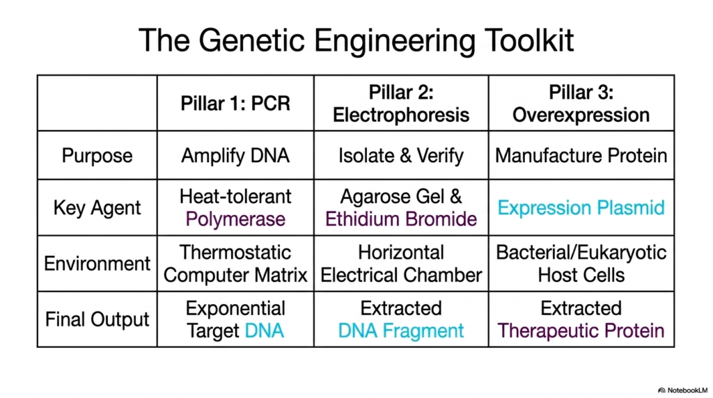
To master modern biological manufacturing, one must see how the individual techniques combine to form a cohesive, unstoppable scientific process. What are the core pillars that uphold the entire industry of synthetic biology? The core purpose of this slide deck is to synthesize the complex workflows into a unified conceptual framework. This slide serves as a summary matrix, breaking down the genetic engineering toolkit into three primary pillars, starting with the crucial polymerase chain reaction.
The first pillar of the toolkit is the polymerase chain reaction, whose primary purpose is to exponentially amplify a specific target DNA sequence. This process relies on a heat-tolerant polymerase enzyme operating within a thermostatic computer matrix environment. The final biological output of the polymerase chain reaction is exponential amounts of target DNA. Without this massive initial amplification, the subsequent pillars would completely lack the genetic raw materials required for further testing and development.
The second pillar is electrophoresis, dedicated to the critical purpose of isolating and verifying the genetic material. This stage uses the agarose gel and the chemical ethidium bromide within a horizontal electrical chamber. The key output here is a highly refined, perfectly extracted DNA fragment, guaranteeing that the product generated by the polymerase chain reaction is flawless. It serves as an uncompromising quality control bridge between the initial raw amplification and the final physical manufacturing phase.
The final pillar is overexpression, where the verified DNA is used to physically manufacture the protein. This stage relies heavily on the engineered expression plasmid delivered safely into bacterial or eukaryotic host cells. The final, triumphant output of this entire toolkit is the purified, extracted therapeutic protein. By understanding how the polymerase chain reaction seamlessly feeds into electrophoresis and overexpression, medical students gain a holistic understanding of how raw biological code is transformed into tangible medical treatments.
Slide 15: Clinical Impact: The Medical Value of Polymerase Chain Reaction
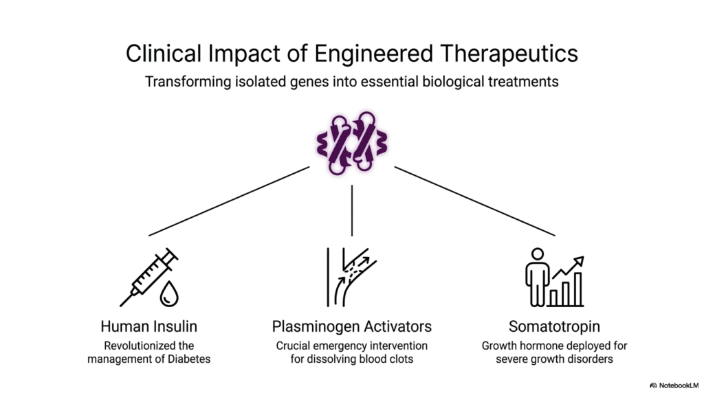
Theoretical biochemistry is fascinating, but its true value is measured by its ability to save human lives and alleviate suffering. How do these complex laboratory techniques actually manifest in the real world of hospitals and pharmacies? The core purpose of this slide deck is to connect microscopic laboratory processes to massive global health outcomes. This final slide showcases the tangible clinical impact of engineered therapeutics, proving that the polymerase chain reaction pipeline revolutionizes patient care.
The most famous example of this technology is the production of synthetic human insulin. Before the polymerase chain reaction and genetic engineering, diabetes management relied on insulin harvested from animal pancreases, which caused allergic reactions and suffered from severe supply shortages. Today, scientists use the genetic pipeline to manufacture flawless human insulin in bacterial vats. This direct application of recombinant technology has completely revolutionized diabetes management, providing a limitless, perfectly safe supply for patients worldwide.
Another life-saving application highlighted on the slide involves plasminogen activators. These specialized proteins serve as a crucial emergency medical intervention for rapidly dissolving lethal blood clots during severe heart attacks and acute strokes. Producing these highly specific enzymes in sufficient clinical quantities is only possible through overexpression systems. By initially isolating the human genes responsible for clot breakdown using the polymerase chain reaction, scientists provide emergency room physicians with vital drugs that prevent permanent organ damage and death.
Finally, the slide notes the production of somatotropin, a synthetic human growth hormone deployed for patients suffering from severe growth disorders and muscle-wasting diseases. Obtaining this hormone naturally was historically dangerous and incredibly rare. Through the targeted amplification of the human growth sequence via the polymerase chain reaction, pharmaceutical companies now provide safe, abundant somatotropin. These three examples powerfully demonstrate that mastering genetic engineering translates directly to creating essential biological treatments that fundamentally alter human medicine.
Please read our Content Disclaimer Statement.
Check out our social media channels: