156. Biochemistry of Cytokines: Signaling, Receptors, and Pathways
Have you ever wondered how your body coordinates billions of immune cells to fight an infection within hours? The answer lies in microscopic molecular messengers called cytokines. This slide deck explores the fundamental biochemistry of cytokines, detailing how these protein signals dock onto cell receptors, activate internal enzymes, and drive targeted gene expression to direct immunity and blood cell formation.
Slide 1: Overview of Cytokine Signaling and the Biochemistry of Cytokines
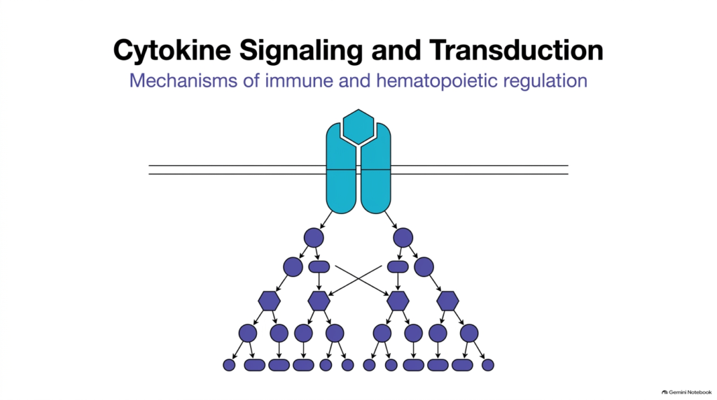
Cellular communication depends on sophisticated signal transduction pathways that translate extracellular cues into precise nuclear responses. When exploring cytokine biochemistry, one immediately notices how these low-molecular-weight proteins regulate fundamental physiological processes throughout the human body, including cell growth, differentiation, and tissue survival. Unlike simple chemical messengers that produce linear effects, cytokines initiate complex intracellular cascades that simultaneously activate multiple downstream targets. This opening slide introduces the overarching blueprint of cytokine signaling, illustrating how a single binding event at the cell membrane triggers an expanding cascade of enzymatic reactions inside the cytoplasm.
Understanding the biochemistry of cytokines requires examining how extracellular signals cross the hydrophobic plasma membrane barrier. Because these signaling molecules are hydrophilic, they cannot pass directly through lipid bilayers. Instead, they rely on specialized membrane-bound receptors to transmit signals across the cell envelope. Once a ligand engages them, these receptor complexes recruit cytoplasmic enzymes that amplify the initial signal. The biochemistry of cytokines reveals an elegant relay in which protein phosphorylation events propagate information directly to the cell nucleus.
This intricate cascade guarantees that immune and blood-forming cells respond dynamically to environmental stress and pathogenic threats. By coordinating intracellular signaling hubs, the cell ensures high-fidelity information transfer. As we move through this slide deck, we will examine how these initial membrane interactions activate specific enzymatic cascades, ultimately driving targeted transcriptional regulation in health and disease.
Slide 2: Defining Molecular Structure in the Biochemistry of Cytokines
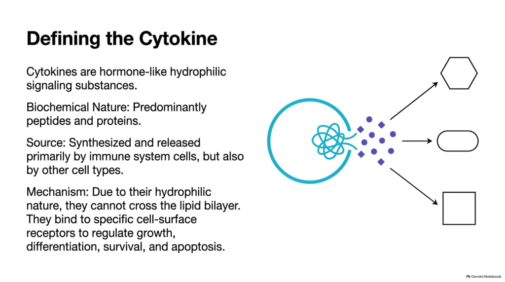
At the molecular level, cytokines are defined as hormone-like, hydrophilic signaling substances composed primarily of low-molecular-weight peptides and proteins. In cytokine biochemistry, researchers categorize these molecules by chemical structure, binding affinity, and cellular origin. Immune cells, such as macrophages, dendritic cells, and lymphocytes, synthesize and secrete these proteins in response to microenvironmental cues or tissue injury. Because their hydrophilic structure prevents them from diffusing across the cell’s fatty plasma membrane, these proteins must interact exclusively with the extracellular domains of specific cell-surface receptors to initiate downstream biological responses.
These protein signals have remarkably diverse physiological impacts across human tissues and organs. Through the lens of the biochemistry of cytokines, we see how these molecules regulate essential cellular decisions, including proliferation, functional maturation, cell survival, and programmed cell death. When a cytokine binds its matching receptor, it induces conformational changes that dictate whether a target cell will divide, specialize, or undergo apoptosis. Understanding cytokine biochemistry helps medical researchers see how these secreted proteins maintain immune balance and prevent uncontrolled cellular proliferation during inflammatory responses.
Furthermore, cytokines act through autocrine, paracrine, and endocrine mechanisms. This means a single secreted protein can act back on the producing cell, alter neighboring target cells, or occasionally enter systemic circulation. Recognizing these distinct operational spheres is crucial to understanding how localized molecular interactions produce coordinated, tissue-wide defenses against pathogens.
Slide 3: Three Physiological Domains in the Biochemistry of Cytokines
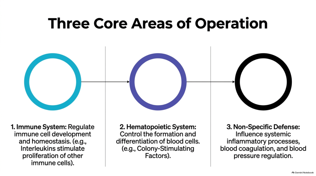
Cytokine signaling operates across three distinct physiological systems in human biology. A foundational concept in the biochemistry of cytokines is pleiotropy, where a single protein messenger acts on multiple cell types to produce varied biological outcomes. Within the immune system, molecules such as interleukins stimulate white blood cell proliferation, regulate lymphocyte development, and maintain baseline immunological homeostasis. Meanwhile, in the hematopoietic system, colony-stimulating factors strictly govern the production, maturation, and differentiation of various blood cell lineages from stem cell precursors in the bone marrow.
Beyond specialized cell maturation, these protein messengers play a crucial role in non-specific host defense mechanisms. In the broader framework of the biochemistry of cytokines, these secreted factors coordinate systemic inflammatory responses, regulate body temperature, and influence blood coagulation pathways during acute tissue trauma. By modulating local immune responses and systemic vascular dynamics, these signaling networks protect human tissues from widespread bacterial and viral infections. Studying the biochemistry of cytokines helps clinical scientists understand how localized cell signaling translates into systemic physiological defense.
This functional division underscores the remarkable versatility of these signaling networks. Whether driving white blood cell differentiation in bone marrow or modulating endothelial permeability during inflammation, cytokines serve as essential integration hubs. Understanding these three domains lays the foundation for analyzing specific cytokine families and their unique biochemical mechanisms.
Slide 4: Functional Classifications in the Biochemistry of Cytokines
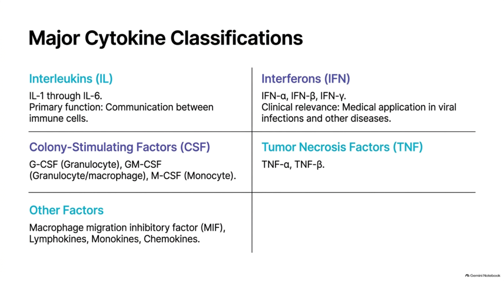
To navigate the immense diversity of signaling molecules, biochemists group them into major functional families based on structural features and biological roles. A central topic in the Biochemistry of Cytokines is distinguishing major groups such as interleukins, interferons, colony-stimulating factors, and tumor necrosis factors. Interleukins, designated IL-1 through IL-6 and beyond, primarily mediate direct communication between immune cells. Interferons, including IFN-alpha, IFN-beta, and IFN-gamma, provide vital antiviral defenses, while colony-stimulating factors direct the selective growth of granulocytes, monocytes, and macrophages.
Beyond these families, tumor necrosis factors like TNF-alpha drive inflammatory cascades and programmed cell death in damaged or transformed tissues. Deepening our knowledge of the Biochemistry of Cytokines also involves analyzing specialized factors like chemokines, monokines, and macrophage migration inhibitory factor. Each subcategory uses unique structural motifs and tertiary folds to perform distinct biological tasks. By classifying these molecules by structural homology, the Biochemistry of Cytokines provides a logical framework for understanding immune cell communication and targeted therapeutic interventions.
These functional groups do not operate in isolation; instead, they form an interconnected network in which one cytokine can stimulate or inhibit another’s production. This intricate balance ensures robust immune responses while guarding against excessive inflammatory damage. Mapping these functional families enables researchers to target specific disease pathways without disrupting broader immune homeostasis.
Slide 5: Diagnostic Differences from Hormones in the Biochemistry of Cytokines
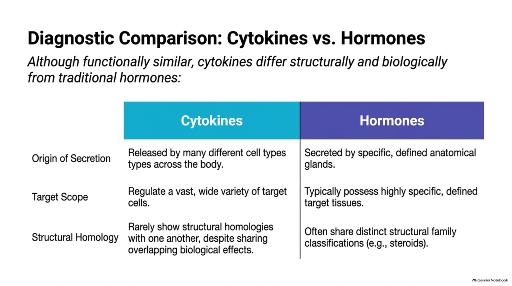
Although cytokines and classic endocrine hormones share functional similarities as circulating chemical messengers, key biological and structural differences set them apart. A comparative study in the Biochemistry of Cytokines shows that traditional hormones are secreted by specialized, anatomically defined endocrine glands, whereas a wide variety of cell types throughout the body produce cytokines. Furthermore, endocrine hormones usually target specific distant tissues, while these immune messengers have broad target-cell specificity, acting locally on neighboring cells or back on the secreting cell itself.
Structural architecture provides another clear line of distinction between these two signaling categories. In the specialized discipline of the Biochemistry of Cytokines, analysis shows that these signaling proteins rarely share structural homology, despite producing overlapping biological activities. In contrast, traditional hormones often fall into distinct structural families, such as steroids, polypeptides, or thyroid hormones. Recognizing these molecular distinctions within the Biochemistry of Cytokines is essential for pharmacologists designing targeted biological therapies that bypass classic endocrine feedback loops.
Additionally, cytokines typically function at nanomolar to picomolar concentrations and exhibit transient, tightly regulated expression patterns. By comparison, hormones often maintain steady basal plasma levels to regulate long-term metabolic stability. Distinguishing between these regulatory paradigms helps medical students comprehend the unique kinetics and systemic impact of cytokine-mediated signaling pathways.
Slide 6: Receptor Architecture and STPs in the Biochemistry of Cytokines
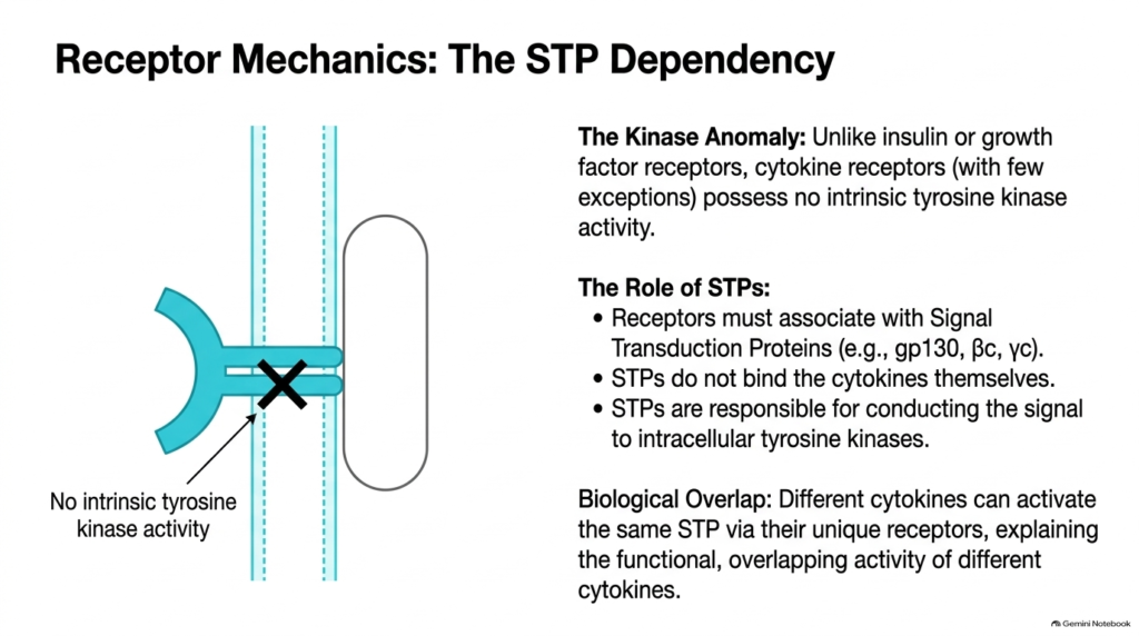
A unique structural feature of cytokine receptors is their total lack of intrinsic enzymatic activity. When exploring the Biochemistry of Cytokines, students encounter what biochemists call the kinase anomaly: unlike insulin or receptor tyrosine kinases, cytokine receptors lack catalytic tyrosine kinase domains in their intracellular tails. To transmit signals inward, these transmembrane receptors must non-covalently pair with specialized membrane proteins known as Signal Transduction Proteins, such as gp130, beta-common, or gamma-common signaling chains.
Signal Transduction Proteins do not bind extracellular cytokine ligands directly; instead, they primarily conduct intracellular signals to cytoplasmic enzymes. Within the conceptual framework of the Biochemistry of Cytokines, this modular arrangement explains functional redundancy in the human immune system. Different cytokine receptors can share the exact same Signal Transduction Protein subunit, allowing distinct extracellular ligands to trigger identical intracellular signaling pathways. This architectural insight from the biochemistry of cytokines shows how nature achieves remarkable functional economy by sharing receptor subunits.
By utilizing shared signal transducers, cells minimize the genomic hardware required to process diverse environmental cues. This assembly mechanism guarantees that multiple extracellular signals converge on a unified intracellular pathway. In the next slides, we will examine how this structural dependency directly leads to the recruitment and activation of non-receptor tyrosine kinases in the cytoplasm.
Slide 7: Ligand Binding and Dimerization in the Biochemistry of Cytokines
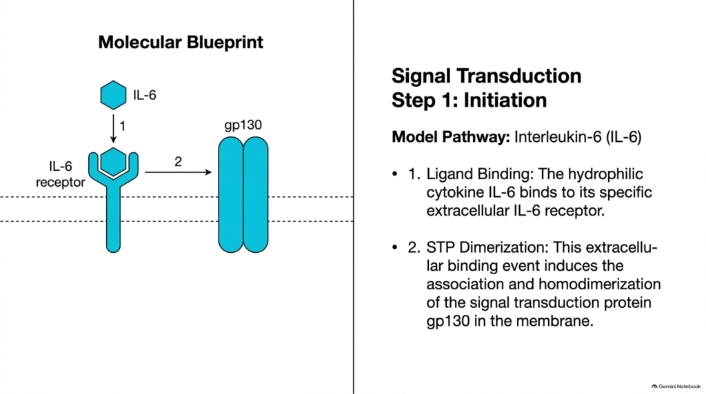
Signal transduction begins with high-affinity ligand binding at the outer surface of the plasma membrane. In the standard model of cytokine biochemistry, exemplified by Interleukin-6, a single hydrophilic cytokine molecule binds specifically to its extracellular receptor-binding pocket. This extracellular binding event alters the receptor subunit’s physical orientation, preparing it to engage partner proteins anchored within the cell-surface lipid bilayer.
After initial ligand engagement, the cytokine-receptor complex recruits and associates with the Signal-Transduction Protein gp130. As detailed in the Biochemistry of Cytokines, this interaction drives the homodimerization of two gp130 proteins within the cell membrane. Dimerization brings the cytoplasmic domains of the signal-transducing subunits into close physical proximity inside the cell. This spatial arrangement is the critical molecular trigger for recruiting internal signaling machinery, showing how the biochemistry of cytokines converts extracellular docking into a physical rearrangement of membrane proteins.
The formation of this stable hexameric or dimeric complex alters the local electrostatic potential along the inner membrane face. This conformational rearrangement exposes previously hidden binding motifs on the receptor’s cytoplasmic tails. Consequently, this step serves as the essential bridge connecting extracellular cytokine recognition to intracellular enzymatic activation.
Slide 8: Recruitment of Janus Kinases in the Biochemistry of Cytokines
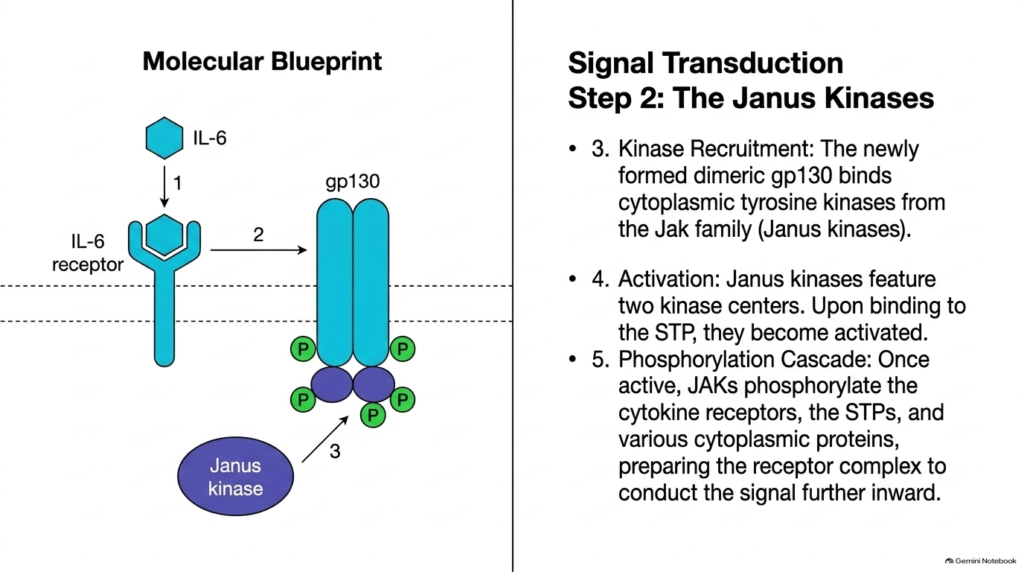
Once the signal-transducing subunits dimerize, they recruit non-receptor tyrosine kinases from the cytoplasm to continue the signaling sequence. A pivotal stage in cytokine biochemistry is the docking of Janus kinase family enzymes, commonly known as JAKs, onto the intracellular tails of dimeric gp130. Janus kinases have two distinct kinase domains, named for the two-faced Roman god Janus. When they bind the receptor complex, these enzymes undergo conformational shifts that activate their catalytic centers.
Activated Janus kinases immediately phosphorylate adjacent target proteins in a process called trans-phosphorylation. As described in the Biochemistry of Cytokines, active JAKs add phosphate groups to specific tyrosine residues on cytokine receptor tails, signal transduction proteins, and surrounding cytoplasmic substrates. These newly added phosphotyrosine residues serve as high-affinity docking sites for downstream signaling molecules. This phosphorylation cascade in the biochemistry of cytokines transforms an inactive membrane complex into a fully operational enzymatic platform.
Trans-phosphorylation increases JAK catalytic efficiency while creating specific binding pockets along the receptor tail. This phase converts a mechanical docking event into chemical energy stored in high-energy phosphate bonds. These localized phosphotyrosines act as beacon signals for latent transcription factors circulating nearby in the cytoplasm.
Slide 9: Structure of STAT Proteins in the Biochemistry of Cytokines
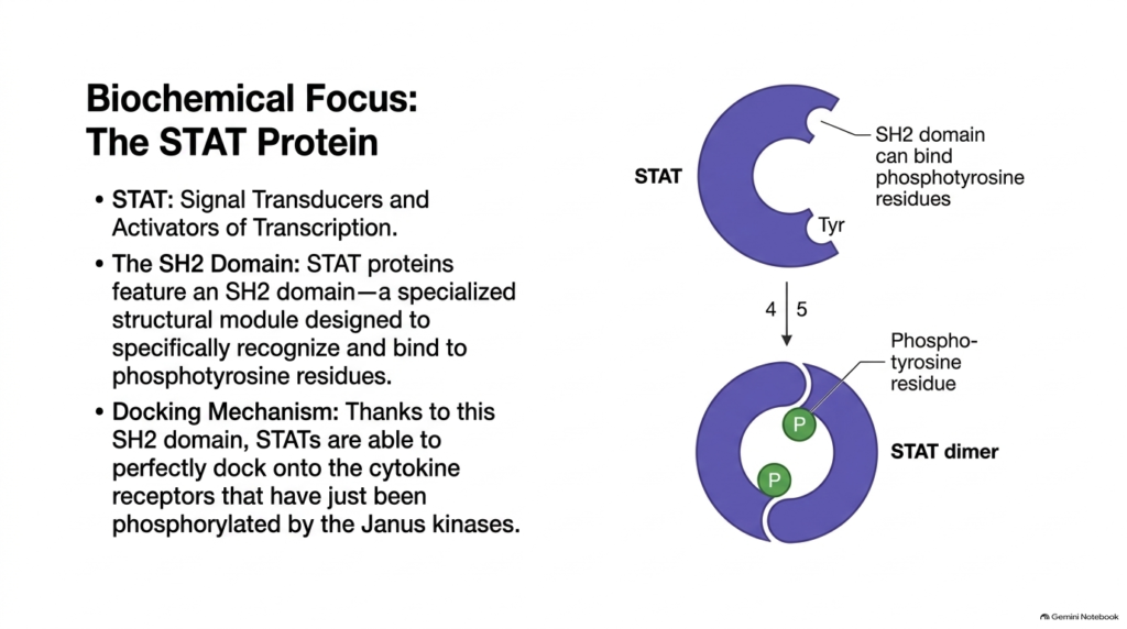
Downstream signal transmission relies heavily on a specialized family of cytoplasmic transcription factors known as STAT proteins. In the Biochemistry of Cytokines, STAT stands for Signal Transducers and Activators of Transcription. These cytosolic proteins exist as inactive monomers until activated membrane receptor complexes recruit them. Their primary structural hallmark is the Src Homology 2 (SH2) domain, a specialized protein module that recognizes and binds phosphotyrosine motifs.
The SH2 domain gives STAT proteins a precise molecular docking mechanism. In the broader context of the Biochemistry of Cytokines, this domain allows latent STAT monomers in the cytoplasm to recognize and bind phosphorylated tyrosine residues on receptor tails created by Janus kinases. By docking directly onto these phosphorylated sites, STAT proteins are positioned adjacent to active kinases. This physical localization highlights how cytokine biochemistry uses domain-specific interactions to organize rapid, highly specific intracellular signaling.
In addition to the SH2 domain, STAT proteins feature a central DNA-binding domain and a carboxyl-terminal transactivation domain. This modular architecture lets them act as both spatial sensors for receptor activation and direct regulators of gene expression. Their recruitment to the membrane complex prepares them for crucial enzymatic modifications in the next step.
Slide 10: STAT Phosphorylation and Dimerization in the Biochemistry of Cytokines
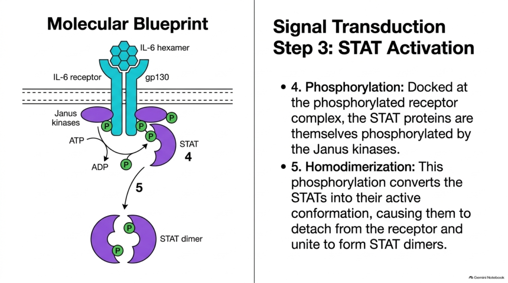
With STAT proteins successfully docked at the receptor complex, the stage is set for their chemical activation and structural transformation. In the step-by-step sequence of the biochemistry of cytokines, adjacent Janus kinases transfer a high-energy phosphate group from ATP to a key tyrosine residue near the carboxyl terminus of the docked STAT protein. This phosphorylation alters the local charge and conformation of the STAT monomer, triggering its immediate release from the membrane receptor docking site.
Once released into the cytoplasm, two phosphorylated STAT monomers interact through reciprocal SH2-phosphotyrosine bindings to form a stable homodimer complex. According to the Biochemistry of Cytokines, this dimerization event masks hydrophobic surfaces and exposes nuclear localization signals on the active dimer. By converting monomeric transcription factors into functional dimers, the Biochemistry of Cytokines ensures that only fully activated, phosphorylated STAT proteins gain the structural conformation required to enter the nucleus and bind target DNA.
This dimerization switch represents a critical biochemical checkpoint in cytokine signaling pathways. Without reciprocal SH2 domain interactions, STAT monomers remain trapped in the cytoplasm and cannot pass through nuclear pore complexes. Phosphorylation thus provides the precise chemical energy and structural shift required to drive this transition and license gene transcription.
Slide 11: Transcriptional Control in the Biochemistry of Cytokines
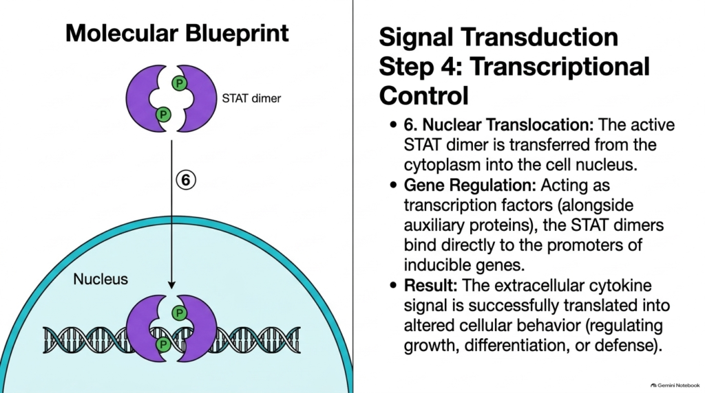
The intracellular signaling cascade culminates in the nucleus, where active gene transcription occurs. A final regulatory step in the biochemistry of cytokines involves the active nuclear translocation of STAT dimers through nuclear pore complexes via importin transport proteins. Once inside the nucleoplasm, these dimerized proteins act as potent transcription factors, binding directly to specific promoter sequences on inducible target genes alongside essential coactivator proteins and chromatin-remodeling complexes.
By binding to target DNA elements, STAT dimers stimulate the transcription of specific genes that control cell proliferation, lineage differentiation, and immune defense. Analyzing the Biochemistry of Cytokines demonstrates how an extracellular signal at the plasma membrane is faithfully translated into altered genomic output and modified cellular behavior. This transcriptional reprogramming illustrates the Biochemistry of Cytokines’ primary objective: converting brief membrane-binding events into long-lasting physiological changes within target cells.
The duration of transcriptional activation depends on how long STAT dimers remain bound to promoter regions before nuclear phosphatases deactivate them. Once dephosphorylated, STAT monomers exit the nucleus via exportin proteins and return to the cytoplasm to be recycled. This continuous nuclear-cytoplasmic shuttling ensures that gene expression dynamically tracks the real-time presence of extracellular cytokine signals.
Slide 12: Integrated Intracellular Cascades in the Biochemistry of Cytokines
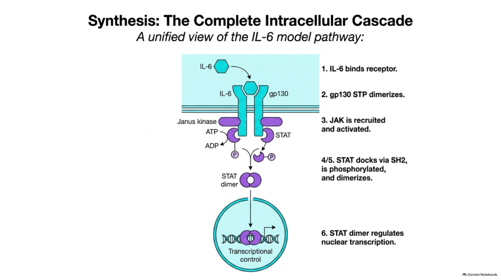
Synthesizing the individual steps of cytokine signaling reveals a beautifully integrated intracellular pathway connecting the cell exterior directly to the genome. Reviewing the complete model in the Biochemistry of Cytokines highlights the seamless transition from Interleukin-6 binding to receptor dimerization, JAK recruitment, and STAT activation. Each step in this biological pathway relies strictly on the successful execution of the preceding molecular event, ensuring high signaling fidelity and minimizing accidental background activation.
This unified pathway model underscores the elegance of intracellular signal transduction in human biology. Through the comprehensive framework of the Biochemistry of Cytokines, students can trace a single phosphate group as it moves from ATP through Janus kinases to STAT transcription factors and into the nucleus. Mastery of the Biochemistry of Cytokines enables researchers to identify potential therapeutic targets along this pathway, supporting the development of specific enzyme inhibitors to treat chronic autoimmune diseases and cellular malignancies.
From membrane docking to chromatin modification, this linear cascade represents one of the most efficient signaling mechanisms in cell biology. By mastering this unified roadmap, medical students gain a holistic perspective on how extracellular cues dictate immune cell fates. In the final slides, we examine how cells safely turn off this powerful signaling machinery.
Slide 13: Signal Termination Mechanisms in the Biochemistry of Cytokines
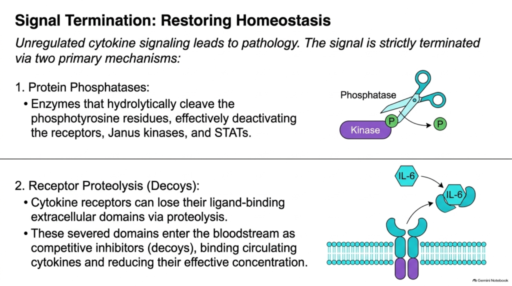
Because persistent or uncontrolled cytokine signaling causes severe tissue damage and inflammatory pathology, cells maintain robust signal-termination mechanisms. Within the Biochemistry of Cytokines, protein phosphatases serve as primary negative regulators by hydrolytically removing phosphate groups from active receptors, Janus kinases, and STAT proteins. Dephosphorylation rapidly neutralizes the enzymatic activity of these signaling components, resetting the cell to its basal resting state.
A second crucial mechanism for restoring cellular homeostasis involves receptor proteolysis and the generation of soluble decoy receptors. As discussed in the Biochemistry of Cytokines, specific proteases cleave the extracellular binding domains of cytokine receptors from the cell surface. These freed extracellular fragments enter the bloodstream and act as decoy receptors, binding circulating cytokines before they can reach cell-surface complexes. Through these dual regulatory pathways, the Biochemistry of Cytokines highlights how cells actively restore biochemical equilibrium.
Additionally, suppressor of cytokine signaling (SOCS) proteins provide a classic negative feedback loop by directly inhibiting JAK enzymatic activity and marking signaling proteins for proteasomal degradation. Together, phosphatases, receptor cleavage, and SOCS proteins prevent hyper-inflammatory states such as cytokine storms. Understanding these shutdown pathways is vital for designing drugs that modulate immune overactivity in clinical settings.
Slide 14: Systems Efficiency and Integration in the Biochemistry of Cytokines
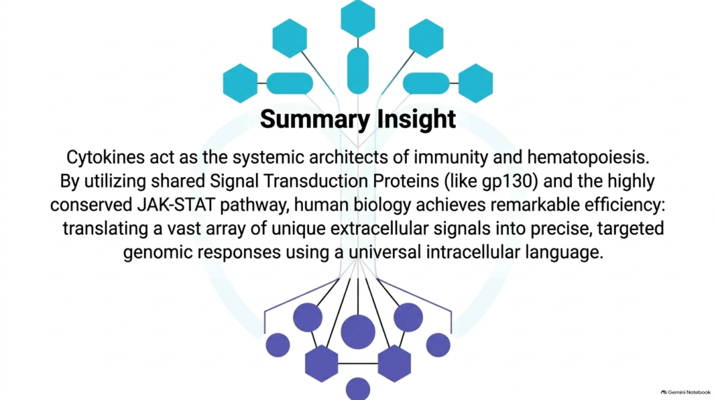
Reflecting on the overarching architecture of cytokine networks reveals an extraordinarily efficient molecular design refined by evolution. In this final overview of the biochemistry of cytokines, we see how nature utilizes shared Signal Transduction Proteins like gp130 alongside the highly conserved JAK-STAT signaling pathway to process diverse cellular messages. By employing a universal intracellular language, human cells process dozens of unique extracellular inputs using a standardized set of core enzymes and transcription factors.
This biological modularity allows complex multicellular organisms to achieve precise, targeted genomic responses without requiring millions of unique intracellular proteins. Examining the Biochemistry of Cytokines illustrates how shared receptors, kinase cascades, and STAT transcription factors work together to direct immunity, inflammation, and hematopoiesis. Ultimately, studying the Biochemistry of Cytokines provides students and clinicians with a foundational blueprint for understanding health, disease, and modern targeted immunotherapies.
As immunology research advances, targeting specific nodes in this signaling network continues to revolutionize clinical medicine. From Janus kinase inhibitors used in autoimmune disorders to engineered cytokines in cancer immunotherapy, basic molecular insights translate directly into transformative treatments. Mastering these biochemical principles empowers the next generation of medical scientists and clinicians to design safer, highly targeted therapeutic interventions for autoimmune and neoplastic diseases.
Please read our Content Disclaimer Statement.
Check out our social media channels: