107. Blood Coagulation Explained: Pathways and The Coagulation Cascade
Have you ever wondered how a tiny paper cut does not lead to catastrophic blood loss? The secret lies in a beautifully orchestrated molecular defense system. This blog post explores the elegant biochemistry of how our body seals vascular wounds. We will break down our latest academic slide deck piece by piece. This comprehensive guide is designed to help college and medical students master the complex chemical reactions that keep our circulatory system secure, transforming a liquid stream into a solid protective shield when injury strikes.
Slide 1: The Molecular Foundations of Blood Coagulation
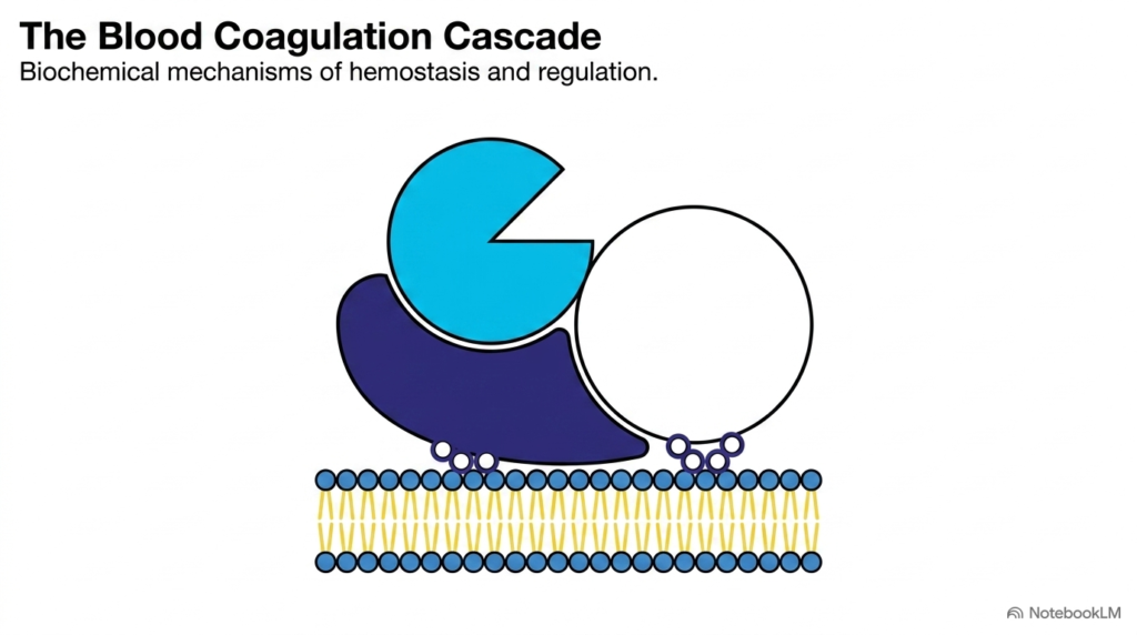
At the heart of cardiovascular preservation lies a highly coordinated sequence of biochemical events. When a blood vessel is injured, the body must respond immediately to prevent excessive bleeding while maintaining blood flow throughout the rest of the body. This elegant physiological balance is maintained by blood coagulation. This introductory slide illustrates these crucial molecular foundations, showing how various geometric shapes representing clotting factors anchor to biological surfaces. This organized grouping of components serves as a vital regulatory mechanism, preventing random systemic activation and ensuring that repair is strictly localized to the wound site. By focusing on this localized assembly, we can appreciate the beautiful design of human physiology.
The coagulation cascade is not a simple linear path, but rather a web of enzymatic reactions. This system is highly sensitive and tightly regulated because any imbalance can lead to severe clinical consequences. If the clotting process triggers too easily, dangerous clots can block blood vessels, leading to strokes or heart attacks. On the other hand, a sluggish cascade can cause severe bleeding. To prevent these issues, the biochemical components of Blood Coagulation are designed to remain inactive in normal circulation. They only assemble into active complexes on specific phospholipid membranes exposed by injury, which acts as a spatial control system. This ensures that clotting occurs only at the wound.
As we progress through this academic slide deck, we will analyze the precise structural mechanisms that drive this process. Medical students must appreciate how individual proteins transition from inactive zymogens into functional enzymes. This slide highlights that wound repair is not a chaotic event but a highly organized, surface-dependent assembly of proteins. This spatial and temporal control is the defining feature of Blood Coagulation, showcasing how molecular biology prevents hemorrhage. By mastering these foundational assemblies, you will be well prepared to understand the complex pathways and clinical interventions we will discuss in the upcoming slides. This foundational knowledge is key.
Slide 2: Primary Hemostasis and Secondary Blood Coagulation
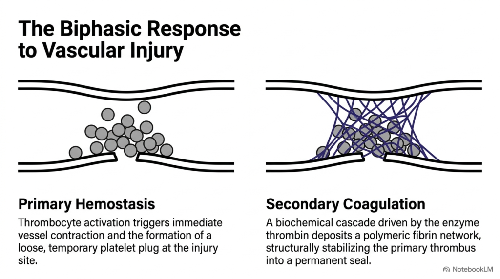
When a blood vessel is damaged, the body initiates a rapid, dual-stage repair process to restore vascular integrity. The first phase, known as primary hemostasis, involves the immediate activation of platelets, which gather to form a temporary, fragile plug. This initial physical barrier is highly effective at preventing the immediate escape of blood. However, this platelet plug is structurally weak and cannot withstand the high hydraulic pressure of the active circulatory system. Therefore, secondary Blood Coagulation must quickly follow to reinforce this delicate structure, forming a durable seal that remains intact during healing. This transition is vital for long-term health and tissue recovery.
During secondary Blood Coagulation, a complex cascade of chemical reactions occurs, culminating in the conversion of soluble fibrinogen into insoluble fibrin. This insoluble fibrin forms a durable, cross-linked network that acts like a structural scaffold, weaving directly through the aggregated platelets. This biochemical reinforcement locks the fragile platelet cluster securely in place, creating a permanent seal that supports long-term tissue repair. Without this crucial protein mesh, the primary plug would be quickly washed away by the shear forces of flowing blood, leading to recurrent bleeding at the site of injury. This step is indispensable for success and prevents dangerous blood loss during active vascular repair.
This coordinated sequence highlights the cooperative nature of biological defense systems. Platelets and clotting factors do not work in isolation; instead, they operate as mutual partners. The activated platelet membrane provides the essential lipid platform required for the assembly of key enzymatic complexes. By localizing these chemical reactions directly on the platelet plug, the body achieves rapid and localized Blood Coagulation. This elegant integration ensures that the permanent fibrin seal forms precisely where the vessel was damaged, preserving systemic blood volume and protecting the organism from hemorrhaging. This collaboration is a marvel of biological engineering that every student must study.
Slide 3: Fibrin Polymerization in Blood Coagulation
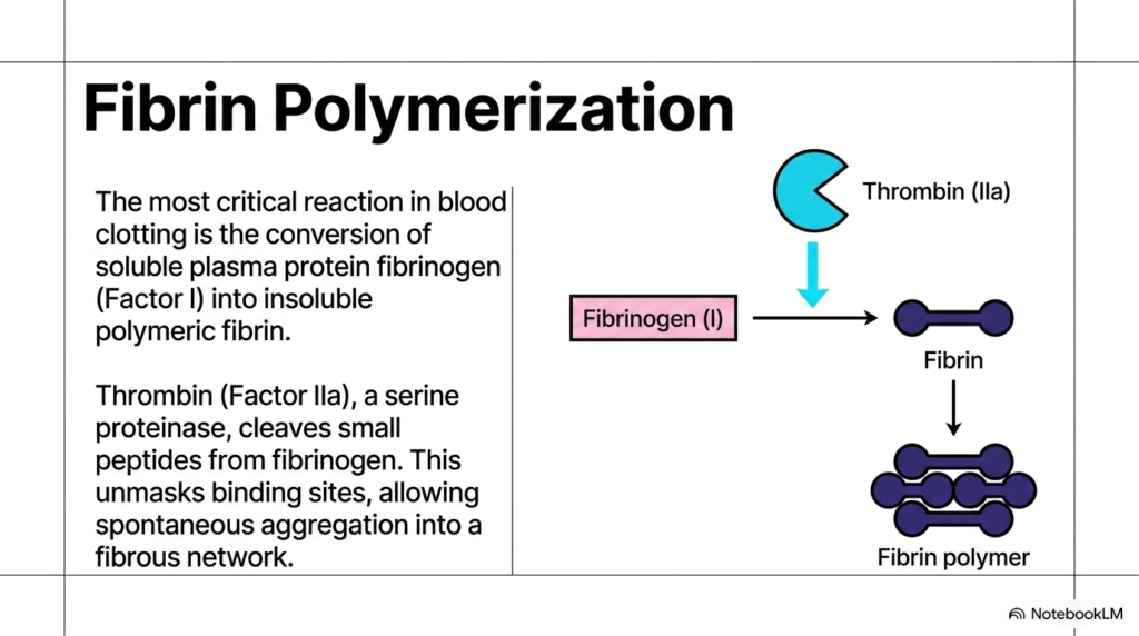
The physical transition of blood from a liquid to a solid gel relies on the polymerization of fibrin. Under normal conditions, the precursor protein fibrinogen is highly soluble and circulates harmlessly in the plasma. This high solubility is essential to prevent unwanted clotting in healthy vessels. However, when injury occurs, the active enzyme thrombin is generated. Thrombin acts as molecular scissors, chemically trimming specific peptide fragments from the central domain of the soluble fibrinogen. This precise proteolytic cleavage is the critical first step in the biochemistry of Blood Coagulation, converting soluble molecules into active fibrin monomers that can readily interact with one another in space.
This cleavage removes negative charges that previously caused the fibrinogen molecules to repel one another. With these electrical barriers gone, the newly formed fibrin monomers spontaneously aggregate. They align in a half-staggered, end-to-end and side-by-side fashion, forming protofibrils. This spontaneous self-assembly initiates fibrin polymerization. As these protofibrils grow and branch, they transform from dissolved proteins into a vast, insoluble fibrous network that traps blood cells. This dramatic physical phase transition is the ultimate goal of Blood Coagulation: to physically plug the leak in the vascular wall and stop further fluid loss from the vascular compartment.
For biochemistry and medical students, this process is an exceptional demonstration of structural biology in action. The conversion of a soluble monomer into an insoluble polymeric mesh highlights how minor proteolytic modifications can completely alter a protein’s physical state. Although this initial polymerized network is highly efficient, it is held together only by weak, non-covalent bonds. As we will see in the next slide, further enzymatic steps are required to strengthen this delicate web. Mastering this polymerization mechanism is fundamental to understanding the structural basis of Blood Coagulation and its role in human wound healing. This helps us grasp pathology at the molecular level.
Slide 4: Factor XIII and Clot Stabilization in Blood Coagulation
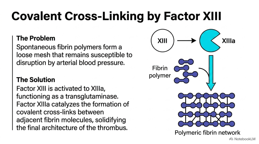
Although spontaneous fibrin polymerization is highly efficient, the initial clot is structurally fragile. Held together solely by non-covalent forces, this temporary mesh cannot withstand the severe mechanical stress of pulsatile blood flow. To prevent the premature breakdown of this essential barrier, the body must reinforce the clot’s structural integrity. This critical stabilization step is performed by Factor XIII, a specialized transglutaminase. Once activated by thrombin, Factor XIIIa fundamentally alters the internal chemistry of the clot, ensuring that Blood Coagulation produces a permanent, durable seal that withstands stress under physiological conditions. This process represents a fascinating subject for any clinical biochemist studying clot mechanics.
Factor XIIIa catalyzes the formation of strong, covalent cross-links between neighboring fibrin monomers. Specifically, it creates isopeptide bonds between the side chains of specific glutamine and lysine residues. These covalent bonds transform the fragile web of fibers into a solidified polymeric network. This chemical reinforcement is a vital phase of Blood Coagulation because it ensures the thrombus remains intact under physiological pressure. Without this enzymatic cross-linking, the clot would remain highly susceptible to mechanical displacement and rapid enzymatic breakdown by fibrinolytic proteins circulating in the bloodstream. This represents a major failure mode that we must avoid in clinical scenarios.
By establishing these covalent bridges, Factor XIIIa secures the thrombus, allowing the blood vessel to heal safely over time. This step is a prime clinical example of how covalent modification regulates macromolecular structures. A deficiency in this cross-linking enzyme leads to severe bleeding disorders, as patients can form an initial clot, but it quickly breaks down. This underscores how the mechanical strength of the clot is just as important as its formation. Ultimately, Factor XIIIa activity represents the final, fortifying touch in the biochemistry of Blood Coagulation. It makes the clot truly permanent and reliable for cardiovascular healing and long-term recovery.
Slide 5: Zymogens and Cascade Amplification in Blood Coagulation
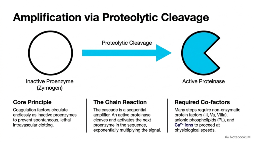
How does the human body translate a microscopic vascular injury into a massive physical clot within seconds? The secret lies in a series of highly amplified chemical reactions. Most of the proteins involved in clotting circulate as inactive precursors called zymogens. These zymogens are designed to be completely harmless during normal circulation. However, when an upstream enzyme cleaves a specific peptide bond in the zymogen, the zymogen undergoes a conformational change that exposes its active catalytic site. This transition is the fundamental mechanism driving the biochemistry of Blood Coagulation. This activation ensures rapid protective responses that occur only when required.
This structural activation allows for rapid signal amplification. A single active enzyme molecule can quickly cleave and activate dozens of downstream zymogen molecules, which in turn activate hundreds more. This molecular domino effect is the defining feature of Blood Coagulation pathways. To achieve the extreme reaction speeds required to save lives, these activation steps require non-enzymatic cofactors, calcium ions, and phospholipid membranes. These components act as physical scaffolds, accelerating enzymatic cleavage by several orders of magnitude and ensuring that clotting is both rapid and localized. This speed is crucial during trauma and vascular disruption.
This cascade design allows the body to maintain a highly sensitive system that remains completely silent until a threshold stimulus is met, preventing dangerous systemic clotting. Once triggered, the cascade generates an explosive, localized burst of active enzymes, culminating in fibrin formation. For students of biochemistry, understanding how zymogen activation drives signal amplification is essential to mastering the pathways of Blood Coagulation. It is this multi-tiered enzymatic engine that makes the clotting cascade one of the most responsive physiological systems in the body, protecting us daily from minor and major injuries alike. This highly organized pathway exemplifies the remarkable efficiency of biological regulatory mechanisms.
Slide 6: Extravascular Initiation of Blood Coagulation
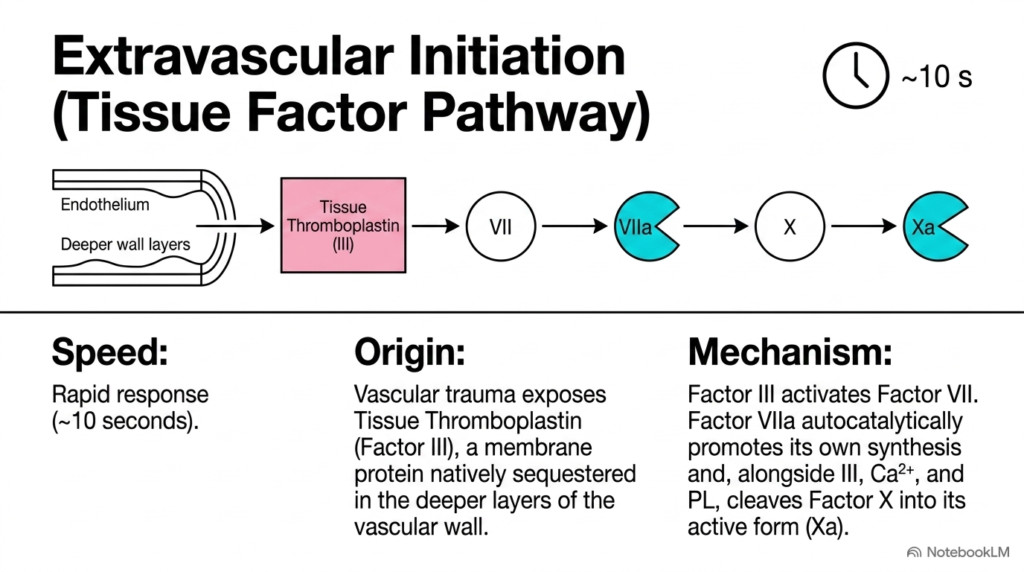
When physical trauma tears open a blood vessel, the body must act almost instantly to prevent catastrophic blood loss. To achieve this, the primary initiator of clotting in vivo is the extravascular, or extrinsic, pathway. Under normal physiological conditions, the transmembrane receptor known as Tissue Factor (Factor III) is sequestered on cells outside the bloodstream. This spatial isolation prevents accidental clotting. However, when vascular injury occurs, circulating blood immediately comes into contact with these tissue-factor-bearing extravascular cells, initiating a rapid response in Blood Coagulation. This contact acts as the primary trigger for the entire downstream pathway.
Once exposed, Tissue Factor binds to circulating Factor VII, which is quickly cleaved to form active Factor VIIa. This newly formed TF-VIIa complex, in the presence of calcium ions and phospholipid membranes, acts as a highly potent enzymatic engine. It directly cleaves and activates Factor X into its active form, Factor Xa. Remarkably, this entire extravascular initiation sequence takes only about ten seconds to transform Factor X into Xa. This rapid speed makes it the primary responder in Blood Coagulation, addressing the bleeding almost instantly at the site of impact. This speed prevents excessive hemorrhage in damaged tissues.
The simplicity and speed of this pathway are vital for surviving acute physical injuries. By using a pre-synthesized extracellular receptor as the trigger, the body bypasses the need for complex, time-consuming intravascular steps during the initial phase of injury. This rapid activation highlights the evolutionary efficiency of Blood Coagulation mechanisms. For medical students, mastering the Tissue Factor-initiated pathway is fundamental, as it is the spark that ignites the systemic clotting cascade and ensures rapid clot formation at the wound site. It is a vital clinical concept in trauma medicine. This rapid response is a critical evolutionary adaptation that protects complex life.
Slide 7: Intravascular Initiation of Blood Coagulation
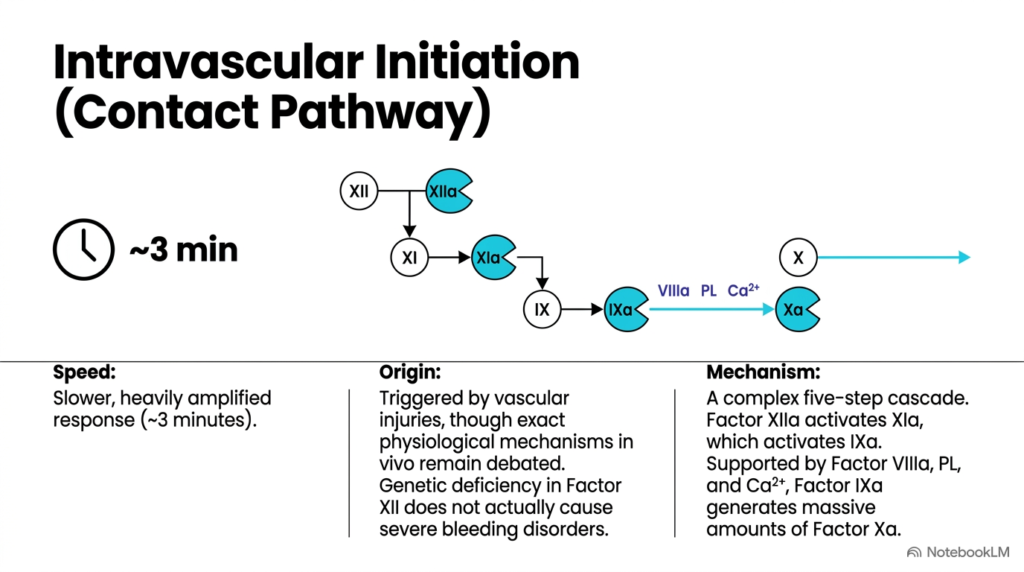
While physical trauma triggers the extravascular pathway, the body also possesses an internal mechanism known as the contact activation, or intrinsic, pathway. This pathway is initiated entirely within the blood vessels when circulating proteins encounter negatively charged surfaces, such as collagen exposed by endothelial damage, or foreign materials like medical implants. Unlike the rapid extravascular pathway, this internal sequence is a five-step cascade that takes approximately three minutes to complete. This makes it a much slower, though heavily amplified, response in Blood Coagulation. This slower speed reflects its complex molecular nature and multi-layered design.
The pathway begins when Factor XII binds to a charged surface, undergoing a conformational change that activates it to Factor XIIa. This enzyme then sequentially cleaves and activates Factor XI, which in turn activates Factor IX. Finally, active Factor IXa, together with its cofactor, active Factor VIIIa, on the membrane surface cleaves and activates Factor X to Factor Xa. This multi-step process is a crucial contributor to the overall amplification of Blood Coagulation, producing a massive reservoir of active enzymes downstream. This pathway provides a highly structured cascade of molecular events and feedback regulation that ensures robust enzyme production.
Interestingly, clinical studies show that patients lacking Factor XII do not suffer from severe bleeding disorders. This suggests that while the contact activation pathway is vital for intravascular clotting, the extrinsic pathway remains the primary driver of normal wound healing. However, the intrinsic pathway is highly relevant in inflammatory diseases and medical device design, where artificial materials can trigger unwanted Blood Coagulation. Thus, understanding both pathways is essential for managing thrombotic risks and appreciating the full, elegant complexity of human physiological systems. This knowledge helps clinicians prevent strokes and thrombosis, providing multiple points of regulatory control. These complex enzymatic dynamics are vital to hemodynamics, providing a deeper academic perspective.
Slide 8: The Prothrombinase Complex in Blood Coagulation
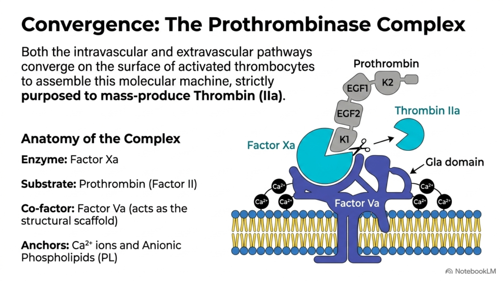
As we trace the clotting pathways, we find that both the extrinsic and intrinsic systems converge at a single, critical junction: the activation of Factor X. Once Factor Xa is generated, it must perform its primary role of converting prothrombin into active thrombin. However, free Factor Xa floating in solution is a highly inefficient enzyme. To achieve the explosive rate of thrombin generation required to seal a wound, Factor Xa must assemble into a highly specialized molecular machine known as the prothrombinase complex, a core driver of Blood Coagulation. This assembly is essential for physiological survival under stress.
This multi-protein engine assembles on the negatively charged phospholipid membrane of activated platelets. The complex consists of the active enzyme Factor Xa and its non-enzymatic cofactor, Factor Va, which are held together securely by calcium ions. By organizing these components on the membrane surface, the prothrombinase complex increases the catalytic efficiency of prothrombin activation by several hundred thousand-fold. This incredible speed is what makes the complex the core engine of Blood Coagulation, producing the massive burst of thrombin needed to create a clot. This is a vital physiological mechanism that is central to life. It represents a masterpiece of natural engineering.
For medical and biochemistry students, the prothrombinase complex is a classic textbook model of membrane-localized enzymology. It illustrates how the structural assembly of an enzyme, a cofactor, a substrate, and a membrane surface can dramatically accelerate a chemical reaction. Without this localized assembly, the process of Blood Coagulation would be far too slow to prevent severe hemorrhage. Thus, the prothrombinase complex represents the vital biochemical turning point where the regulatory signals of the cascade are focused into a powerful, localized clotting response. It is a beautiful example of metabolic control in biological systems, ensuring clotting is strictly restricted to wounds. It is essential to medical study.
Slide 9: Gla Domains and Calcium Bridging in Blood Coagulation
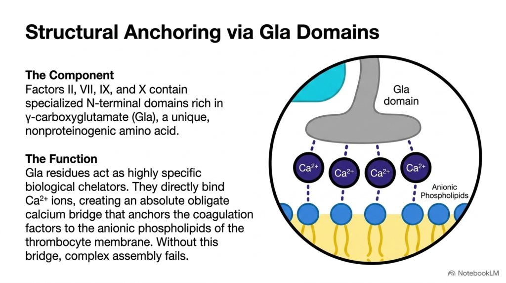
To understand how clotting factors assemble so precisely on membrane surfaces, we must look at their molecular structures. Several key proteins in the clotting cascade, including Factors VII, IX, X, and prothrombin, share a highly conserved N-terminal region known as the Gla domain. This domain contains unique gamma-carboxyglutamic acid residues, formed by a post-translational modification that adds a second carboxyl group to specific glutamic acid residues, creating a strong double negative charge. This domain is critical for the mechanics of Blood Coagulation. It allows selective surface binding only when required. Without this specific modification, the process would fail.
This unique chemical structure allows the Gla domain to function as a highly specialized biological magnet. The negatively charged carboxyl groups bind to positively charged divalent calcium ions. These bound calcium ions then act as a physical molecular bridge, linking the clotting factor to the negatively charged headgroups of phospholipids on the surface of activated platelet membranes. This calcium-mediated tethering is a fundamental requirement for localized Blood Coagulation, ensuring that the enzymatic complexes are anchored exactly where clotting is needed. This prevents systemic clotting and localized bleeding. The negative surface charge acts as a localization signal.
Without this structural anchor, the clotting factors would float freely in the general circulation, rendering them unable to assemble into active complexes like the prothrombinase engine. This would completely disrupt the orderly process of Blood Coagulation, leading to systemic bleeding and clinical instability. This molecular bridging mechanism beautifully illustrates how precise post-translational modifications dictate the physical localization and function of essential proteins, showing that the physical chemistry of membrane binding is absolutely vital to the success of clotting. It is a key pharmaceutical target for modern therapies, demonstrating how molecular structure influences physiology. Therefore, cell membranes are active participants in clotting.
Slide 10: Pharmacological Control and Inhibition of Blood Coagulation
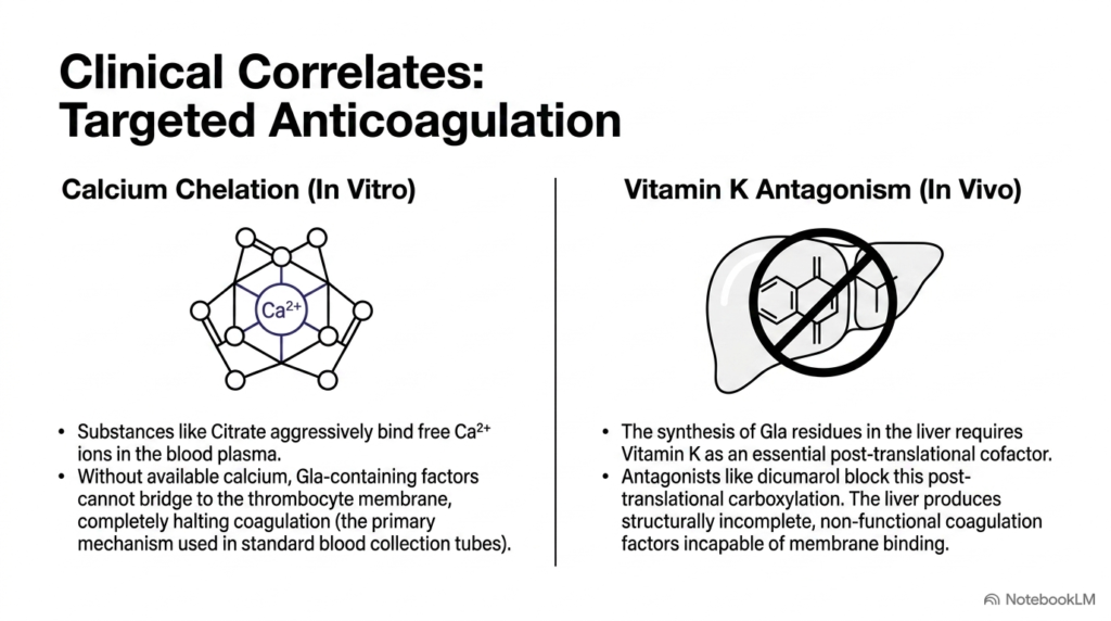
Given the life-saving nature of clotting, it is equally important to understand how we can manipulate this system medically. In both clinical medicine and laboratory diagnostics, controlling the rate of clot formation is essential. By targeting the specific chemical requirements of the clotting cascade, scientists and physicians have developed precise methods to prevent blood from solidifying. These interventions are vital for managing thrombotic risks, performing diagnostic tests, and preserving drawn blood samples, representing important applications of Blood Coagulation science. This control is highly beneficial for patients worldwide, as managing clotting rates remains a major focus in cardiovascular care.
One common laboratory method is calcium chelation. Since calcium ions are required to bridge Gla domains to platelet membranes, adding substances such as EDTA or citrate to a test tube binds free calcium ions. This effectively prevents the assembly of clotting complexes, halting Blood Coagulation in vitro. This demonstrates how simple ionic interactions can have profound physiological effects. Inside the living body, a different strategy is used: Vitamin K antagonism. Vitamin K is a required cofactor for the hepatic carboxylase enzyme that creates the Gla domains. By administering Vitamin K antagonists such as warfarin, the liver synthesizes inactive factors that cannot bind calcium. Thus, drugs like heparin or warfarin are highly effective.
These therapeutic strategies demonstrate how fundamental biochemistry translates directly into clinical therapies. By disrupting either calcium ion availability or Gla domain synthesis, we can render essential clotting proteins inactive. This provides precise pharmacological control of blood coagulation, preventing dangerous strokes or deep vein thrombosis in patients. Understanding these chemical requirements helps medical students appreciate how altering molecular structures can lead to powerful, life-saving clinical outcomes. It allows us to grasp how anticoagulants manage thrombosis and protect patients from stroke. This biological mechanism represents a triumph of translational research, uniting basic chemistry and clinical intervention.
Slide 11: Thrombin and Positive Feedback in Blood Coagulation
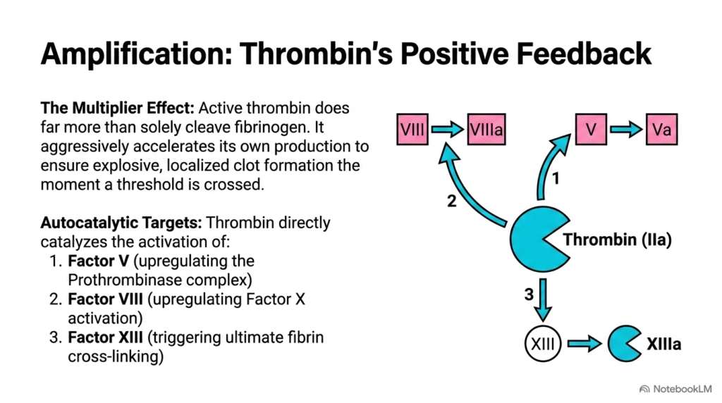
Once a small amount of thrombin is generated, how does the body ensure that a robust, stable clot is formed before the blood carries the active enzymes away? The system solves this challenge through a highly elegant positive feedback loop. Thrombin is not merely the final enzyme that cuts fibrinogen; it also acts as a powerful autocatalytic engine that accelerates its own production. This feedback mechanism creates a rapid multiplier effect, which is essential to the dynamics of Blood Coagulation. This ensures a prompt response that is tightly restricted to the area of damage, converting a minor stimulus into a robust physical barrier.
Specifically, active thrombin travels upstream to activate its own essential cofactors. It cleaves and activates Factor V into Va and Factor VIII into VIIIa. These active cofactors then assemble into the tenase and prothrombinase complexes, accelerating thrombin generation by several orders of magnitude. Additionally, thrombin activates Factor XI, which boosts the intrinsic pathway, and Factor XIII, which secures the final clot. This coordinated feedback loop ensures that once a threshold of injury is reached, Blood Coagulation proceeds rapidly. This prevents delayed clotting and ensures robust vascular defense through a self-amplifying system.
This positive feedback loop is a classic biological example of a non-linear response. It guarantees that the clotting defense is all-or-nothing, preventing weak, incomplete clots that would fail to stop bleeding. By concentrating this explosive enzymatic activity directly on the activated platelet membrane, the body achieves localized and rapid Blood Coagulation exactly where the vascular wall is breached. For students of medicine, mastering these autocatalytic targets of thrombin is essential to understanding the dynamic, self-amplifying nature of this cascade. It represents elegant enzymatic control that prevents extensive blood loss. Ultimately, positive feedback is the physiological key to rapid, localized clot formation. This is an important concept for medical licensure exams.
Slide 12: Endogenous Regulation of Blood Coagulation
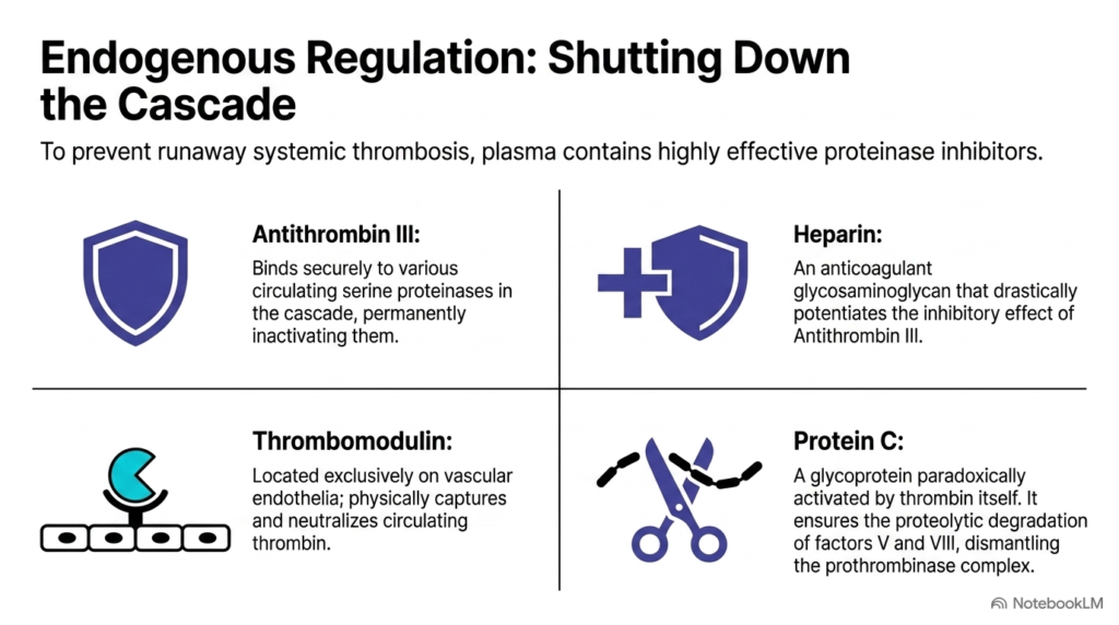
While the positive feedback loops of thrombin are essential for rapid wound sealing, an unchecked clotting cascade would quickly block the entire circulatory system. To prevent dangerous systemic thrombosis, the body must employ highly sophisticated chemical brakes. These endogenous regulatory mechanisms are designed to neutralize active enzymes and dismantle clotting complexes, ensuring that clot formation remains strictly confined to the immediate site of vascular damage, maintaining proper Blood Coagulation control. This maintains healthy homeostasis, prevents ischemia, and stops the spread of clot formation throughout the network.
One primary regulator is Antithrombin III, a circulating proteinase inhibitor that binds to and inactivates thrombin and Factor Xa. This inhibitory action is accelerated thousands of times by heparin, which is present on endothelial surfaces. Another vital pathway involves the vascular-bound receptor Thrombomodulin. When thrombin binds to Thrombomodulin, it undergoes a conformational change. Instead of clotting fibrinogen, this modified thrombin activates Protein C, which, along with Protein S, cleaves and inactivates the cofactors Va and VIIIa, halting further Blood Coagulation. This limits unwanted clot expansion and tissue damage. This receptor acts as a biological switch, converting a pro-coagulant enzyme into an anti-coagulant catalyst.
This elegant system of chemical brakes illustrates the delicate balance required to maintain health. The body must constantly balance pro-coagulant forces with anti-coagulant regulators. A deficiency in any of these regulatory proteins leads to severe hypercoagulable states, where patients suffer from recurrent, life-threatening blood clots. Thus, mastering these endogenous brakes is just as critical as learning the cascade’s activating steps. This ensures Blood Coagulation remains a localized, protective defense rather than a widespread disease. This balance preserves life and ensures healthy endothelial function. Without these mechanisms, localized clotting would progress, leading to systemic thrombosis and widespread vessel occlusion. Thus, the balance between activation and regulation is vital for survival. These chemical brakes protect our vascular network.
Slide 13: A Biochemical Reference of Blood Coagulation Factors
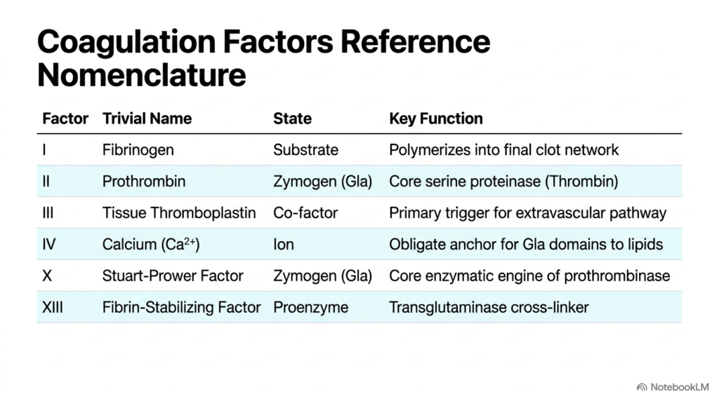
To fully master this physiological defense system, one must become familiar with the individual molecular components that drive the cascade. This final slide serves as a comprehensive reference table, organizing the essential clotting proteins by their formal Roman numeral designations, common names, and specific biochemical roles. It categorizes these substances into inactive zymogens, active serine proteases, non-enzymatic cofactors, substrates, and essential structural enzymes, providing a complete map of Blood Coagulation. This table simplifies a complex biological network for easier digestion, consolidating a massive amount of clinical information.
For example, the table traces the journey of Fibrinogen (Factor I) as the primary substrate, and Thrombin (Factor IIa) as the primary activator. It highlights how calcium ions (Factor IV) are necessary for membrane anchoring, and how Factor XIII acts as the transglutaminase responsible for cross-linking the final fibrin network. By mapping out these specific interactions, the table clarifies how the individual pieces fit together to form the powerful molecular engine that drives Blood Coagulation, allowing students to visualize the entire system. This aids conceptual understanding enormously, showing how elements work in unison. It serves as a perfect map for understanding clotting pathology. This simplifies complex pathway details.
This reference chart is an invaluable study aid for college and medical school students. It consolidates complex pathways into an organized, digestible format, making it easier to memorize the various factors and their functions. Understanding these individual components is the foundation of clinical hematology, as many genetic bleeding disorders, such as hemophilia, are caused by a deficiency in a single factor listed here. Ultimately, this structural map brings our study of Blood Coagulation to a satisfying conclusion, showing how molecular details unite to protect human life. It is a cornerstone of medical biochemistry and hematological science. This detailed summary provides students with a solid foundation for clinical practice.
Please read our Content Disclaimer Statement.
Check out our social media channels: