157. Mammalian Cell Cycle Regulation: Molecular Mechanisms and Control
Every single human body begins as a microscopic, single-celled zygote that must divide trillions of times with near-flawless biological precision. How do living organisms coordinate this monumental growth process while preventing catastrophic cellular errors? The answer lies within the elegant biochemical machinery of cellular division. This slide deck breaks down the essential molecular engines, enzymatic switches, and surveillance pathways that govern human proliferation. By examining these structural mechanisms step by step, college and medical school students will gain a clear foundational understanding of cellular control, tumor suppression, and the biochemical origins of human cancer.
Slide 1: Cell Cycle Regulation and the Phases of Division
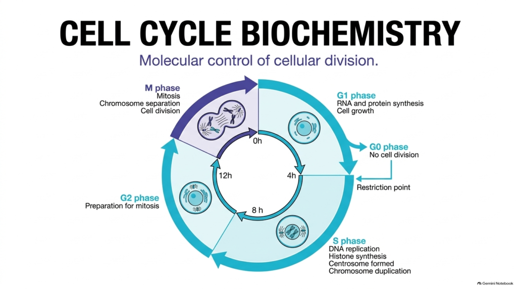
The growth and duplication of eukaryotic cells depend upon precise temporal coordination across distinct functional stages. Cell Cycle Regulation ensures that nuclear replication and cellular growth proceed in a strict, orderly sequence. The standard eukaryotic cycle spans roughly twenty-four hours and is divided into four main functional phases. First, the cell enters the G1 phase, where it synthesizes RNA, produces proteins, and grows. After growth, the cell enters the S phase, during which nuclear DNA replicates, histone proteins are synthesized, centrosomes duplicate, and chromosomes double. Next, during the G2 phase, the cell conducts final biochemical checks and preparations before entering the M phase, where mitosis completes chromosome separation and physical cell division.
Central to this overarching architectural framework is the ability of cells to exit active division into a resting state known as G0. The machinery governing cell cycle regulation allows non-dividing cells to pause indefinitely or re-enter division in response to specific extracellular cues. At the restriction point near the end of G1, the cell evaluates external growth factors and nutrient availability before committing to complete an entire round of division. If environmental conditions remain unfavorable, the cell safely halts progression. This baseline architecture shows how Cell Cycle Regulation integrates external signals with internal biochemical timers to maintain cellular homeostasis, prevent premature nuclear division, and support healthy tissue development throughout the organism.
Slide 2: Cell Cycle Regulation during Macro-Phases of Proliferation
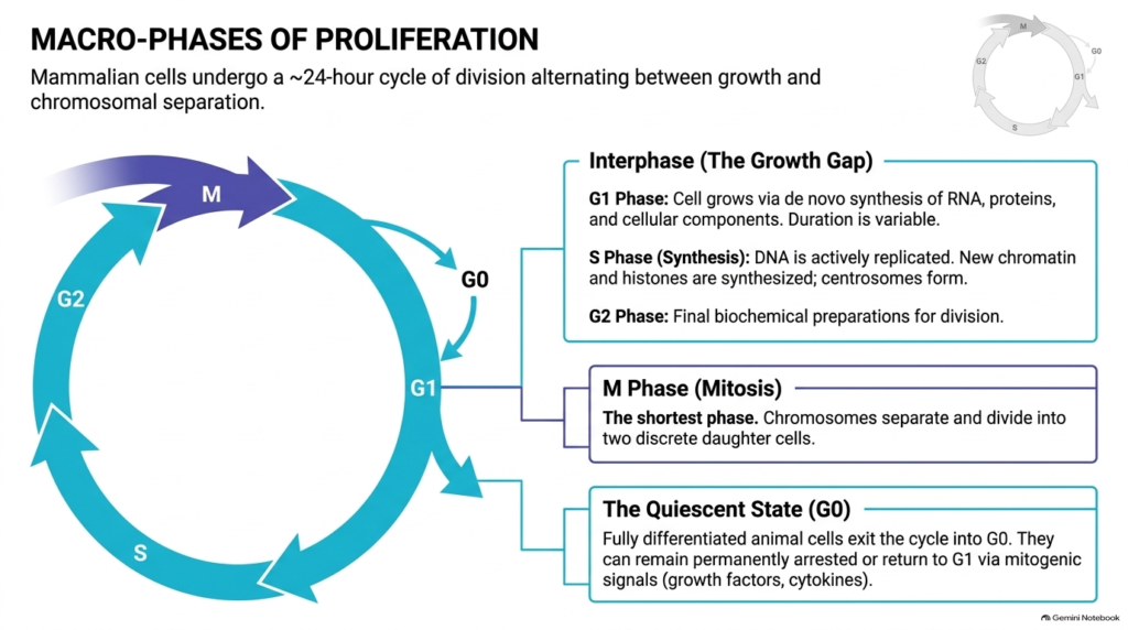
Eukaryotic cellular proliferation splits into two principal functional categories: interphase, which encompasses cell growth, and the M phase, which executes physical division. Proper Cell Cycle Regulation requires the cell to spend most of its lifecycle in interphase. During the G1 phase of interphase, the cell increases its volume through continuous de novo synthesis of proteins, structural organelles, and functional RNA molecules. This initial growth phase exhibits variable duration depending on cell type and metabolic conditions. Next, the cell enters the S phase to copy its genetic material, assemble new chromatin complexes, and organize spindle poles. Finally, the G2 phase completes the remaining metabolic preparations needed to support nuclear envelope breakdown and chromosome movement.
In stark contrast to interphase, the M phase represents the shortest stage of proliferation, rapidly separating duplicated chromosomes into two daughter cells. Stringent cell cycle regulation ensures that nuclear division occurs only after interphase is complete. Fully differentiated mature tissues often withdraw from active cycling into the G0 quiescent state. Cells in G0 maintain active metabolism without dividing unless stimulated by external growth factors or cytokines. Mitogenic signaling pathways prompt quiescent cells to re-enter the G1 phase and resume growth. Through these macro-phase controls, Cell Cycle Regulation protects genomic stability while allowing tissues to adaptively regenerate in response to physiological demand.
Slide 3: Cell Cycle Regulation via CDKs and Cyclin Complexes
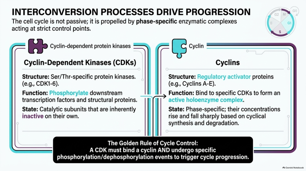
At the enzymatic core of cell division are specialized protein complexes that propel the cell through each critical boundary. Effective Cell Cycle Regulation relies on two main classes of regulatory molecules: cyclin-dependent kinases (CDKs) and regulatory proteins called cyclins. CDKs are serine- and threonine-specific protein kinases that phosphorylate target proteins to drive structural changes. However, CDK catalytic subunits remain completely inactive on their own. To become active, CDKs must associate with specific cyclin partners, forming active heterodimeric holoenzymes. While CDK concentrations remain relatively constant throughout the cell, cyclin levels fluctuate dramatically through cycles of gene transcription and protein degradation.
The functional partnership between CDKs and cyclins follows a strict biochemical rule. Successful Cell Cycle Regulation dictates that binding to a cyclin subunit is necessary, but not sufficient, to fully activate CDK enzymatic function. The assembled CDK-cyclin complex must also undergo specific phosphorylation and dephosphorylation events on key amino acid residues to unlock kinase activity. Once fully activated, these holoenzyme complexes phosphorylate downstream transcription factors and structural substrates. Different cyclin-CDK pairings orchestrate distinct transitions, ensuring unidirectional movement through each stage. By linking enzymatic activity to cyclin availability, cell cycle regulation turns gradual protein synthesis into sharp, unambiguous biochemical transitions.
Slide 4: Cell Cycle Regulation at the G1-S Phase Restriction Point
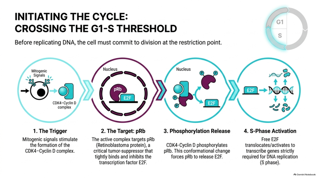
Committing to duplicate nuclear DNA requires passing the critical restriction point late in G1. Precise Cell Cycle Regulation ensures that cells do not initiate genomic replication until extracellular conditions support successful division. When mitogenic signals bind surface receptors, intracellular signaling cascades stimulate the expression and assembly of the CDK4-Cyclin D complex. This active kinase complex targets the retinoblastoma protein, a potent tumor suppressor that acts as a molecular brake. In its unphosphorylated state, retinoblastoma protein binds tightly to the E2F transcription factor, sequestering E2F and preventing the expression of crucial genes needed for DNA synthesis.
To overcome this inhibition, active CDK4-Cyclin D phosphorylates the retinoblastoma protein at specific amino acid positions. This targeted phosphorylation event induces a major conformational change that forces retinoblastoma protein to release E2F. Liberated E2F then translocates within the nucleus to initiate transcription of essential S-phase genes, including enzymes required for DNA synthesis, centrosome duplication, and histone assembly. Through this multi-step cascade, Cell Cycle Regulation creates an irreversible molecular switch that converts external growth factors into nuclear gene activation. Defects in this restriction point pathway undermine Cell Cycle Regulation, enabling unconstrained cellular division and driving the rapid progression of malignant human cancers.
Slide 5: Cell Cycle Regulation through Temporal Cyclin B Dynamics
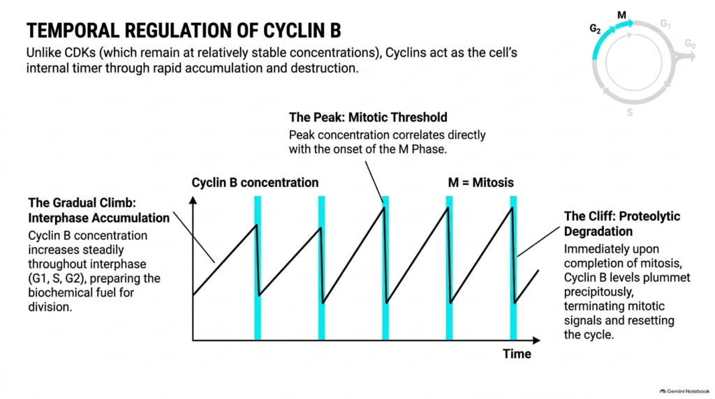
While catalytic CDK levels remain stable across the lifecycle, regulatory cyclin concentrations act as an internal biological clock. Robust Cell Cycle Regulation depends on the precise temporal synthesis and destruction of Cyclin B to manage entry into mitosis. Throughout interphase, including the G1, S, and G2 phases, Cyclin B concentration rises steadily as the cell synthesizes new proteins. This gradual accumulation provides the biochemical fuel needed to prepare the cell for division. As interphase nears completion, accumulating Cyclin B molecules bind CDK1 catalytic subunits, building a large pool of latent mitotic complexes that await specific chemical activation.
Mitosis begins when Cyclin B reaches a critical threshold concentration inside the nucleus. Once this peak concentration is reached, active complexes initiate the structural events of mitotic division. However, Cell Cycle Regulation requires that this powerful signal be shut off rapidly once chromosomes align. As mitosis finishes, specialized proteolytic machinery tags Cyclin B for immediate, precipitous destruction. Cyclin B levels drop off a sharp cliff, terminating mitotic kinase signaling and resetting the cell back to interphase. This dramatic rise and fall shows how Cell Cycle Regulation uses regulated protein degradation to drive unidirectional, irreversible cell cycle transitions across eukaryotes.
Slide 6: Cell Cycle Regulation and Inactive MPF Assembly
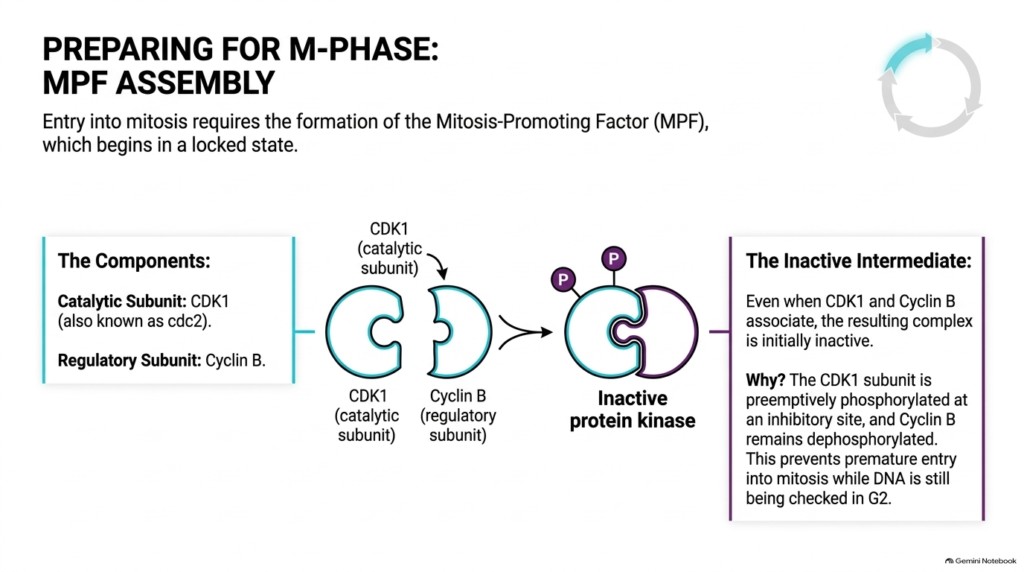
Entry into mitosis requires the precise assembly of a master protein complex known as the mitosis-promoting factor, or MPF. MPF triggers chromosome condensation, spindle formation, and nuclear membrane breakdown. MPF consists of two core subunits: the catalytic kinase subunit CDK1 (historically called cdc2) and the regulatory subunit Cyclin B. As Cyclin B accumulates during G2, it binds directly to CDK1 to form the binary MPF complex. Paradoxically, the initial assembly of this heterodimeric complex does not immediately produce kinase activity in the cytoplasm or nucleus.
Instead, the newly formed MPF complex remains held in an inactive intermediate state. Cell Cycle Regulation enforces this vital safety delay by preemptively phosphorylating the CDK1 subunit at an inhibitory amino acid site. While Cyclin B stays unphosphorylated, inhibitory phosphate groups on CDK1 keep the catalytic cleft locked, preventing premature enzymatic activity. This spatial and chemical inhibition prevents the cell from rushing into mitosis while nuclear DNA is still replicating or undergoing error checking during G2. By keeping assembled MPF locked, Cell Cycle Regulation establishes an essential safety pause, ensuring all interphase preparations finish before triggering nuclear breakdown.
Slide 7: Cell Cycle Regulation and the MPF Biochemical Switch
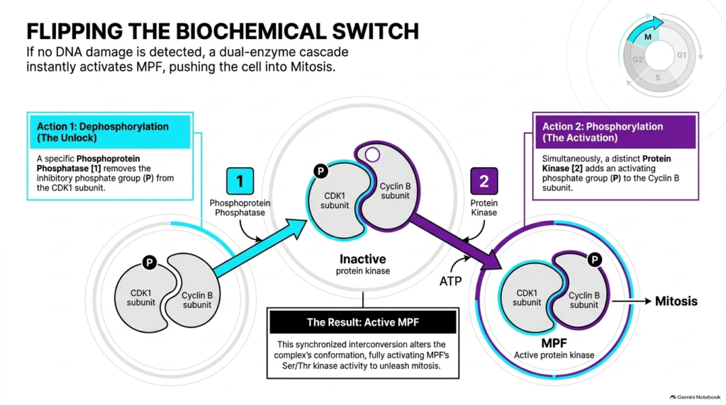
Once the cell completes DNA replication and verifies genomic integrity, a rapid dual-enzyme cascade transforms locked MPF into an active kinase engine. Robust Cell Cycle Regulation relies on this instantaneous biochemical switch to push the cell past the G2-M threshold into active mitosis. The activation pathway involves two coordinated enzymatic reactions that occur simultaneously within the cell. First, a specific phosphoprotein phosphatase removes the inhibitory phosphate group from the CDK1 subunit, unlocking the catalytic kinase site. Second, a distinct protein kinase adds an activating phosphate group to a specific amino acid residue on the Cyclin B regulatory subunit.
These simultaneous modifications alter the MPF holoenzyme’s conformation, fully activating its serine and threonine kinase activity. In the presence of adenosine triphosphate, active MPF rapidly phosphorylates key downstream structural targets throughout the nucleus and cytoplasm. By coupling dephosphorylation and phosphorylation, Cell Cycle Regulation converts a gradual buildup of precursor molecules into an all-or-none molecular trigger. This switch-like behavior ensures synchronous mitotic entry across the entire cell. Consequently, Cell Cycle Regulation prevents partial or asynchronous division, safeguarding structural components and maintaining overall genomic fidelity during nuclear division across proliferating tissue systems in human biology.
Slide 8: Cell Cycle Regulation during Early Mitotic Reactions
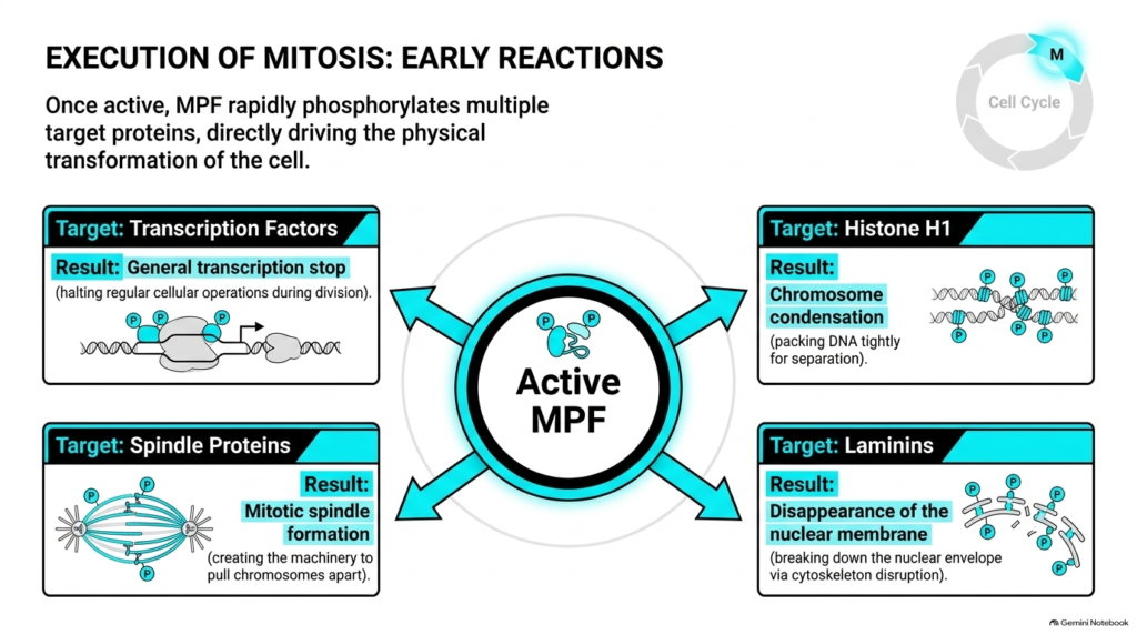
After full activation, MPF triggers a rapid wave of substrate phosphorylation that drastically alters the cell’s physical structure. Efficient Cell Cycle Regulation depends on active MPF to modify four key protein targets during prophase. First, MPF phosphorylates transcription factors, immediately shutting down general gene transcription so the cell can focus energy exclusively on division. Second, MPF phosphorylates histone H1 proteins, promoting tight chromosome condensation so chromatin can be safely organized and moved. Third, MPF phosphorylates spindle-associated proteins, stimulating dynamic microtubule assembly to form the mitotic spindle apparatus that will pull sister chromatids apart cleanly during metaphase.
Fourth, MPF directly phosphorylates nuclear lamins, disrupting the supportive intermediate filament network and causing the nuclear envelope to disassemble completely. Through these coordinated enzymatic actions, Cell Cycle Regulation converts chemical kinase signals into dramatic morphological changes throughout the cytoplasm and nucleus. Each phosphorylation event targets structural components necessary to prepare chromosomes for equal distribution into daughter cells. By synchronizing gene silencing, chromatin packing, spindle construction, and nuclear envelope breakdown, Cell Cycle Regulation ensures the cell smoothly reorganizes its internal machinery. This downstream phosphorylation cascade illustrates how Cell Cycle Regulation transforms local enzymatic activity into cell-wide structural division during prophase across proliferating tissues.
Slide 9: Cell Cycle Regulation and Signal Termination via Proteolysis
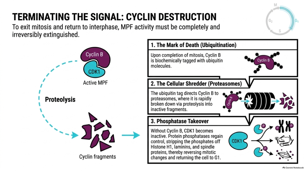
Completing mitosis and returning the cell to a stable interphase state requires completely extinguishing MPF kinase activity. Effective Cell Cycle Regulation relies on irreversible protein degradation to terminate mitotic signals and prevent cells from remaining trapped in active division. After chromosome segregation, specialized ubiquitin ligases attach ubiquitin tags to Cyclin B. These ubiquitin tags mark Cyclin B for destruction, directing it to the proteasome, a cylindrical protein-degrading complex. Inside the proteasome, enzymes rapidly shred Cyclin B into small, inactive peptide fragments through targeted enzymatic proteolysis in the cell cytoplasm.
Destroying Cyclin B removes the required regulatory activator of CDK1, leaving the catalytic kinase subunit inert. Without CDK1 kinase activity, cellular phosphatases regain control, rapidly stripping phosphate groups from histone H1, laminins, and spindle proteins. This dephosphorylation decondenses chromosomes, reforms the nuclear envelope, and disassembles the mitotic spindle apparatus cleanly and completely. Through this ubiquitin-proteasome pathway, Cell Cycle Regulation converts temporary phosphorylation signals back into stable baseline architecture. This mechanism shows how Cell Cycle Regulation uses targeted proteolysis to enforce unidirectional progress, extinguish mitotic signals, and return daughter cells safely to G1 to start a new division cycle.
Slide 10: Cell Cycle Regulation by Tumor Suppressors and Checkpoints
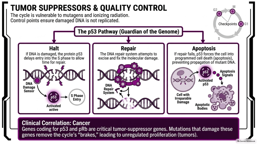
During cellular growth, environmental mutagens, oxidative stress, and ionizing radiation continuously threaten the chemical structure of nuclear DNA. Stringent Cell Cycle Regulation uses surveillance checkpoints to detect genetic lesions and prevent the propagation of damaged chromosomes into daughter cells. Central to this quality control system is p53, a critical tumor-suppressor protein known as the guardian of the genome. When DNA damage sensors detect double-strand breaks or modified bases, p53 rapidly accumulates and activates. Activated p53 halts cell-cycle progression before S phase, giving the cell time to mobilize specialized enzymatic repair machinery before copying DNA.
If the DNA repair system fixes the lesions, p53 activity subsides, allowing the cell to resume normal division. However, if the genetic damage proves irreparable, p53 forces the cell to undergo apoptosis, or programmed cell death, thereby eliminating the compromised cell completely. In human clinical oncology, somatic mutations in p53 or retinoblastoma genes severely disrupt Cell Cycle Regulation, removing vital molecular brakes. Without functional checkpoints, mutated cells divide unchecked, directly leading to tumor formation. Understanding checkpoint pathways highlights how Cell Cycle Regulation preserves genomic integrity, prevents oncogenesis, and protects multicellular organisms against malignant transformation in clinical medical settings.
Slide 11: Cell Cycle Regulation Master Framework and Phase Comparisons
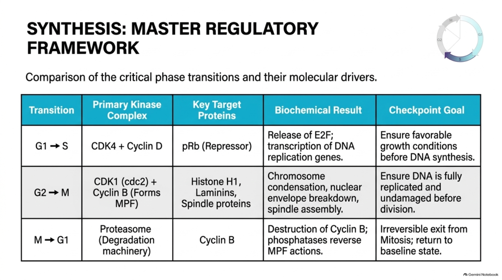
A comprehensive overview of cellular division highlights the specific kinase complexes, substrate targets, and overarching molecular goals that define each major phase transition across the cellular lifecycle. Master Cell Cycle Regulation depends on distinct regulatory pairings acting at defined control points throughout interphase and mitosis. During the G1-to-S transition, the CDK4-Cyclin D complex phosphorylates the retinoblastoma repressor protein, releasing E2F to transcribe genes required for DNA replication. This crucial step in Cell Cycle Regulation ensures that growth conditions and energy reserves remain favorable before committing the cell to genomic synthesis.
At the G2-to-M boundary, the CDK1-Cyclin B complex, acting as MPF, phosphorylates histone H1, laminins, and spindle proteins. This phosphorylation cascade drives chromosome condensation, nuclear envelope breakdown, and spindle assembly, ensuring that DNA is fully replicated and undamaged before physical division begins. Finally, during the M-to-G1 transition, proteasomal machinery degrades Cyclin B, allowing cellular phosphatases to reverse MPF activity and reset nuclear structures. Comparing these three boundaries reveals how Cell Cycle Regulation coordinates specialized molecular drivers to maintain genomic stability, providing a clear foundation for studying cancer biology, cellular physiology, and modern therapeutic development across medical sciences and healthcare disciplines worldwide.
Please read our Content Disclaimer Statement.
Check out our social media channels: