132. Calcium Metabolism: Homeostasis and Bone Remodeling
Have you ever wondered how your heart beats reliably, your muscles contract instantly, and your bones stay remarkably strong throughout life? The secret lies in a tightly controlled physiological process. Calcium ions serve as indispensable chemical messengers and structural building blocks inside the human body. This educational slide deck explores the foundational principles of calcium metabolism, walking you through systemic pools, cellular mechanisms, endocrine signaling circuits, and clinical disorders. By the end of this guide, you will master how your body maintains ionic equilibrium and safeguards skeletal integrity every single second.
Slide 1: Introduction to Calcium Metabolism and Systemic Signaling
Calcium serves as a fundamental inorganic ion required for human life, playing critical roles in cell signaling, structural stability, and enzymatic activation. Understanding calcium metabolism begins with appreciating how the body maintains a delicate equilibrium of divalent calcium cations across distinct physiological compartments. The central sphere depicted in the opening slide symbolizes the unified ion pool that circulates throughout systemic fluids, surrounded by a complex structural lattice representing cell membranes and bone matrices.
Every organ system relies heavily on calcium metabolism to sustain vital functions. Intracellular concentrations are kept exceptionally low at rest, enabling rapid calcium influx to act as a powerful second messenger. Meanwhile, extracellular concentrations remain tightly regulated within a narrow range to prevent metabolic dysfunction or tissue calcification. Consequently, calcium metabolism represents a masterclass in homeostatic regulation, where molecular sensors, transport channels, and endocrine networks interact seamlessly across tissues.
For college and medical students, studying calcium metabolism provides a vital framework for understanding human physiology and clinical biochemistry. As students progress through this slide deck, they examine how the body coordinates dietary intake, bone storage, and renal excretion. Mastering calcium metabolism helps future healthcare professionals and scientists understand muscle contraction, nerve signal transmission, hormone secretion, and metabolic disorders encountered in clinical practice.
Slide 2: The Total Body Pool in Calcium Metabolism
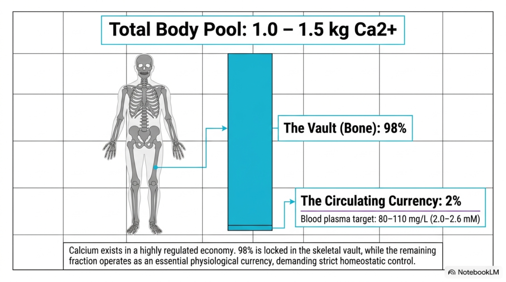
The human body contains approximately 1.0 to 1.5 kilograms of calcium, establishing a highly regulated physiological economy. In calcium metabolism, this total body pool is divided into two main compartments: the skeletal reservoir and the circulating plasma pool. As shown on the slide, an overwhelming 98 percent of total body calcium is locked within the bone vault, providing mechanical rigidity while serving as an expansive ionic reserve.
The remaining fraction represents the circulating currency of calcium metabolism, constituting roughly 2 percent of total body calcium. In blood plasma, the target concentration is strictly maintained between 80 and 110 milligrams per liter, corresponding to an ionized concentration of 2.0 to 2.6 millimolar. This circulating fraction is essential for sustaining neuromuscular excitability, cardiac pacemaking, vascular smooth muscle tone, and systemic enzymatic reactions across tissues.
Maintaining this narrow plasma range is a primary objective of calcium metabolism. Because extracellular fluid contains only a small fraction of total body calcium, even minor shifts in ionized calcium levels can trigger profound physiological consequences, such as neuromuscular tetany or lethal cardiac arrhythmias. Therefore, calcium metabolism relies on rapid exchange pathways between the skeletal vault and blood plasma to cushion against daily dietary fluctuations, physical exertion, and physiological stress.
Slide 3: Essential Physiological Functions in Calcium Metabolism
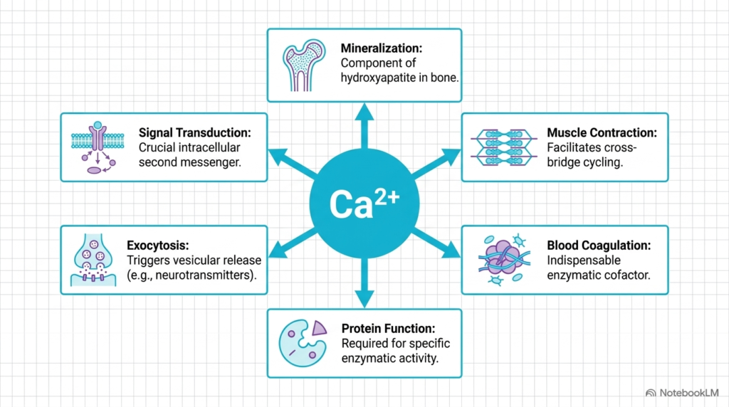
Divalent calcium ions act as versatile biological workhorses, driving diverse biochemical pathways across multiple organ systems. A central theme in calcium metabolism is the multifunctional nature of ionized calcium, as highlighted by six major physiological roles on this slide. In skeletal tissue, calcium provides structural mineralization by combining with inorganic phosphate to form hydroxyapatite crystals, giving bones their compressive strength, mechanical resilience, and load-bearing capacity during daily movements.
Beyond structural support, calcium metabolism governs cellular signaling and mechanical motion. Intracellular calcium functions as a pivotal second messenger in signal transduction cascades, altering enzyme activities upon membrane depolarization. In skeletal and cardiac muscle, calcium binding to troponin C exposes myosin-binding sites on actin filaments, facilitating cross-bridge cycling and contraction. Furthermore, calcium triggers exocytosis, enabling neurotransmitter release at neuronal synapses and hormone secretion from endocrine glands.
Finally, calcium metabolism plays indispensable roles in enzymatic regulation and blood coagulation. Calcium acts as an essential cofactor for several clotting factors in the coagulation cascade, including Factor X and prothrombin. Additionally, specific extracellular and membrane-bound enzymes require bound calcium to maintain active tertiary conformations. These diverse roles show why precise homeostatic control of calcium metabolism is vital for human life and cellular integrity.
Slide 4: Molecular Coordination and Protein Binding in Calcium Metabolism
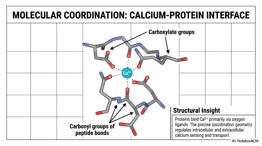
At the molecular level, calcium-binding specificity depends on defined coordination geometry within specialized protein motifs. In the broader context of calcium metabolism, calcium ions interact with target proteins primarily through oxygen-containing ligands. This slide illustrates how a central divalent calcium cation coordinates with negative carboxylate groups from aspartate or glutamate side chains, alongside neutral carbonyl groups from peptide backbones within the protein structure.
This precise coordination geometry is a hallmark of protein interactions within calcium metabolism. Because calcium has a flexible coordination sphere that usually accommodates six to eight oxygen atoms, it can induce significant conformational changes upon binding. Signaling proteins such as calmodulin, troponin C, and the calcium-sensing receptor utilize these structural shifts to transduce intracellular signals, activate downstream enzymes, and regulate ion transport channels with exceptional speed, precision, and fidelity.
Understanding this molecular interface clarifies how cells differentiate calcium from other abundant cations like magnesium in physiological fluids. In calcium metabolism, the spatial arrangement of oxygen ligands enables selective, high-affinity binding even as intracellular ion concentrations change. This atomic-level coordination regulates everything from membrane channel gating to enzyme activation, showing that global homeostatic balance ultimately rests on precise molecular interactions between specialized proteins and inorganic ions across tissues.
Slide 5: Cellular Dynamics of Osteoblasts and Osteoclasts in Calcium Metabolism
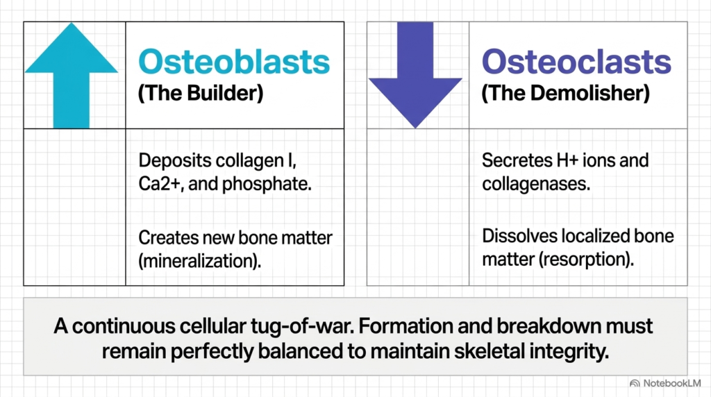
Skeletal integrity depends on a continuous cellular balance between bone formation and bone resorption. Two specialized cell types drive this ongoing remodeling process: osteoblasts and osteoclasts. Osteoblasts, often called the builders, secrete type I collagen and deposit calcium and phosphate ions into the extracellular matrix, facilitating new bone formation through matrix mineralization, osteoid deposition, and structural assembly within skeletal tissue.
Conversely, osteoclasts serve as the demolishers in calcium metabolism, specializing in localized bone resorption. These multinucleated cells secrete hydrogen ions and proteolytic collagenases into isolated resorption pits, dissolving the hydroxyapatite lattice and digesting organic matrix components. This continuous cellular tug-of-war ensures that old or damaged bone matter is systematically replaced while simultaneously supplying calcium ions to blood plasma when systemic concentrations drop below baseline requirements.
Maintaining equal osteoblast and osteoclast activity is essential for calcium homeostasis. When bone formation matches resorption, skeletal mass remains stable and systemic ion concentrations stay within homeostatic boundaries. However, chronic imbalances in this cellular dynamic can lead to metabolic bone diseases like osteopenia or osteoporosis. Understanding these opposing cellular functions prepares students to analyze how hormonal signals modulate bone remodeling to preserve systemic equilibrium, structural stability, and mineral availability throughout the body.
Slide 6: The Bone Remodeling Circuit in Calcium Metabolism
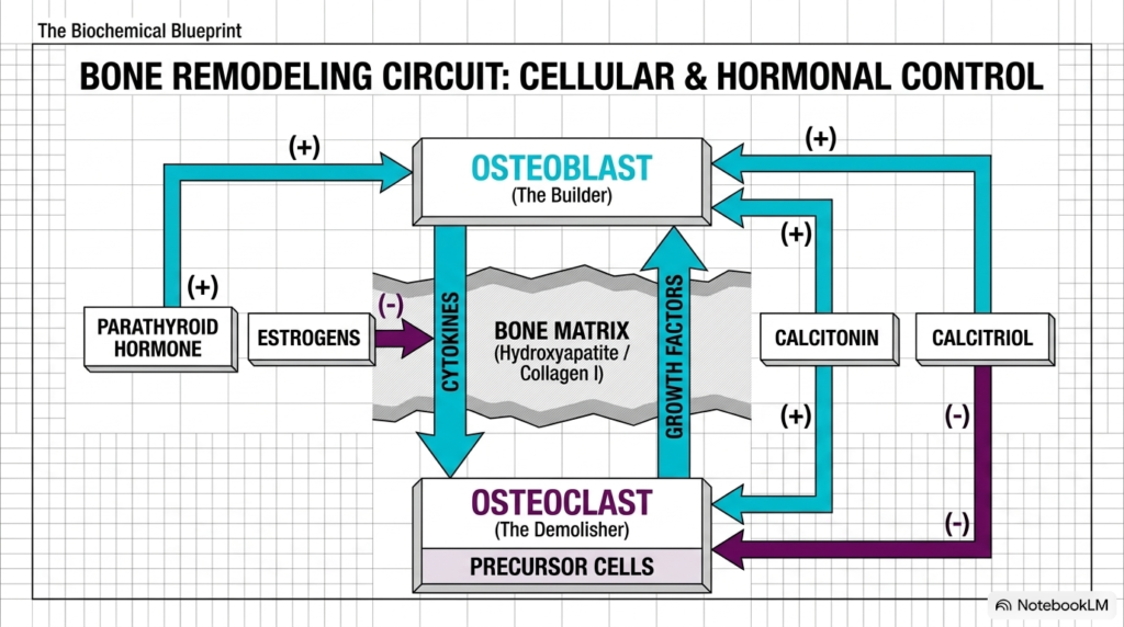
The bone remodeling circuit integrates cellular activity with endocrine and local signal networks. In calcium metabolism, this circuit coordinates communication among osteoblasts, osteoclasts, and bone matrix components such as hydroxyapatite and collagen. Hormonal drivers such as parathyroid hormone, calcitriol, calcitonin, and estrogens modulate this cellular communication network to balance structural integrity with systemic ion demands across different physiological states and environmental challenges.
Parathyroid hormone stimulates osteoblasts to express cytokines, such as RANK ligand, which bind to precursor cells and drive osteoclast differentiation and activation. Thus, in calcium metabolism, parathyroid hormone indirectly promotes bone resorption to liberate calcium into circulation. Estrogens protect bone by inducing osteoclast apoptosis and suppressing cytokine release, thereby preserving skeletal mass. Concurrently, matrix-bound growth factors released during resorption stimulate osteoblast activity, directly coupling breakdown to new formation.
Calcitriol and calcitonin add further layers of regulation to calcium metabolism. Calcitriol enhances both bone formation and osteoclastic mobilization depending on systemic requirements, whereas calcitonin directly inhibits osteoclast activity to restrict bone breakdown. This intricate cellular and endocrine loop shows that bone is not a static scaffold but a dynamic, highly responsive metabolic organ dedicated to homeostatic control, structural adaptation, and mineral storage throughout the human lifespan.
Slide 7: Osteoclast Acidification Mechanisms in Calcium Metabolism
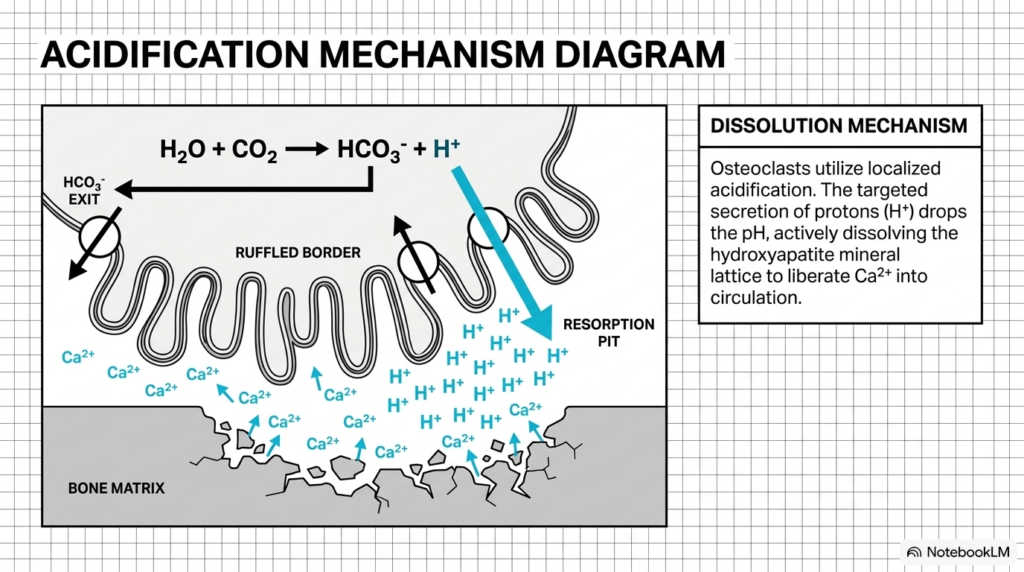
To liberate calcium from the dense hydroxyapatite matrix, osteoclasts employ a highly specialized chemical mechanism. In calcium metabolism, understanding osteoclast acidification highlights how cellular bioenergetics directly affects systemic ion homeostasis. During active bone resorption, osteoclasts attach tightly to the bone surface, forming a sealed microenvironment called a resorption pit beneath a specialized, highly folded cell membrane known as the ruffled border.
Intracellular carbonic anhydrase catalyzes the hydration of carbon dioxide into carbonic acid, which rapidly dissociates into bicarbonate and protons. In calcium metabolism, osteoclasts actively pump these protons across the ruffled border into the resorption pit using ATP-driven vacuolar proton pumps. This targeted proton secretion significantly lowers the localized extracellular pH, dissolving the mineralized hydroxyapatite lattice into free calcium and phosphate ions within the sealed resorption pit.
Simultaneously, bicarbonate ions exit the cell across the basolateral membrane via chloride-bicarbonate exchangers, maintaining intracellular pH stability and ionic balance. The liberated calcium ions then cross the osteoclast body via transcytosis and enter systemic circulation. This localized acidification shows how single-cell transport mechanics drive large-scale ion release in calcium metabolism, ensuring rapid physiological responses to systemic calcium deficits, endocrine cues, and metabolic challenges.
Slide 8: Endocrine Regulation of Calcium Metabolism
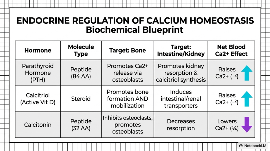
Systemic calcium balance relies on three key endocrine hormones: parathyroid hormone, calcitriol, and calcitonin. In calcium metabolism, these hormones act on primary target tissues including bone, kidneys, and intestines to modulate ion fluxes. Parathyroid hormone, an 84-amino-acid peptide, responds directly to hypocalcemia by promoting renal calcium reabsorption, stimulating calcitriol synthesis, and enhancing osteoclast-mediated bone release, ultimately raising blood calcium levels back to normal physiological ranges.
Calcitriol, the active steroid form of vitamin D, operates alongside parathyroid hormone in calcium metabolism to elevate systemic ion concentrations. Calcitriol acts primarily on the intestine and kidneys, inducing the expression of specific epithelial calcium channels and transport proteins to maximize dietary absorption and renal recovery. Additionally, calcitriol supports bone mineralization while facilitating controlled mineral mobilization when circulating calcium levels fall below optimal thresholds during prolonged dietary deprivation.
In contrast, calcitonin acts as a physiological antagonist within calcium metabolism. Secreted by thyroid parafollicular cells as a 32-amino-acid peptide, calcitonin responds to hypercalcemia by directly binding osteoclast receptors to inhibit bone resorption. It also promotes renal calcium excretion, helping lower elevated plasma concentrations. Together, this triad of endocrine hormones establishes a robust homeostatic feedback system that stabilizes circulating calcium concentrations in blood plasma across varying physiological states.
Slide 9: Daily Flux and Mass Balance in Calcium Metabolism
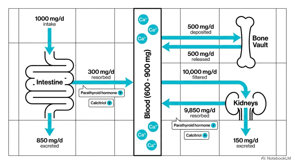
Quantifying daily ion movements provides a clear picture of systemic mass balance across organ systems. In calcium metabolism, an average daily dietary intake of 1000 milligrams enters the intestinal tract. Under the influence of calcitriol and parathyroid hormone, approximately 300 milligrams are absorbed into the blood plasma, while 850 milligrams are excreted in feces due to unabsorbed dietary intake and digestive secretions.
The central blood pool, containing roughly 600 to 900 milligrams of calcium, constantly exchanges ions with skeletal and renal tissues in calcium metabolism. Under steady-state conditions, bone tissue deposits 500 milligrams of calcium daily during bone formation and releases an equal 500 milligrams through resorption. This internal kinetic equilibrium keeps total bone mass constant while supporting plasma buffering needs, neuromuscular activity, and physiological demands across tissues.
The kidneys process vast quantities of calcium, filtering about 10,000 milligrams per day through the glomeruli. Tubular reabsorption recovers about 9,850 milligrams, leaving a net excretion of 150 milligrams in urine. Regulated by parathyroid hormone and calcitriol, renal reabsorption serves as the final fine-tuning mechanism, ensuring total daily output matches net intestinal absorption to maintain systemic equilibrium throughout the day and prevent unwanted mineral loss from systemic circulation.
Slide 10: Clinical Correlates and Pathophysiology in Calcium Metabolism
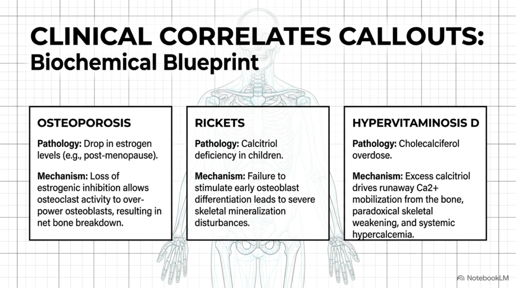
Disruptions in hormonal signaling or mineral availability lead to significant clinical disorders. In calcium metabolism, osteoporosis exemplifies the consequences of altered cellular regulation. Following menopause, declining estrogen levels remove a crucial inhibitory signal on osteoclasts. Consequently, osteoclasts outpace osteoblasts, driving net bone breakdown, microarchitectural deterioration, and increased fracture risk despite normal serum calcium concentrations across physiological compartments.
Rickets represents another major pathological manifestation within calcium metabolism, primarily affecting developing children. Caused by calcitriol deficiency or inadequate dietary vitamin D, rickets impairs early osteoblast differentiation and matrix mineralization. Without sufficient calcium deposition, unmineralized osteoid accumulates, resulting in soft, pliable bones, skeletal deformities, and delayed growth, underscoring the necessity of calcitriol for proper skeletal development during early childhood, bone remodeling, and physical growth.
Conversely, hypervitaminosis D highlights the dangers of excessive hormonal drive in calcium metabolism. Overdosing on cholecalciferol leads to uncontrolled calcitriol production, which excessively stimulates intestinal absorption and bone mobilization. This runaway calcium release causes severe hypercalcemia, tissue calcification, and paradoxical skeletal weakening. Analyzing these clinical conditions reinforces how precise homeostatic regulation in calcium metabolism is vital for maintaining human health, preventing systemic disease, and avoiding severe metabolic pathologies in modern clinical practice.
Slide 11: The Homeostatic Cascade in Calcium Metabolism
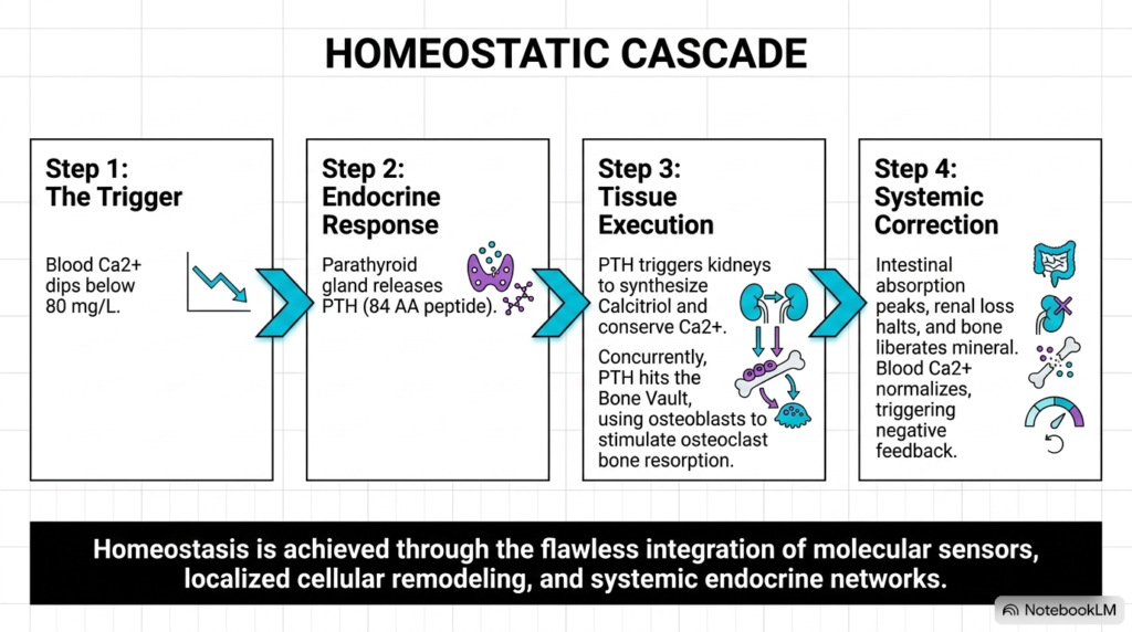
When blood calcium levels drop below the normal threshold of 80 milligrams per liter, the body initiates a rapid homeostatic cascade. In calcium metabolism, membrane receptors on parathyroid chief cells sense this acute hypocalcemic trigger. Upon detecting decreased ionic concentration, the parathyroid glands immediately release parathyroid hormone into circulation to initiate an integrated multi-organ physiological response across targeted organs and tissues.
The circulating peptide hormone acts swiftly across key target tissues in calcium metabolism. In the kidneys, parathyroid hormone increases tubular calcium reabsorption and activates the enzyme 1-alpha-hydroxylase to produce active calcitriol. At the same time, parathyroid hormone acts on bone tissue, signaling osteoblasts to release cytokines that stimulate osteoclast activity, accelerating bone resorption and releasing stored calcium into the blood plasma to restore baseline concentrations rapidly and effectively.
As calcitriol levels rise, intestinal absorption peaks, while renal conservation and bone mobilization restore circulating calcium to normal physiological levels. In calcium metabolism, this normalization of blood calcium shuts down further parathyroid hormone release through negative feedback inhibition. This elegant cascade illustrates how molecular sensors, endocrine signals, and tissue effectors work synchronously to maintain homeostatic equilibrium across the human body during metabolic challenges and fluctuating nutrient availability throughout the human life cycle.
Please read our Content Disclaimer Statement.
Check out our social media channels: