140. Biochemistry of Vision: Photoreceptors and Signal Cascades
Imagine stepping out of a dark movie theater into bright midday sun. Within a split second, the human eye adapts and converts light into crisp visual images in the brain. How does this remarkable process occur at the cellular level? This slide deck breaks down the visual transduction pathway step by step. Designed specifically for college and medical students, this guide explores the molecular cascade, membrane dynamics, and metabolic recycling loops that make visual perception possible.
Slide 01: Biochemistry of Vision – Molecular Mechanisms of Phototransduction
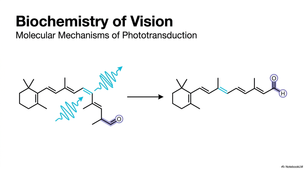
The field of the biochemistry of vision examines how physical light is converted into electrical signals inside the human retina. Phototransduction begins when a light photon collides with a specialized light-absorbing chromophore housed inside photoreceptor cells. As illustrated on the opening slide, the foundational event in the biochemistry of vision is the photochemical isomerization of 11-cis-retinal into all-trans-retinal. This light-driven reaction occurs with astonishing speed and precision, instantly altering the double bond geometry of the conjugated polyene carbon chain.
Understanding the biochemistry of vision requires tracing how a simple molecular shape change propagates across cellular membranes. When light strikes 11-cis-retinal, the molecule shifts from a bent configuration to an extended linear arrangement. This structural shift alters physical interactions between the chromophore and its surrounding opsin protein, initiating a domino effect of enzymatic events inside the rod cell segment. These initial conformational changes trigger a complex signaling cascade across the outer segment disc membrane.
Students studying the biochemistry of vision will see how this initial photochemical switch drives G-protein signaling, enzyme activation, and ion channel gating across the disc membrane. By mastering this primary event, learners lay the essential groundwork for comprehending how photoreceptor cells convert raw photon energy into precise neural signals sent directly to the brain. This initial molecular mechanism forms the backbone of visual sensory perception, demonstrating the profound efficiency of biological photon detection in humans.
Slide 02: Biochemistry of Vision – Photoreceptor Cell Structure and Organization

Anatomy and molecular organization form the structural baseline for the biochemistry of vision. The retina contains two principal types of photoreceptor cells: rods and cones. Rods are extremely sensitive to low light, making them essential for night vision. Inside each rod outer segment, stacked membrane discs house the enzymatic machinery required for phototransduction. The biochemistry of vision relies on these dense disc membranes to concentrate visual pigments and maximize photon capture across the retinal tissue layer.
Cones operate at higher light intensities and mediate bright daylight vision and color perception. While rods rely on a single visual pigment, cones express distinct opsin proteins tuned to different wavelengths. Studying the biochemistry of vision reveals how the anatomical isolation of phototransduction within membrane discs enhances signaling efficiency across the cell. This specialized compartmentalization prevents signal dissipation and accelerates enzymatic interactions during light absorption.
As light photons pass through retinal tissue, they encounter these specialized cellular compartments designed specifically to capture light and initiate downstream biochemical pathways. Comparing rod and cone photoreceptors helps students appreciate how subtle variations in opsin protein structure tailor different cells for specific visual tasks in the biochemistry of vision. This structural hierarchy ensures robust visual performance across diverse lighting environments, enabling seamless adaptation from dim starlight to bright sunlight.
Slide 03: Biochemistry of Vision – Rhodopsin Architecture and Chromophore Binding
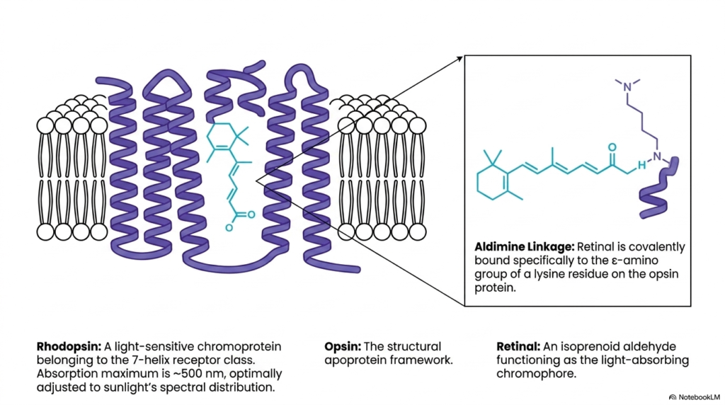
At the heart of rod phototransduction sits rhodopsin, a classic G-protein-coupled receptor. In the biochemistry of vision, rhodopsin consists of a protein apoprotein called opsin bound to the light-sensitive chromophore 11-cis-retinal. Opsin contains seven transmembrane alpha-helices that traverse the disc membrane bilayer. The chromophore binds specifically to the epsilon-amino group of a conserved lysine residue through a covalent protonated Schiff base, also known as an aldimine linkage. This specific linkage is fundamental to the biochemistry of vision.
The covalent linkage between opsin and 11-cis-retinal tunes the chromophore’s light-absorption properties. Free 11-cis-retinal absorbs ultraviolet light, but its binding environment within opsin shifts its absorption maximum to approximately 500 nanometers. This spectral tuning aligns perfectly with sunlight’s peak distribution in natural aquatic environments, optimizing light capture for dim-light vision. This chromophore-protein interaction showcases the structural elegance found in the biochemistry of vision.
In the biochemistry of vision, rhodopsin remains completely inactive until light strikes. The surrounding opsin protein acts as a protective conformational cage, holding the bent chromophore securely in place until photon absorption triggers its dramatic molecular unlocking and subsequent signal propagation. Understanding this architecture allows medical students to grasp how GPCRs transduce physical stimuli into chemical activity across cellular membranes, providing a key template for broader pharmacology studies.
Slide 04: Biochemistry of Vision – Photochemical Cis-Trans Isomerization
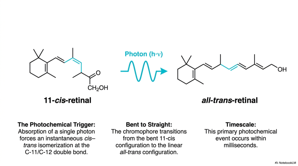
The primary event in light detection is a rapid photochemical reaction. In the biochemistry of vision, absorbing a single photon forces an instantaneous cis-to-trans isomerization at the carbon-11 and carbon-12 double bond of 11-cis-retinal. This converts the bent chromophore into rigid, elongated all-trans-retinal within milliseconds. This light-driven transition serves as the universal photochemical trigger across visual systems, illustrating the exquisite efficiency of vision biochemistry.
The chromophore’s physical transformation creates massive steric strain inside the opsin binding pocket. The bent 11-cis configuration fits snugly, but the straight all-trans isomer pushes against adjacent amino acid side chains. In the biochemistry of vision, this mechanical force drives structural rearrangements in the surrounding protein helices. The spatial conflict between the ligand and protein framework forces the receptor helices to tilt outward dynamically, opening binding sites on the cytoplasmic surface.
Students will note that this ultrafast chemical event effectively stores photon energy as mechanical strain inside the protein structure. This stored strain then drives the conformational shifts needed to activate downstream signaling partners in the visual cascade, turning physical light energy into chemical communication. Studying this step in the biochemistry of vision highlights the atomic precision of biological energy conversion across sensory cells, showing how physical forces translate directly into biological work.
Slide 05: Biochemistry of Vision – Opsin Activation and Transducin Recruitment
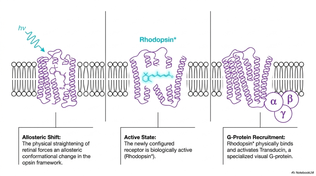
Straightening the retinal chromophore forces an allosteric conformational shift throughout the opsin protein framework. Within the biochemistry of vision, this structural transition generates the active receptor state known as Metarhodopsin II, or active Rhodopsin. Active Rhodopsin exposes specific cytoplasmic loops that interact directly with intracellular signaling proteins. This receptor activation step demonstrates how the biochemistry of vision translates an intramolecular physical push into a functional biological signal across the cell membrane.
Activated Rhodopsin physically recruits Transducin, a specialized visual G-protein heterotrimer composed of alpha, beta, and gamma subunits. In the biochemistry of vision, Transducin resides on the cytoplasmic surface of the disc membrane, positioned to interact with activated receptors. When active Rhodopsin binds Transducin, it alters the G-protein structure and initiates signal transmission across the cell membrane, starting the enzymatic relay efficiently to amplify incoming sensory information.
This physical association links light perception at the membrane chromophore to secondary messenger pathways inside the cell cytoplasm. Medical students should recognize this mechanism as a textbook example of G-protein-coupled receptor activation in human physiology, establishing a central theme in the biochemistry of vision. The interaction between active receptors and G-proteins forms the core of many physiological signal pathways throughout human biology, making this mechanism a vital cornerstone of medical biochemistry.
Slide 06: Biochemistry of Vision – Physiological State Matrix in Photoreceptors

Photoreceptor physiology differs fundamentally from typical sensory neurons. In the biochemistry of vision, dark and light states exhibit contrasting biochemical profiles. During the resting dark state, cyclic GMP concentrations remain high at approximately 70 micromolar. This elevated secondary messenger level holds cyclic nucleotide-gated cation channels open, allowing sodium and calcium ions to flow continuously into the cell. Consequently, the biochemistry of vision maintains a depolarized membrane potential in complete darkness, creating a steady electrical baseline.
Light exposure completely reverses these physiological parameters. Activated enzymes rapidly destroy cyclic GMP, causing cation channels to close and blocking ion influx. As a result, the cell hyperpolarizes and halts continuous glutamate release at the synaptic terminal. Studying the biochemistry of vision shows that light acts as an inhibitory signal that suppresses neurotransmitter output rather than directly stimulating it across the synapse, reversing standard neuronal patterns.
The adjoining bipolar neurons register this sudden drop in synaptic glutamate as a light signal, demonstrating how the biochemistry of vision turns cellular quietness into neural information. Understanding this inverse signaling mechanism is crucial for students analyzing visual processing in health and clinical pathology. This unique physiological feature distinguishes photoreceptors from standard excitatory neurons found throughout the human central nervous system, providing a fascinating example of evolutionary adaptation.
Slide 07: Biochemistry of Vision – Rod Outer Membrane Dynamics in the Dark
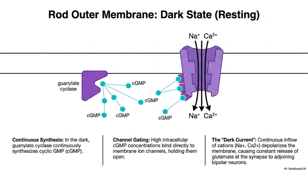
To understand light activation, students must first examine photoreceptors’ resting dark state. In the biochemistry of vision, membrane-bound guanylate cyclase continuously synthesizes cyclic GMP from GTP in darkness. High cytoplasmic cyclic GMP concentrations bind directly to cation channels in the outer-segment membrane, keeping them open. This steady channel gating is a hallmark of vision biochemistry in dark-adapted rod cells, establishing a continuous influx of cations across the cell membrane.
Continuous influx of sodium and calcium ions through open cation channels creates the classic dark current. This steady inward ion current depolarizes the rod cell membrane to approximately minus 40 millivolts. In the biochemistry of vision, depolarization maintains voltage-gated calcium channels open at the synaptic terminal, driving continuous glutamate release onto adjoining bipolar cells and interneurons across the retinal synapse, maintaining baseline synaptic communication.
Maintaining this dark current consumes substantial metabolic energy, requiring active sodium-potassium pumps to maintain ion gradients continuously across the outer membrane. Students analyzing the biochemistry of vision will appreciate how the dark state represents an active, highly energized resting baseline rather than a passive cellular state. This high energy investment ensures maximum responsiveness to sudden light changes in the surrounding environment, preparing the cell for instantaneous photon detection.
Slide 08: Biochemistry of Vision – Transducin Activation and Signal Amplification
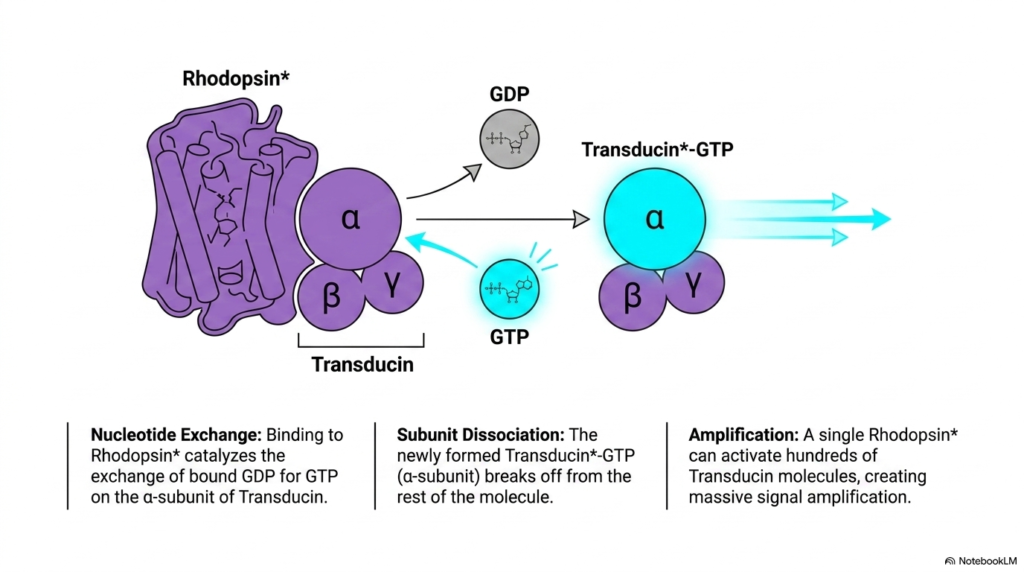
Signal amplification is a defining characteristic of the phototransduction cascade. In the biochemistry of vision, active Rhodopsin functions as a guanine nucleotide exchange factor. Binding to active Rhodopsin catalyzes the exchange of bound GDP for GTP on Transducin’s alpha subunit. Upon binding GTP, the alpha subunit dissociates from the beta-gamma complex and the receptor. This reaction showcases the enzymatic efficiency inherent in the biochemistry of vision, initiating a powerful secondary messenger cascade.
A single activated Rhodopsin molecule does not bind just one G-protein; it catalyzes nucleotide exchange on hundreds of Transducin molecules before inactivation. In the biochemistry of vision, this catalytic multi-step process creates massive signal amplification at the very first stage of the cascade. As activated Transducin alpha subunits diffuse along the disc membrane, they rapidly and efficiently carry the light signal to downstream target enzymes, broadening the scope of cellular signal propagation.
Students studying the biochemistry of vision will recognize that this early amplification enables photoreceptors to detect even single photons with high fidelity. This remarkable sensitivity allows the human eye to perceive faint light sources in pitch-black environments. High catalytic turnover ensures that small physical inputs yield large chemical outputs inside the cell, demonstrating a key principle in the biochemistry of vision that underlines human sensory capability.
Slide 09: Biochemistry of Vision – cGMP Phosphodiesterase Activation and Hydrolysis

Amplified G-protein signals directly target secondary messenger degradation pathways. In the biochemistry of vision, activated Transducin alpha subunits bind to inhibitory subunits of membrane-bound cyclic GMP phosphodiesterase, or PDE. This binding relieves PDE inhibition and activates the enzyme’s catalytic core. Active PDE rapidly hydrolyzes cyclic GMP into inactive 5′-GMP, demonstrating the rapid enzymatic speed underlying the biochemistry of vision, enabling rapid signal propagation across the cytoplasm.
Because each active PDE enzyme turns over thousands of cyclic GMP molecules per second, cytoplasmic messenger levels plunge dramatically within milliseconds. In the biochemistry of vision, this sudden drop removes the ligand required to keep outer membrane cation channels open. By destroying cyclic GMP faster than guanylate cyclase can synthesize it, PDE converts a G-protein signal into a sharp reduction in secondary messenger concentration across the cytoplasm, rapidly altering channel gating dynamics.
Studying the biochemistry of vision highlights how enzymatic destruction of secondary messengers provides an extremely fast, sensitive mechanism for sensory signal transduction, ensuring photoreceptor cells respond almost instantaneously to incoming photons. This efficient enzymatic breakdown ensures high temporal resolution in human visual perception, illustrating a vital concept in the biochemistry of vision that connects molecular kinetics with perceptual speed and optical clarity.
Slide 10: Biochemistry of Vision – Cation Channel Closure and Hyperpolarization
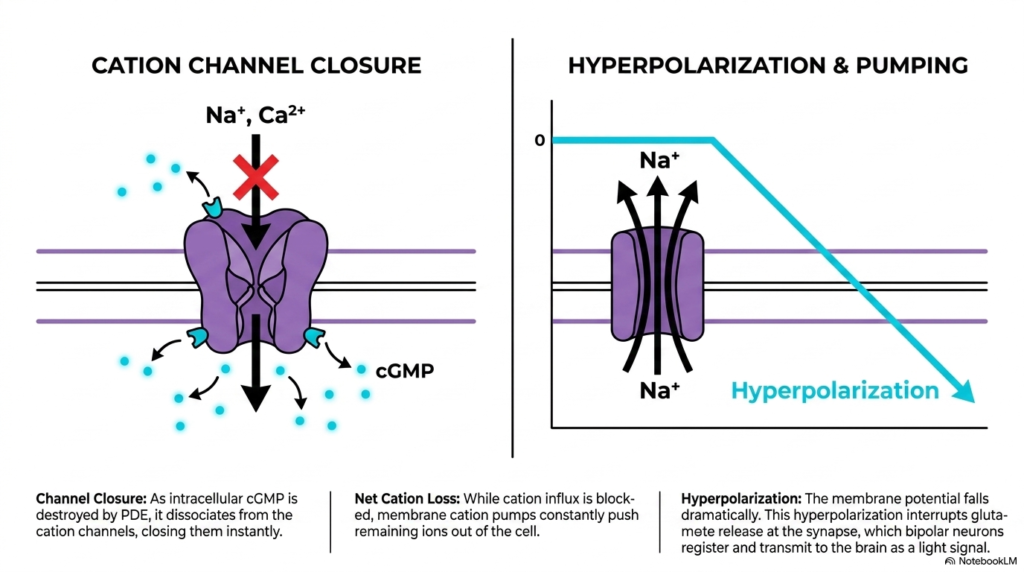
Depletion of intracellular cyclic GMP drives dramatic electrical changes across the cell membrane. In the biochemistry of vision, as cyclic GMP levels plunge, the messenger dissociates from cation channels, causing them to close immediately. Cation channel closure halts inward sodium and calcium flow, completely breaking the dark current. This channel-gating mechanism is central to understanding electrical responses in the biochemistry of vision, demonstrating direct ligand-gated ion-channel control during sensory reception.
While inward cation flow stops, membrane ion pumps continue exporting positive ions from the cytoplasm efficiently. In the biochemistry of vision, this net loss of positive charge hyperpolarizes the rod membrane from minus 40 millivolts toward minus 70 millivolts. Membrane hyperpolarization spreads smoothly to the synaptic terminal, closing voltage-gated calcium channels and interrupting glutamate release into the synaptic cleft between cells, altering synaptic transmission dynamically across the retinal circuit.
Adjoining bipolar neurons detect this sudden decrease in neurotransmitter release and transmit visual signals toward the optic nerve. This key step demonstrates how the biochemistry of vision bridges biochemical enzyme reactions and macroscopic electrical neural activity. The resulting change in membrane potential translates chemical dynamics into electrical signaling, highlighting the integrative nature of vision biochemistry across complex biological circuits and synapses in human neurobiology.
Slide 11: Biochemistry of Vision – Transducin Self-Inactivation and Recovery
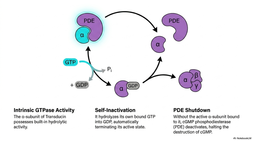
Sensory systems must turn off active signals rapidly to prepare for subsequent light stimuli. In the biochemistry of vision, Transducin’s alpha subunit possesses built-in GTPase activity. This intrinsic enzyme activity hydrolyzes bound GTP into GDP and inorganic phosphate, automatically terminating Transducin’s active state. This self-limiting timer is a vital protective control in the biochemistry of vision, ensuring signals do not persist indefinitely and allowing cells to reset efficiently for continuous photon reception.
GTP hydrolysis causes Transducin’s alpha subunit to release active phosphodiesterase and reassociate with its beta-gamma complex. In the biochemistry of vision, releasing phosphodiesterase allows its inhibitory subunits to rebind, shutting down cyclic GMP destruction. Without active phosphodiesterase present, cyclic GMP breakdown ceases instantly across the disc membrane, allowing secondary messenger levels to recover and prepare for subsequent light absorption events in the photoreceptor cytoplasm.
Students studying the biochemistry of vision will see that this GTPase mechanism keeps visual signals brief and tightly regulated. This precise temporal control prevents prolonged signaling from a single light flash and allows the retina to track rapidly changing visual environments. Intrinsic enzymatic timers maintain signaling fidelity, showcasing an essential control mechanism in the biochemistry of vision that governs signal duration and reset speed in human visual physiology.
Slide 12: Biochemistry of Vision – Calcium Feedback and Cellular Recovery
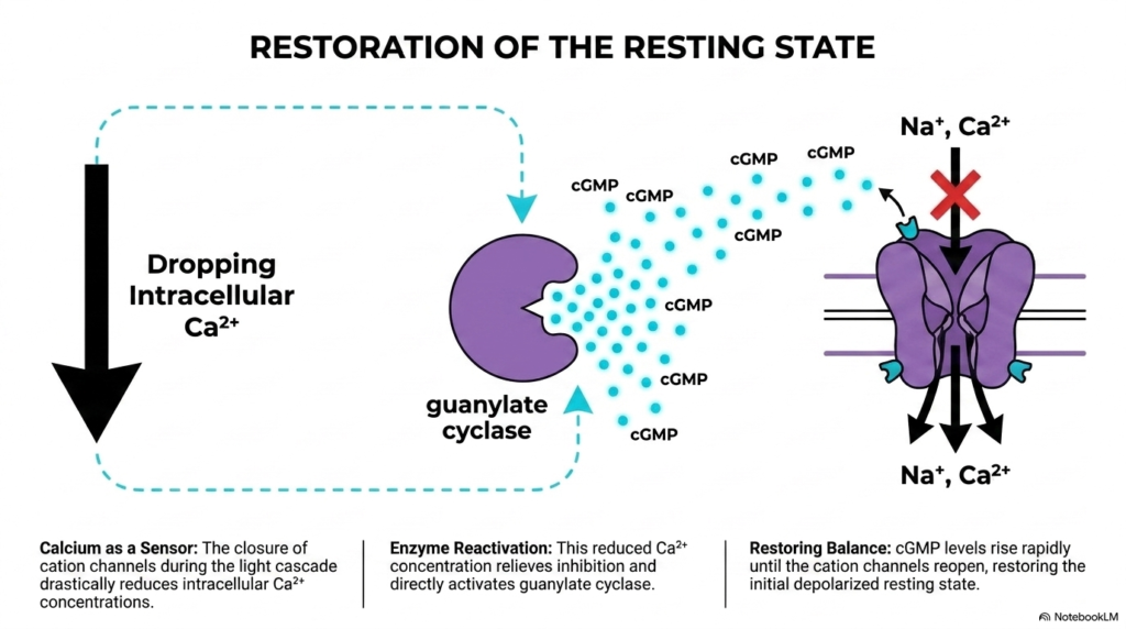
Restoring the resting state requires rebuilding cyclic GMP pools and reopening cation channels. In the biochemistry of vision, calcium ions act as a key intracellular sensor for recovery. Closing cation channels blocks calcium entry, but sodium-calcium exchangers continue pumping calcium out. Consequently, intracellular calcium levels drop precipitously during light exposure. This calcium drop plays a crucial role in regulating recovery in the biochemistry of vision, triggering essential feedback loops inside outer segments dynamically.
Falling intracellular calcium levels relieve inhibition of guanylate cyclase-activating proteins, stimulating guanylate cyclase to rapidly synthesize fresh cyclic GMP. In the biochemistry of vision, rising cyclic GMP concentrations rebind to outer membrane cation channels, forcing them open and restoring the dark current. This negative feedback loop restores resting membrane potential and synaptic glutamate release smoothly across the entire outer segment compartment, resetting the cell’s electrical baseline for future activation.
Medical students analyzing the biochemistry of vision will see how calcium feedback grants photoreceptors extraordinary sensitivity while allowing rapid adaptation to changing background light intensities, protecting the visual system from saturation. This homeostatic mechanism ensures operational stability under varying environmental illumination, demonstrating the adaptability inherent in the biochemistry of vision during continuous daily visual processing and perception under dynamic ambient conditions across diverse environments.
Slide 13: Biochemistry of Vision – The Retinoid Cycle and Molecular Recycling
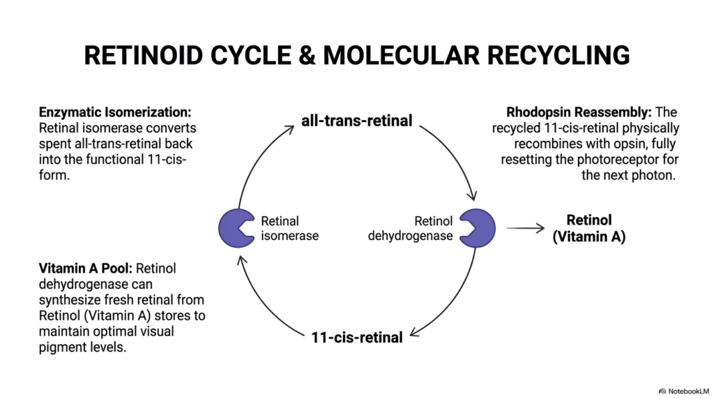
After phototransduction, spent chromophores must be chemically regenerated to sustain vision. In the biochemistry of vision, activated all-trans-retinal detaches from opsin and enters the retinoid cycle. Specialized enzymes in the adjacent retinal pigment epithelium, including retinal isomerase, convert all-trans-retinal back into functional 11-cis-retinal. This metabolic pathway is indispensable for the continuous functioning of the biochemistry of vision, sustaining visual pigment availability over extended light exposure across the retina.
When necessary, retinol dehydrogenase synthesizes additional chromophores from Vitamin A stores to maintain optimal visual pigment levels. In the biochemistry of vision, newly regenerated 11-cis-retinal returns to rod outer segments and recombines with opsin to reform functional rhodopsin. Without this multi-step enzymatic recycling pathway, visual pigments would deplete rapidly under continuous light exposure in daily life, causing visual exhaustion and functional loss across the retinal photoreceptor layer in humans.
Students of the biochemistry of vision can appreciate how metabolic cooperation between photoreceptors and pigment epithelial cells maintains long-term visual function and prevents clinical conditions like night blindness, emphasizing the clinical relevance of retinoid metabolism. This enzymatic cycle guarantees a steady supply of active visual pigments, demonstrating intercellular coordination in the biochemistry of vision across distinct tissue layers and metabolic compartments in the human eye throughout life.
Slide 14: Biochemistry of Vision – Integrated Blueprint of Visual Perception

The complete phototransduction loop unites photon absorption, enzymatic amplification, membrane hyperpolarization, and metabolic recycling into a single continuous circuit. In the biochemistry of vision, every component works in perfect harmony to balance sensitivity and response speed. From initial chromophore isomerization to calcium-mediated recovery, the biochemistry of vision demonstrates remarkable evolutionary optimization across species, yielding incredible sensory precision and perceptual power in human vision across diverse environments.
By integrating fast G-protein cascades with robust retinoid recycling, photoreceptors detect single photons while continuously adapting to bright sunlight. In the biochemistry of vision, this cyclic blueprint allows the retina to process millions of visual inputs every second without overwhelming cellular resources. The system balances high gain with fast recovery, ensuring uninterrupted visual signaling across dynamic environmental changes in natural illumination throughout daily human activities and complex visual challenges in real-world settings.
For medical and graduate students, mastering the biochemistry of vision provides a complete molecular framework for understanding sensory physiology, neurobiology, and clinical disorders of the human visual pathway. This knowledge prepares future scientists to explore novel therapeutic targets for retinal degenerative diseases, building a strong foundation in the biochemistry of vision for advanced clinical research and neuroscience applications in modern medicine and ophthalmology worldwide.
Please read our Content Disclaimer Statement.
Check out our social media channels: