147. Lipophilic Hormones: Mechanisms, Types and Biological Function
Hormonal communication governs human life, driving growth, metabolism, and reproductive health. While many chemical messengers act at the surface of target cells, lipid-soluble signaling molecules can cross hydrophobic cell membranes directly. This blog post explores an essential slide deck detailing the biochemical properties, transport mechanisms, and physiological actions of lipophilic hormones. Designed for college and medical students, this comprehensive guide breaks down how steroid, thyroid, and vitamin derivatives alter gene transcription, maintain homeostasis, and coordinate complex biological functions across human organs.
Slide 01: Introduction to Lipophilic Hormones and Cell Membrane Permeability
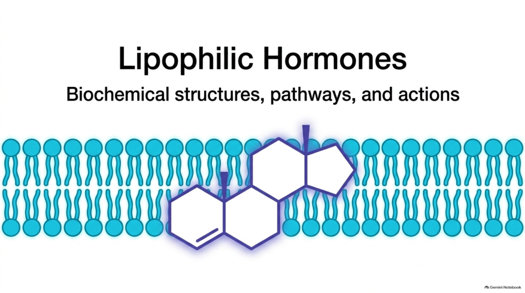
Biological signaling relies heavily on chemical messengers that transmit physiological instructions throughout the organism. Among these signaling agents, lipophilic hormones represent a distinct class of lipid-soluble molecules designed to cross cellular boundaries seamlessly. Unlike water-soluble peptide hormones that bind cell-surface receptors, lipophilic hormones diffuse directly across the hydrophobic phospholipid bilayer of target cell membranes. This structural capability stems from their nonpolar chemical framework, which integrates smoothly with the fatty acid tails of plasma membrane lipids.
The basic architecture of lipophilic hormones typically features a compact steroid ring framework or an iodinated amino acid backbone. Because these chemical messengers easily penetrate lipid membranes, target tissues do not rely on cell-surface signal transduction proteins to perceive the message. Instead, lipophilic hormones freely enter internal cellular compartments where they encounter specific intracellular binding proteins. Once inside the cytosol or nucleus, these hydrophobic signaling molecules initiate cascades that modify cellular behavior directly at the level of genomic transcription.
Mastering this entry mechanism is foundational for college and medical students studying endocrine physiology. The direct plasma membrane permeation of lipophilic hormones establishes a unique signaling paradigm. By bypassing traditional surface-bound second messenger pathways, lipophilic hormones orchestrate sustained, long-term adjustments in systemic metabolism, tissue differentiation, and physiological homeostasis across diverse organ systems.
Slide 02: General Biochemical Characteristics of Lipophilic Hormones
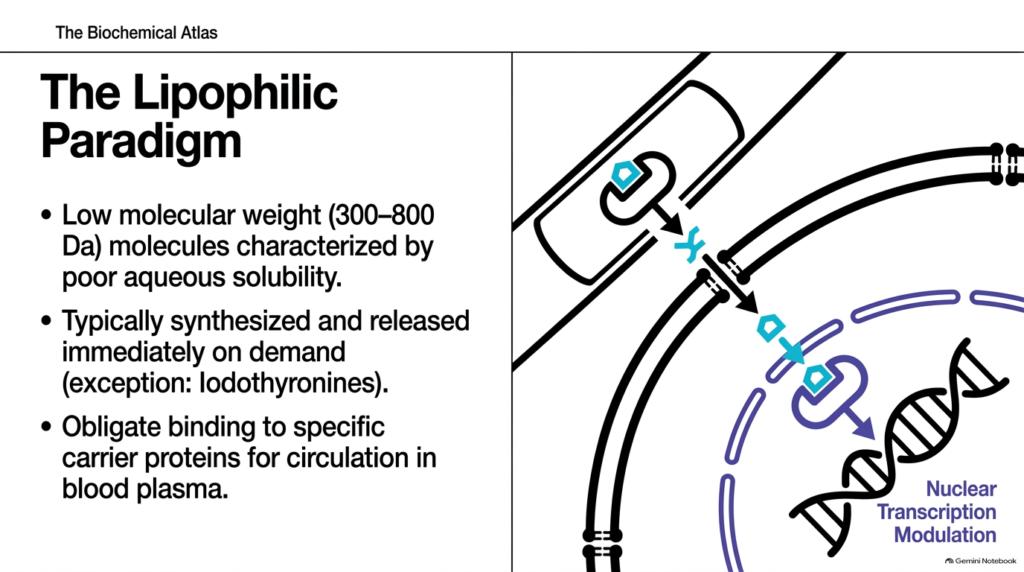
The foundational biochemical properties of lipophilic hormones distinguish them from hydrophilic signaling molecules. With molecular weights of 300 to 800 Daltons, lipophilic hormones have poor aqueous solubility in plasma. Consequently, these hydrophobic messengers cannot travel freely through blood. Instead, lipophilic hormones require obligate binding to specialized plasma carrier proteins, such as globulins and albumin, which keep them solubilized during circulatory transport toward target tissues.
At the cellular level, the physiological storage strategy for lipophilic hormones differs markedly from peptide signals. Endocrine cells generally do not store steroid-based lipophilic hormones in membrane-bound vesicles; instead, cells synthesize and release them immediately upon physiological demand. A notable biochemical exception occurs with iodothyronines, which undergo unique post-translational storage within thyroid follicle tissue as thyroglobulin before enzymatic cleavage and secretion into the bloodstream.
Upon reaching target cells, free lipophilic hormones dissociate from carrier proteins and cross the plasma membrane. They bind specific intracellular or nuclear receptors, forming hormone-receptor complexes that bind hormone response elements on genomic DNA. This nuclear transcription modulation alters mRNA synthesis, directly dictating cellular protein production. Medical students must recognize that this genomic mechanism accounts for the characteristically delayed yet long-lasting physiological responses observed across endocrine systems.
Slide 03: Progesterone Functions Among Lipophilic Hormones
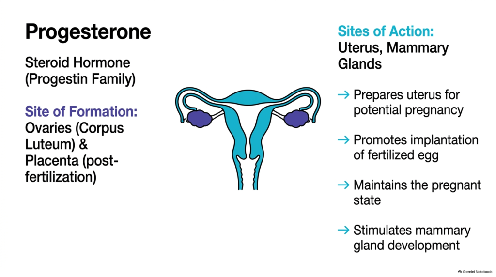
Progesterone represents an essential female sex steroid belonging to the progestin family of lipophilic hormones. The corpus luteum of the ovaries synthesizes it during the luteal phase of the menstrual cycle, and the placenta produces it after successful fertilization. Because progesterone is a lipophilic hormone, its hydrophobic structure allows it to diffuse readily into target cells in reproductive tissues to regulate gene expression.
The primary target organs for progesterone are the uterus and the mammary glands. Within the uterine wall, progesterone prepares the endometrium for potential blastocyst implantation by promoting secretory maturation and vascular remodeling. If fertilization occurs, sustained progesterone secretion maintains pregnancy by inhibiting uterine smooth muscle contractions and supporting embryonic development. In breast tissue, progesterone stimulates the growth and differentiation of alveoli within mammary glands, preparing the organ for eventual lactation.
Understanding the physiological timing of progesterone action helps medical students comprehend reproductive endocrinology. As classic lipophilic hormones, progestins coordinate genomic transcriptomic changes that align uterine physiology with embryonic needs. Without adequate progesterone activity, the endometrial lining cannot support pregnancy, demonstrating how single steroid molecules profoundly impact human reproductive success and fetal survival.
Slide 04: Estradiol Signaling in the Family of Lipophilic Hormones
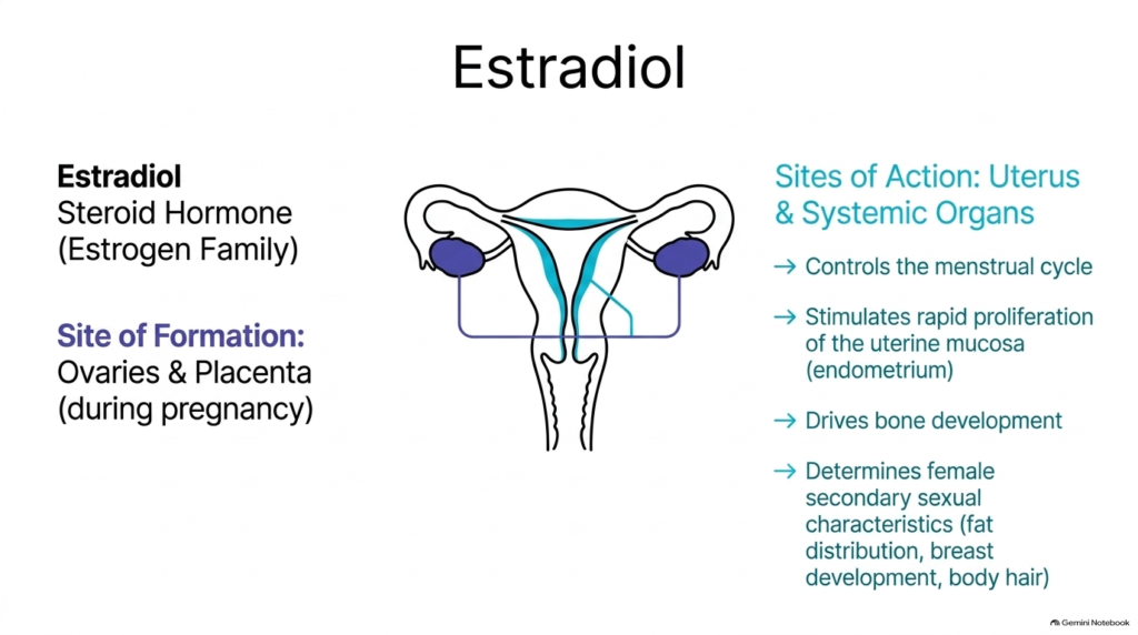
Estradiol is the primary and most potent estrogenic steroid in the broad family of lipophilic hormones. Biosynthesized predominantly by the granulosa cells of ovarian follicles and later by the placenta during gestation, estradiol plays an indispensable role in female reproductive physiology. Like other steroid-derived lipophilic hormones, estradiol freely crosses cell membranes and binds intracellular estrogen receptors to modulate nuclear gene transcription in target tissues.
Systemically, estradiol exerts wide-ranging physiological effects across reproductive and non-reproductive organs. Within the uterine cavity, estradiol stimulates the rapid follicular-phase proliferation of the endometrial mucosa, preparing the tissue for the menstrual cycle. Additionally, estradiol acts on skeletal tissue to promote bone development and maintain bone mineral density by suppressing osteoclasts. It is also the primary driver of secondary female sexual characteristics, including specific adipose tissue distribution, breast tissue maturation, and body hair patterns.
From a clinical perspective, studying estradiol provides students with clear insight into nuclear receptor signaling. Because estradiol is a key lipophilic hormone in human physiology, disruptions in its circulating levels can cause menstrual irregularities, osteoporosis, or developmental anomalies, illustrating the critical balance required in endocrine feedback loops across female life stages.
Slide 05: Testosterone Actions and Endocrine Roles of Lipophilic Hormones
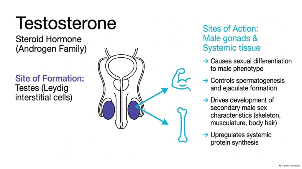
Testosterone is the principal male sex steroid and serves as a classic representative of androgenic lipophilic hormones. Produced within the Leydig interstitial cells of the testes under the influence of luteinizing hormone, testosterone governs male reproductive maturation and systemic anabolic processes. As a lipophilic hormone, testosterone crosses target cell membranes to bind androgen receptors, directly influencing genomic transcription pathways.
During embryonic development, testosterone directs the sexual differentiation of undifferentiated gonad structures toward the male phenotype. In adult males, it regulates spermatogenesis within the seminiferous tubules and drives the formation of ejaculate components. Systemically, testosterone exerts profound anabolic actions across target tissues, upregulating systemic protein synthesis. This metabolic drive stimulates skeletal growth, increases muscle mass, and promotes the development of secondary male sex characteristics, including facial hair growth and vocal cord thickening.
Medical students analyzing musculoskeletal and reproductive health must appreciate how testosterone functions. As essential lipophilic hormones, androgens show how nuclear gene regulation translates into macro-level physiological changes, from spermatogenesis to muscle protein accretion. The interplay between testosterone and intracellular receptors highlights the immense power of steroid-mediated gene control in human physiology.
Slide 06: Cortisol and Metabolic Regulation by Lipophilic Hormones
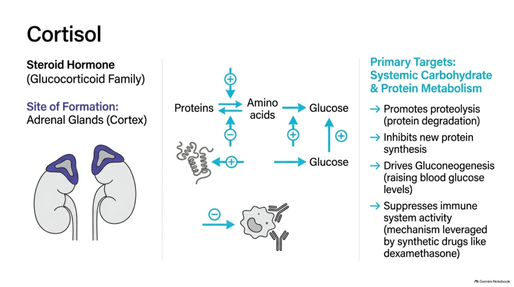
Cortisol is the major glucocorticoid steroid secreted by the adrenal cortex and represents a vital stress-responsive messenger among lipophilic hormones. Synthesized from cholesterol in the zona fasciculata, cortisol regulates systemic carbohydrate, lipid, and protein metabolism. Because cortisol is a lipophilic hormone, it crosses cell membranes easily to bind intracellular glucocorticoid receptors, modulating target gene expression across almost every tissue in the human body.
Metabolically, cortisol elevates blood glucose to maintain energy availability during prolonged physiological stress. It achieves this by promoting proteolysis, breaking down skeletal muscle proteins into free amino acids that serve as substrates for hepatic gluconeogenesis. Concurrently, cortisol inhibits peripheral protein synthesis and glucose uptake in non-essential tissues. Beyond metabolism, cortisol exerts potent anti-inflammatory and immunosuppressive actions by downregulating pro-inflammatory cytokine transcription, a pharmacological mechanism leveraged by synthetic drugs like dexamethasone.
For healthcare students, understanding cortisol provides a foundation for clinical pharmacology and endocrinology. As exemplary lipophilic hormones, glucocorticoids illustrate how cellular signal transduction directly regulates intermediary metabolic pathways. Studying cortisol reveals how steroid gene regulation protects the body during acute challenges while posing health risks during chronic elevation.
Slide 07: Aldosterone and Renal Electrolyte Control by Lipophilic Hormones
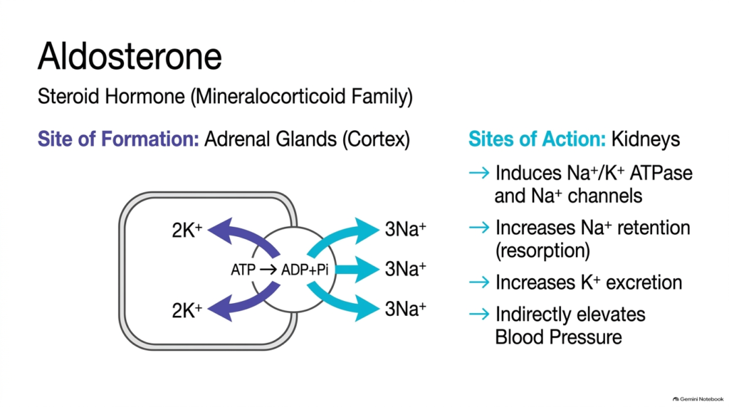
Aldosterone is the primary mineralocorticoid produced by the zona glomerulosa of the adrenal cortex and plays a pivotal role among lipophilic hormones in maintaining fluid balance. Regulated by the renin-angiotensin-aldosterone system, aldosterone targets the renal tubular epithelium. Like other steroid-class lipophilic hormones, aldosterone diffuses through target cell plasma membranes to bind intracellular mineralocorticoid receptors located in the distal convoluted tubules and collecting ducts of the kidney.
Aldosterone binding triggers genomic transcription of key transport proteins, specifically inducing synthesis of sodium-potassium ATPase pumps and epithelial sodium channels. This upregulation increases renal sodium retention while promoting potassium excretion into the tubular lumen. As sodium is reabsorbed, water follows passively via osmotic gradients, expanding extracellular fluid volume and indirectly elevating systemic blood pressure to maintain cardiovascular stability.
Biochemistry and medical students must grasp aldosterone action to understand renal physiology and hypertension. As essential renal-acting lipophilic hormones, mineralocorticoids demonstrate how gene transcription control alters ion channel density. Through these genomic mechanisms, aldosterone dynamically fine-tunes systemic electrolyte balance and maintains arterial pressure homeostasis across varying physiological states.
Slide 08: Calcitriol Synthesis and Calcium Homeostasis via Lipophilic Hormones
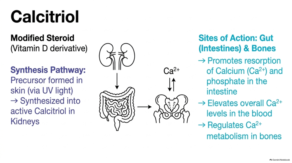
Calcitriol, the active form of vitamin D, is a modified steroid derivative that occupies a specialized position among lipophilic hormones. Unlike classical steroid glands, calcitriol synthesis involves a multi-organ pathway starting in the skin, where ultraviolet light converts 7-dehydrocholesterol into precursor forms. Subsequent hydroxylation in the liver and activation in the kidneys produce functional calcitriol. As a lipophilic hormone, calcitriol penetrates target cells and binds intracellular vitamin D receptors.
The primary physiological target sites for calcitriol are the intestinal epithelium and skeletal bone tissue. In the small intestine, calcitriol stimulates the transcription of calcium-binding proteins, significantly promoting the active resorption of calcium and phosphate ions from food. In bone tissue, calcitriol regulates mineral turnover and osteoblast-osteoclast coordination, maintaining optimal blood calcium concentrations necessary for neuromuscular transmission and skeletal integrity.
Studying calcitriol provides students with a clear model of endocrine coordination across multiple organ systems. As essential lipophilic hormones, vitamin D derivatives link environmental exposure, metabolic conversion, and gene regulation. Without adequate calcitriol, calcium absorption fails, leading to bone mineralization disorders such as rickets or osteomalacia in clinical practice.
Slide 09: Iodothyronines and Metabolic Control by Lipophilic Hormones
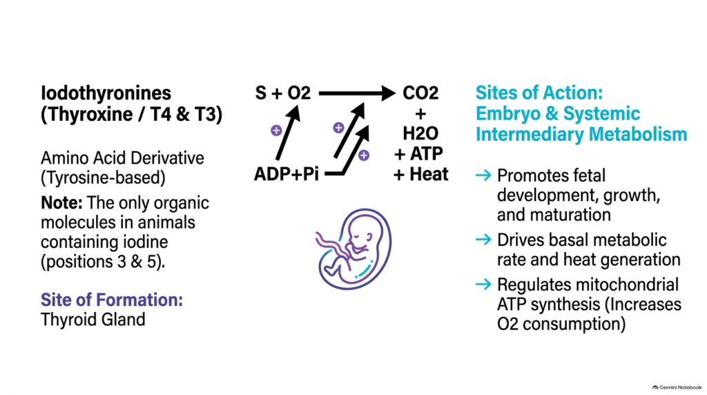
Iodothyronines, including thyroxine (T4) and triiodothyronine (T3), are tyrosine-derived signaling molecules that form a unique class of lipophilic hormones. Synthesized exclusively in the thyroid gland, these compounds are the only organic molecules in the animal kingdom that contain iodine atoms attached to their phenol rings. Although they are amino acid derivatives, their nonpolar nature makes them lipophilic hormones that enter target cells to regulate genomic transcription.
At the organelle level, iodothyronines act on systemic intermediary metabolism and mitochondrial function. Once inside target cells, T4 is converted into active T3, which binds nuclear thyroid hormone receptors. This activation regulates mitochondrial ATP synthesis, stimulates oxygen consumption, and drives heat generation, thereby establishing the basal metabolic rate. Furthermore, iodothyronines are essential during embryonic and fetal development, driving tissue maturation, skeletal growth, and central nervous system development.
For medical students, iodothyronine physiology links fundamental biochemistry to systemic clinical pathology. As non-steroid lipophilic hormones, thyroid signals showcase how iodinated amino acid derivatives govern overall cellular respiration and embryonic growth. Understanding these lipophilic hormones clarifies clinical disorders ranging from congenital hypothyroidism to hypermetabolic states observed in thyroid disease.
Slide 10: On-Demand Synthesis Versus Post-Translational Storage of Lipophilic Hormones
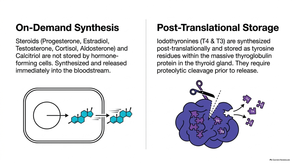
A fundamental distinction in endocrine physiology lies in how different classes of lipophilic hormones are produced and stored prior to secretion. Steroids—including progesterone, estradiol, testosterone, cortisol, and aldosterone—along with calcitriol, are not stored within endocrine cells. Because these hydrophobic molecules diffuse freely through lipid membranes, cells cannot encapsulate them in storage vesicles. Instead, endocrine glands synthesize these lipophilic hormones on demand and release them immediately into circulation upon stimulation.
In stark contrast, iodothyronines exhibit a unique post-translational storage mechanism within the thyroid gland. Thyroxine and triiodothyronine are synthesized post-translationally as tyrosine residues on the massive precursor protein thyroglobulin, which is stored colloidally inside thyroid follicles. When the thyroid is stimulated, thyroglobulin undergoes endocytosis and proteolytic cleavage within lysosomes, releasing free T4 and T3 for immediate release into the bloodstream.
Comparing these biosyntheses offers medical students crucial insight into endocrine cellular architecture. While most lipophilic hormones require immediate enzyme activation for acute synthesis, thyroid tissue uses protein pre-packaging to maintain large hormone reserves. Recognizing these differing storage mechanisms among lipophilic hormones helps explain physiological response times and clinical gland pathologies during hormone deficiency states.
Slide 11: Comparative Summary Table of Lipophilic Hormones
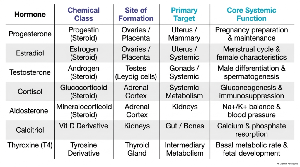
A systematic comparative analysis consolidates the diverse chemical classes, synthesis sites, target organs, and core functions of lipophilic hormones. As summarized across these slides, lipophilic hormones span several major chemical groups: progestins, estrogens, androgens, glucocorticoids, mineralocorticoids, vitamin D derivatives, and iodothyronines. Despite their structural differences, these signaling molecules share the ability to cross cell membranes and modulate genomic transcription in target tissues.
The primary sites of formation for these chemical messengers include the ovaries, placenta, testes, adrenal cortex, kidneys, and thyroid gland. Each tissue synthesizes specific lipophilic hormones designed for specialized physiological targets. For instance, gonadal steroids regulate reproductive cycles and sexual differentiation, adrenal steroids govern glucose metabolism and electrolyte balance, renal calcitriol manages calcium homeostasis, and thyroid iodothyronines control systemic basal metabolic rates.
Mastering this comparative matrix enables college and medical students to integrate endocrinology concepts effectively. Understanding how distinct lipophilic hormones utilize shared nuclear receptor mechanisms while achieving diverse organ-specific actions provides a comprehensive framework for clinical diagnostic reasoning. Analyzing these biochemical pathways reinforces how lipophilic hormone signal transduction shapes human health and systemic homeostasis.
Please read our Content Disclaimer Statement.
Check out our social media channels: