158. Apoptosis: Mechanisms and Regulation of Programmed Cell Death
Every second, millions of cells in the human body quietly destroy themselves to keep the organism healthy and functioning normally. This controlled process prevents damaged or unwanted cells from causing harm. Without this essential self-destruction mechanism, tissues would grow uncontrollably, leading to developmental errors, severe autoimmune disorders, or cancer. This slide deck breaks down the molecular pathways and cellular machinery that govern programmed cell death, providing students with a clear visual blueprint of how cells execute their own demise safely and cleanly.
Slide 1: Introduction to Apoptosis and Cellular Suicide
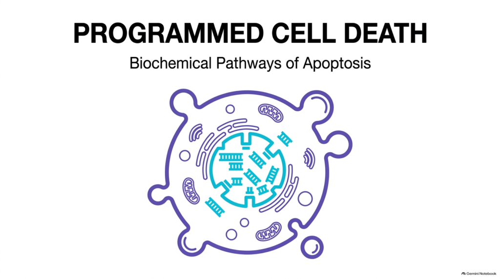
Multicellular life depends on a delicate balance between cell birth and cell death. Apoptosis represents the primary physiological mechanism by which cells undergo genetically controlled self-destruction. Unlike uncontrolled cellular injury or tissue necrosis, Apoptosis operates as a tightly regulated biochemical routine that removes surplus or damaged cells without disrupting neighboring tissues. Understanding Apoptosis is foundational for students in biochemistry, medicine, and cell biology.
The visual representation on this introductory slide highlights a cell initiating internal breakdown while maintaining outer membrane integrity. Inside the cell, key organelles such as mitochondria, endoplasmic reticulum, and nuclear structures remain organized during the early stages of disassembly. This structural organization distinguishes orderly cell death from accidental cell destruction. Specific molecular triggers initiate signaling cascades that direct the cell toward deliberate disassembly.
Through precise enzymatic pathways, Apoptosis ensures that cellular components break down into manageable packages called apoptotic bodies. This process plays a crucial role in maintaining overall tissue health throughout an organism’s life. From shaping delicate anatomical structures during embryogenesis to eliminating potential cancer cells, Apoptosis serves as a vital safeguard for systemic homeostasis. As we advance through this slide deck, we will examine the precise biochemical engines, receptor signals, and regulatory networks that govern this essential process.
Slide 2: Balancing Tissue Homeostasis Through Apoptosis
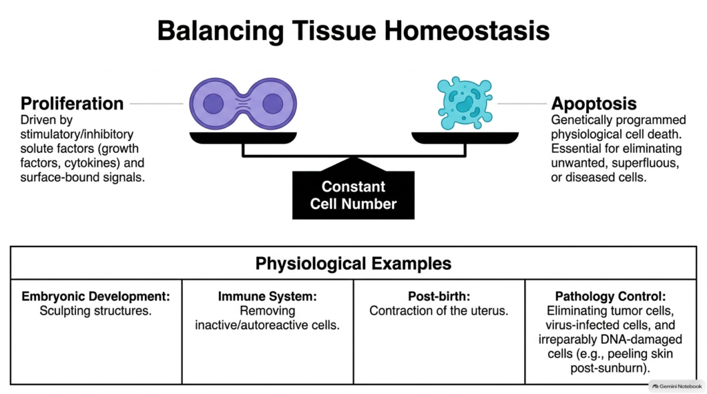
Healthy tissues maintain a constant cell number through a continuous dynamic equilibrium between cell gain and cell loss. Cell proliferation creates new cells in response to growth factors and extracellular signals, while Apoptosis removes older, superfluous, or damaged cells from the population. When these two opposing forces operate in perfect harmony, overall tissue architecture remains stable. Disrupting this equilibrium leads to severe pathology, making apoptosis’s role in maintaining this balance vital for organismal survival.
Physiological examples demonstrate the broad utility of controlled cell death across human biology. During embryonic development, Apoptosis sculpts complex body structures, such as separating individual digits in developing hands and feet by eliminating intervening web tissue. In the immune system, it removes inactive or autoreactive white blood cells, preventing autoimmune attacks on healthy host tissues. Following childbirth, Apoptosis drives the rapid contraction of uterine tissue back to its baseline size.
Furthermore, Apoptosis provides crucial control in pathology by eliminating dangerous cells before they cause systemic harm. Virally infected host cells undergo self-destruction to limit viral replication and spread to adjacent tissues. Similarly, cells bearing severe DNA damage from sunburn or chemical exposure activate Apoptosis to prevent malignant transformation. By eliminating potentially cancerous cells, this controlled death mechanism protects the organism against tumor development.
Slide 3: Comparing Necrosis and Apoptosis Pathways
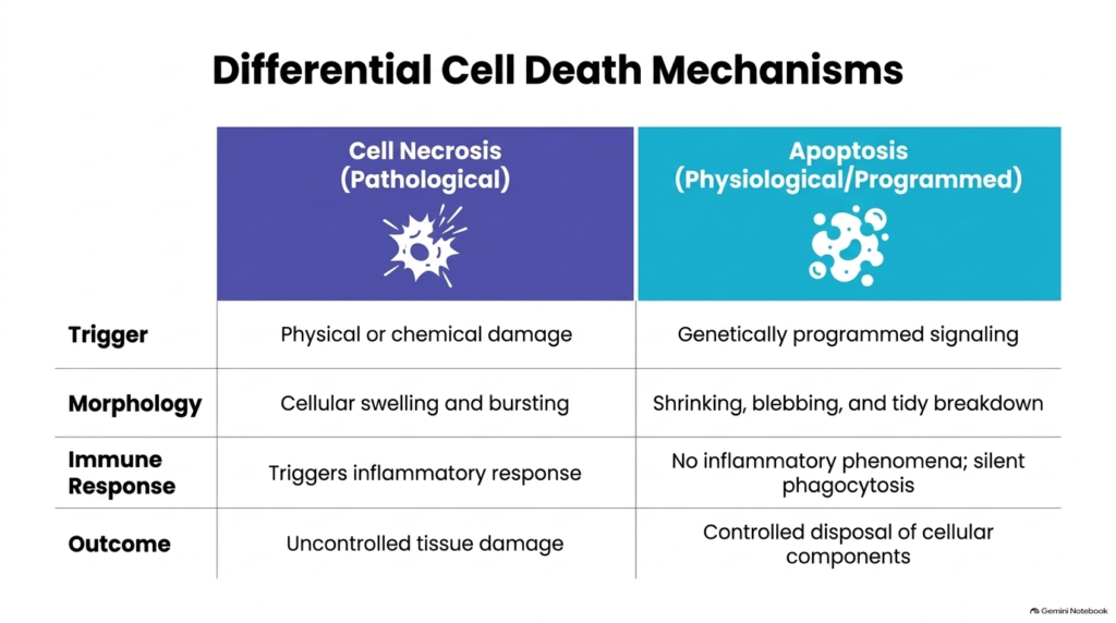
Cells perish through two distinct physiological modes: pathological cell necrosis and physiological Apoptosis. Necrosis results from acute physical trauma, hypoxia, or chemical toxicity, leading to accidental, uncontrolled cell death and cellular lysis. In contrast, Apoptosis follows an orderly, genetically programmed signaling pathway designed to remove cells cleanly. Comparing these two distinct forms of cell death highlights why controlled cellular destruction is essential for maintaining overall tissue health and structural integrity.
Morphologically, necrotic cells swell and burst, spilling their cytoplasmic contents into the surrounding extracellular space. This uncontrolled release of intracellular proteins triggers a robust inflammatory response that damages neighboring healthy tissues. Conversely, Apoptosis causes cells to shrink, condense their nuclear chromatin, and form neat plasma membrane protrusions. The cell breaks down into small, membrane-bound fragments called apoptotic bodies, which are removed quietly without provoking an inflammatory reaction.
The immunological outcomes of these pathways differ significantly across tissues. Because necrosis ruptures the plasma membrane, immune cells invade the damaged region to clear debris, creating localized inflammation, tissue redness, and swelling. Apoptosis avoids inflammation through silent phagocytosis, where nearby macrophages rapidly engulf intact cellular remnants. Controlled disposal ensures that cellular components recycle safely, preventing collateral tissue destruction and maintaining systemic physiological homeostasis.
Slide 4: Morphological Stages of Apoptosis Progression
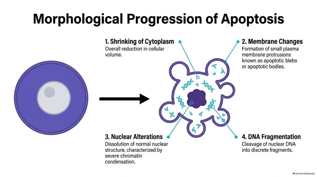
Apoptosis follows a predictable sequence of structural changes that transform the cell’s physical appearance. First, the cell dramatically shrinks its cytoplasm, reducing overall cellular volume. Water and solute ions exit the cell while the internal cytoskeleton reorganizes, causing the cell to lose contact and detach from adjacent matrix structures in the tissue microenvironment.
Second, notable membrane changes occur on the cell surface during Apoptosis. The plasma membrane forms small outward protrusions called blebs, which eventually pinch off to form distinct apoptotic bodies. These membrane-bound packages contain intact organelles and fragmented cellular material. Remarkably, the plasma membrane remains intact throughout these structural transformations, preventing internal enzymes from leaking into surrounding tissues during Apoptosis and causing secondary inflammatory damage to neighboring cells.
Third, internal nuclear changes occur alongside systematic DNA fragmentation. The normal nuclear architecture dissolves as chromatin condenses into dense masses along the nuclear envelope. Specialized cleavage enzymes then digest nuclear DNA into discrete, uniform fragments. These systematic physical steps ensure that the dying cell disassembles into neat, recognizable units ready for rapid cellular disposal, marking a defining morphological hallmark of Apoptosis in living tissues throughout the body.
Slide 5: The Caspase Cascade Engine in Apoptosis
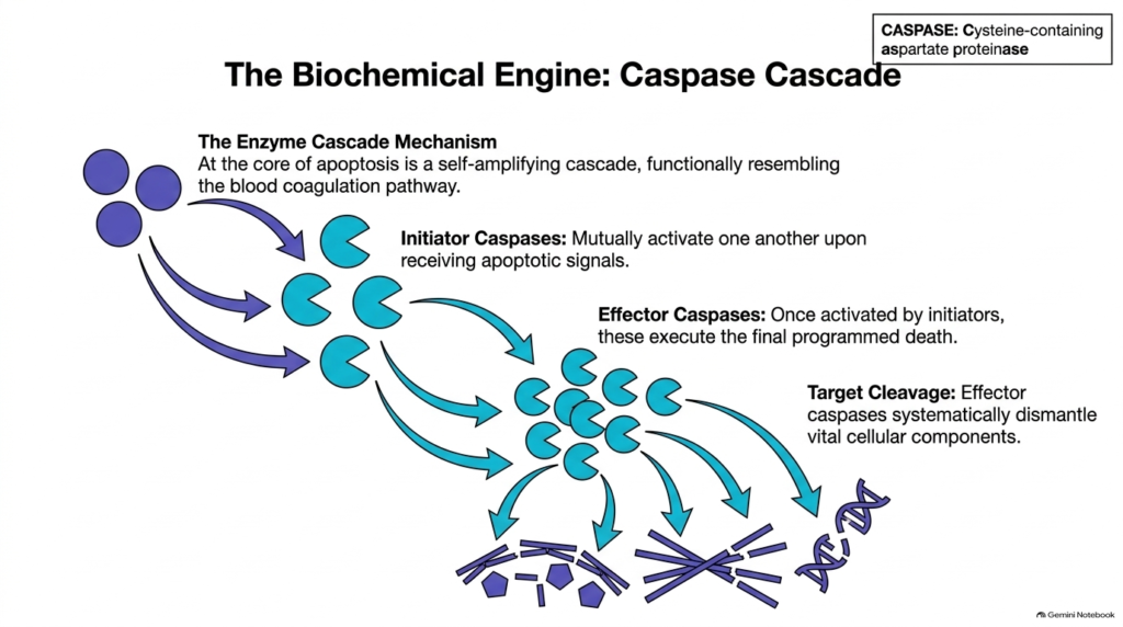
At the heart of the apoptotic machinery is a specialized family of enzymes called caspases. Caspases are cysteine-containing aspartate proteinases that circulate in healthy cells as inactive zymogens. When a cell receives an inductive signal to initiate Apoptosis, these pro-enzymes undergo targeted cleavage to become active proteases. This enzymatic transition initiates a self-amplifying cascade that drives Apoptosis forward with absolute directionality, speed, and precision within the cellular cytoplasm.
The enzyme cascade mechanism operates in two distinct functional tiers during Apoptosis. Initiator caspases sit at the top of the pathway and mutually activate one another upon receiving upstream death signals. Once activated, initiator caspases cleave and activate downstream effector caspases. Effector caspases act as the cell’s primary molecular executioners, amplifying the initial death signal through rapid proteolytic cascades across all cellular compartments.
Once active, effector caspases systematically cleave vital structural and functional proteins across the cytoplasm and nucleus. These targets include cytoskeletal elements, nuclear structural proteins, and regulatory metabolic enzymes. By dismantling essential cellular architecture, effector caspases cause complete structural collapse of the cell. This irreversible enzymatic chain reaction makes Apoptosis an efficient, fast, and tightly controlled physiological mechanism for targeted cellular destruction.
Slide 6: The Fas System as an Extrinsic Trigger for Apoptosis
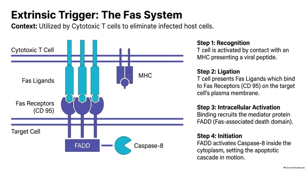
Cells can undergo self-destruction in response to extrinsic signals delivered by neighboring immune cells. Cytotoxic T lymphocytes utilize the Fas ligand system to identify and eliminate virally infected or aberrant host cells. This pathway represents a primary extrinsic mechanism for initiating Apoptosis in response to external physiological threats and cellular pathogens circulating within body tissues during routine immune surveillance.
The signaling sequence begins with recognition, in which a cytotoxic T lymphocyte identifies a target cell presenting foreign viral peptides on its major histocompatibility complex (MHC) surface. After recognition, ligation occurs as Fas ligands on the T cell surface bind directly to Fas receptors (CD95) on the target cell membrane. This binding event clusters the receptors and recruits intracellular signaling proteins directly to the inner plasma membrane to promote Apoptosis.
Recruitment of the mediator adapter protein FADD, or Fas-associated death domain, converts the extracellular binding event into a potent intracellular signal. FADD binds the cytoplasmic tail of aggregated Fas receptors and recruits procaspase-8 molecules. This interaction cleaves procaspase-8 into active caspase-8 inside the cytoplasm. Active caspase-8 then initiates the downstream execution pathway, setting Apoptosis in motion to cleanly eliminate the target cell without injuring adjacent tissue structures.
Slide 7: Tumor Necrosis Factor-alpha Signaling in Apoptosis
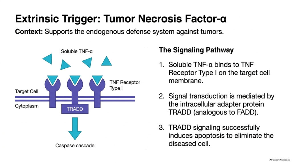
In addition to the Fas pathway, the body uses cytokine signaling to induce cell death from the outside. Tumor Necrosis Factor-alpha, or TNF-alpha, is a soluble signaling protein secreted by active immune cells that supports endogenous defense against malignant tumors and infected tissues. Binding of TNF-alpha to specific surface receptors activates a well-defined extrinsic pathway that leads directly to Apoptosis in targeted cell populations that must be removed from host tissues.
The process begins when soluble TNF-alpha molecules bind to TNF Receptor Type I on the target cell plasma membrane. Receptor engagement causes receptor trimerization and alters the conformation of the intracellular receptor domain. This conformational change attracts the intracellular adapter protein TRADD, known as TNF receptor-associated death domain. TRADD functions analogously to FADD by bridging the membrane receptor to downstream caspase initiators in the cytoplasm, promoting apoptosis execution.
Through TRADD signaling, the target cell recruits initiator caspases to form a functional death-inducing signaling complex inside the cytoplasm. This complex activates initiator proteases, which then trigger the downstream executioner caspase cascade. Ultimately, TNF-alpha signaling successfully induces Apoptosis, ensuring the safe clearance of defective, infected, or transformed cells without disturbing surrounding healthy tissue structures or triggering localized inflammatory reactions in host organs during immune defense.
Slide 8: The Mitochondrial Pathway of Intrinsic Apoptosis
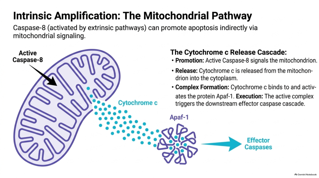
While extrinsic signals initiate cell death from the cell surface, intrinsic pathways monitor internal cellular stress and metabolic damage. However, extrinsic and intrinsic signals often converge through mitochondrial amplification during Apoptosis. Active caspase-8, generated by surface receptor activation, can cleave cytosolic proteins to transmit death signals directly to the mitochondrion, amplifying apoptosis within the cell microenvironment and ensuring complete commitment to cell death.
In response to these signals, the outer mitochondrial membrane becomes permeable, releasing cytochrome c into the cytoplasm. In healthy cells, cytochrome c normally functions in the mitochondrial electron transport chain during ATP synthesis. Once in the cytosol, cytochrome c binds to the adapter protein Apaf-1 (apoptotic protease-activating factor 1), causing Apaf-1 to undergo a dramatic conformational change and multimerize into a specialized complex required for Apoptosis execution.
This binding event promotes the assembly of a multi-protein complex called the apoptosome within the cytosol. The apoptosome recruits and activates procaspase-9, which subsequently cleaves and activates downstream effector caspases. This mitochondrial pathway ensures that internal stress signals and extrinsic death stimuli converge to execute Apoptosis efficiently, demonstrating the interconnected nature of intracellular signaling networks that determine cell survival decisions in response to injury across eukaryotic organisms.
Slide 9: The Role of p53 in DNA Damage-Induced Apoptosis
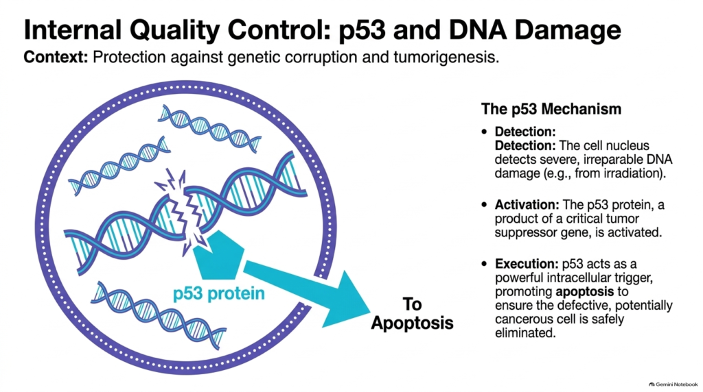
Internal quality-control mechanisms continuously monitor genomic stability to protect the organism from genetic corruption and tumorigenesis. When a cell experiences severe DNA damage from ultraviolet irradiation, ionizing radiation, or chemical mutagens, specialized nuclear sensors detect double-strand breaks. If the genetic damage is irreparable, the cell activates p53 to initiate Apoptosis and prevent damaged genetic material from replicating during cell division.
The p53 protein, produced by a critical tumor suppressor gene, acts as a master transcriptional regulator inside the cell nucleus during Apoptosis signaling. Under normal resting conditions, p53 remains at low intracellular concentrations because the cell rapidly degrades it. When p53 senses irreparable genomic lesions, it stabilizes and accumulates in the nucleus. Active p53 upregulates pro-apoptotic genes that target the outer mitochondrial membrane, inducing cytochrome c release into the cytosol to drive Apoptosis forward cleanly.
As a powerful intracellular trigger, p53 forces defective, mutated, or potentially cancerous cells to undergo self-destruction cleanly and quietly. This protective pathway prevents damaged genetic material from persisting during cell division. Mutations that disable p53 allow damaged cells to survive and divide uncontrollably, underscoring why p53-mediated Apoptosis is vital for tumor suppression, genomic integrity, and overall health across all organ systems.
Slide 10: Effector Caspase Targets During Apoptosis Dismantling
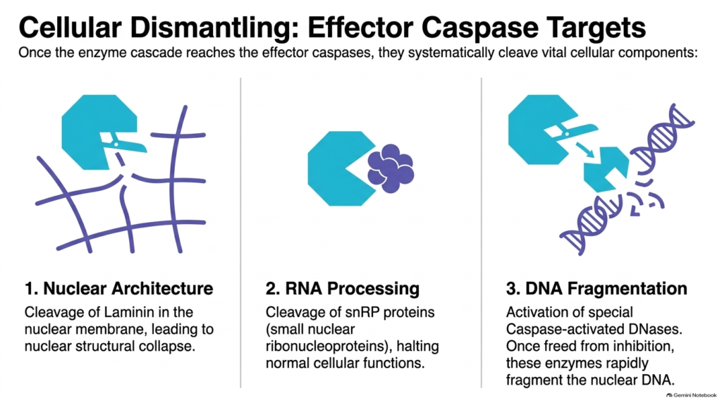
Once the caspase cascade activates executioner caspases, these active proteases systematically dismantle essential cellular components. Effector caspases target specific protein substrates across three critical cellular domains, ensuring the cell collapses in an orderly manner without leaking dangerous enzymes into adjacent tissues during Apoptosis execution throughout the organism.
First, effector caspases target the nuclear architecture by cleaving laminin, an essential structural protein of the nuclear lamina. Cleaving laminin destroys the inner framework supporting the nuclear membrane, causing the nucleus to shrink and collapse rapidly during Apoptosis. Second, caspases disrupt RNA processing by cleaving small nuclear ribonucleoproteins (snRNPs). Halting pre-mRNA splicing shuts down normal protein synthesis and halts cellular metabolism during disassembly and structural breakdown.
Third, effector caspases execute controlled DNA fragmentation in the cell nucleus during apoptosis execution. Under resting conditions, specialized endonucleases remain inactive, bound to inhibitory proteins. Effector caspases cleave these inhibitor proteins, releasing active DNases that rapidly slice genomic DNA into regular fragments between nucleosomes. Through these targeted cleavage events, Apoptosis cleanly dismantles the cell’s internal machinery, enabling orderly disposal by surrounding immune cells and preventing damage to host tissues during development and tissue maintenance.
Slide 11: Phagocytic Clearance and Silent Disposal in Apoptosis
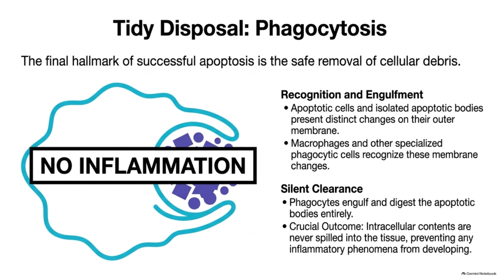
The final milestone of successful Apoptosis is the safe removal of cellular debris from host tissues. During this process, dying cells and isolated apoptotic bodies undergo distinct biochemical changes on their outer plasma membranes. Specifically, phosphatidylserine translocates from the inner membrane leaflet to the outer surface, serving as an eat-me signal for immune surveillance cells in the tissue microenvironment during Apoptosis execution.
Macrophage scavenger receptors and specialized phagocytic cells recognize these exposed surface molecules immediately upon contact. Recognizing these membrane alterations enables phagocytes to bind firmly to apoptotic bodies and engulf them whole through receptor-mediated endocytosis. This swift recognition ensures that dying cellular material is cleared before plasma membrane integrity fails and internal components leak into extracellular spaces during Apoptosis in living organs and tissues throughout the entire body.
Because phagocytes engulf cellular remnants intact, they never spill internal hydrolytic enzymes and toxic metabolites into surrounding tissue spaces. This silent clearance prevents local inflammation, tissue redness, or pain from developing in host organs. By achieving clean, non-inflammatory disposal, Apoptosis allows tissues to clear billions of dead cells daily while maintaining structural integrity, tissue homeostasis, and baseline physiological function across all organ systems throughout human life without immune disruption.
Slide 12: Regulation and Viral Inhibition of Apoptosis
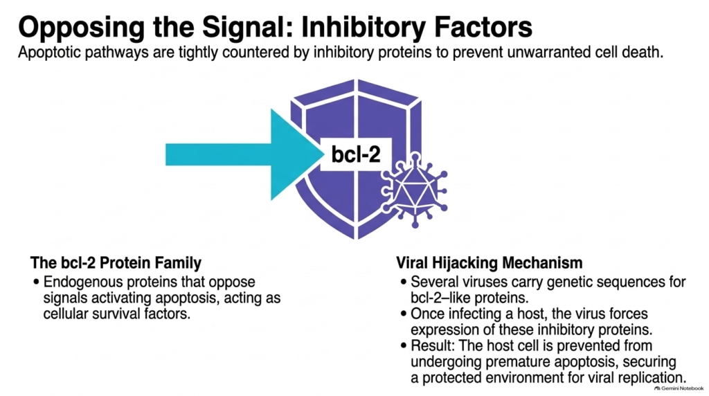
Because cell self-destruction is irreversible, cells tightly regulate death signaling pathways through endogenous inhibitory factors. The bcl-2 protein family plays a central role in this regulatory network. Anti-apoptotic members, such as bcl-2 itself, reside on the outer mitochondrial membrane, where they oppose pro-apoptotic signals and preserve mitochondrial integrity, acting as essential cellular survival factors that protect healthy host tissues throughout the human body during daily life.
Pathogens often target this regulatory checkpoint to ensure their own survival and propagation within the host organism during viral infection. Several viruses have evolved genetic sequences that encode functional bcl-2-like proteins to suppress Apoptosis. After infecting a host cell, the virus forces expression of these inhibitory proteins. These viral proteins neutralize host pro-apoptotic signals, preventing the infected cell from undergoing Apoptosis prematurely before viral assembly completes inside the host cell cytoplasm.
By blocking cell death, the virus secures the protected intracellular environment it needs to complete its replication cycle and safely assemble new viral particles. Understanding how viral proteins hijack bcl-2 regulatory pathways reveals important therapeutic targets. Modulating these inhibitory interactions allows researchers to develop novel pharmacological treatments that restore Apoptosis in persistent viral infections and malignant cancer cells resistant to traditional medical therapies.
Slide 13: The Integrated Signaling Network of Apoptosis
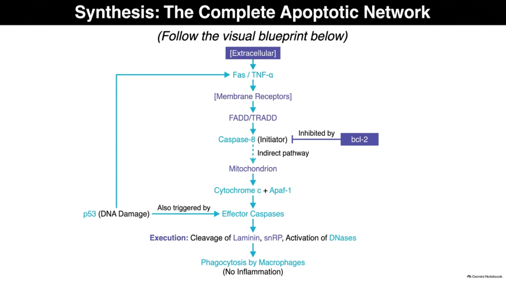
Apoptosis functions as an integrated signaling network in which multiple extracellular and intracellular triggers converge on shared execution machinery. Extracellular signals, such as Fas ligand or TNF-alpha, bind surface membrane receptors to recruit adapter proteins like FADD or TRADD. These adapters activate initiator caspase-8, establishing the extrinsic route toward cell disassembly in response to external physiological cues during Apoptosis in tissues throughout the body.
At the same time, intrinsic stimuli feed into this core network within the cytoplasm. Intracellular triggers like p53 respond to severe DNA damage by promoting permeabilization of the mitochondrial outer membrane. Mitochondria release cytochrome c, which binds Apaf-1 to activate caspase-9. Additionally, active caspase-8 can engage the mitochondrial pathway, creating a cross-talk mechanism that amplifies death signaling when extrinsic signals require intracellular reinforcement during Apoptosis execution in living cells and tissues.
All pathways ultimately activate effector caspases, which cleave vital targets including nuclear laminin, snRPs, and DNases. Opposing this forward signaling, anti-apoptotic proteins like bcl-2 act as regulatory shields to prevent premature destruction. Once execution occurs, macrophages clear cellular remnants via silent phagocytosis without provoking inflammation. This complete network demonstrates how Apoptosis maintains precise control over cellular life and death in health and disease across all human tissue systems and anatomical structures.
Please read our Content Disclaimer Statement.
Check out our social media channels: