79. The Biochemical Mechanisms of Protein Maturation
Imagine building a delicate origami swan inside a turbulent, crowded swimming pool. Newly synthesized molecules face this exact struggle inside the chaotic cellular environment. The core purpose of this slide deck is to break down the biochemical mechanisms that ensure functional molecular structures. From the action of molecular chaperones to the navigation of complex translocation pores, this detailed presentation maps out every critical step. Understanding these biological pathways reveals how life maintains internal order, stabilizes energy landscapes, and prevents fatal cellular aggregation at the microscopic level.
Slide 1: Visualizing Protein Maturation: From Linear Chain to Native Conformation
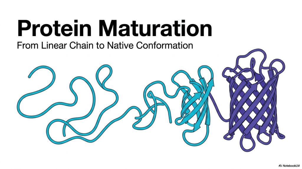
The fundamental goal of Protein Maturation is the successful transition from a simple linear chain to a complex, three-dimensional native conformation. This slide perfectly captures the visual essence of this structural evolution. Initially, the newly synthesized molecule exists as an extended, flexible string of amino acids. In this state, it possesses no biological activity and remains highly vulnerable to the surrounding environment. The visual progression demonstrates how this chaotic string begins to condense, forming localized secondary structures such as alpha-helices and beta-pleated sheets as it seeks energetic stability.
Achieving the correct native architecture during Protein Maturation requires navigating a massive number of potential folding configurations. The linear chain twists and folds upon itself, driven primarily by the intrinsic chemical properties of its constituent amino acids. Hydrophobic residues seek to avoid the aqueous environment, driving the chain to collapse into a more compact, organized state. This spontaneous but guided compaction is a marvel of biophysics, representing a precise optimization problem solved by the cellular machinery in mere milliseconds.
The final folded state depicted on the right side of the illustration represents the functional pinnacle of Protein Maturation. This intricate beta-barrel structure highlights the sheer complexity and beauty of the finished product. In this native conformation, the molecule achieves its lowest possible energy state, locking every atom into a specific spatial arrangement required for its unique biological function. This final geometry is an absolute requirement for life, dictating how the molecule interacts with targets, catalyzes reactions, or provides structural support.
This physical transformation from an unstructured polymer to a highly defined machine is the cornerstone of cellular biology. The visual journey underscores the immense physical changes required to produce functional biology. If the molecule fails to reach this exact native state, it becomes completely useless to the biological system. Understanding this dramatic morphological shift provides vital context for the specific biochemical barriers and the helper molecules required, as explored in the following sections.
Slide 2: The Comprehensive Pipeline of Protein Maturation
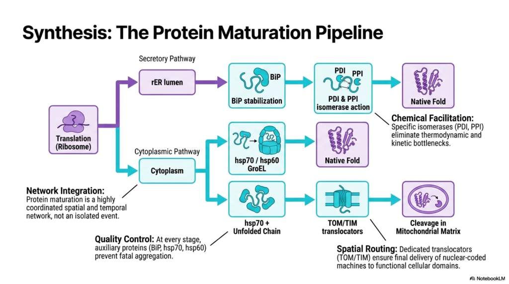
The comprehensive pipeline of Protein Maturation represents a highly coordinated spatial and temporal network rather than a single, isolated event. As illustrated, the journey begins at the ribosome, the cellular factory responsible for translation. Once the raw, linear amino acid sequence emerges, it must immediately navigate distinct developmental routes. The biological system routes the newly synthesized molecules either into the secretory pathway within the rough endoplasmic reticulum lumen or into the busy cytoplasmic pathway. This immediate division of labor ensures that every molecule embarks on the correct biological trajectory right from its inception.
Within these distinct cellular environments, Protein Maturation relies on rigorous quality control mechanisms. Throughout every stage of development, auxiliary molecules act as dedicated guardians. For molecules in the secretory pathway, protein binding stabilizes the immature chain. Meanwhile, the cytoplasmic route utilizes heat-shock proteins like hsp70 and the large hsp60 GroEL complex. These essential components prevent the fatal aggregation of exposed, vulnerable chains. Without such relentless oversight, the highly concentrated cellular matrix would quickly fill with useless, tangled masses of non-functional biological material.
Beyond basic physical stabilization, the later stages of Protein Maturation require precise chemical facilitation to achieve the final native fold. Specific isomerase enzymes, such as protein disulfide isomerase and peptidyl proline isomerase, act as molecular catalysts. They work tirelessly to eliminate severe thermodynamic and kinetic bottlenecks that would otherwise halt the folding process. The elimination of these barriers allows the structural architecture to lock into its most stable, functional conformation, preparing the molecule for its ultimate physiological role.
The final phase involves dedicated spatial routing to ensure delivery to specific functional cellular domains. Nuclear-coded machines destined for the mitochondria utilize the specialized TOM and TIM translocators. These intricate transport channels navigate molecules across strict membrane barriers, often terminating in precise cleavage within the mitochondrial matrix. The seamless integration of quality control, chemical facilitation, and precise spatial routing guarantees that life processes proceed without critical interruptions.
Slide 3: The Crowded Matrix and the Challenges of Protein Maturation
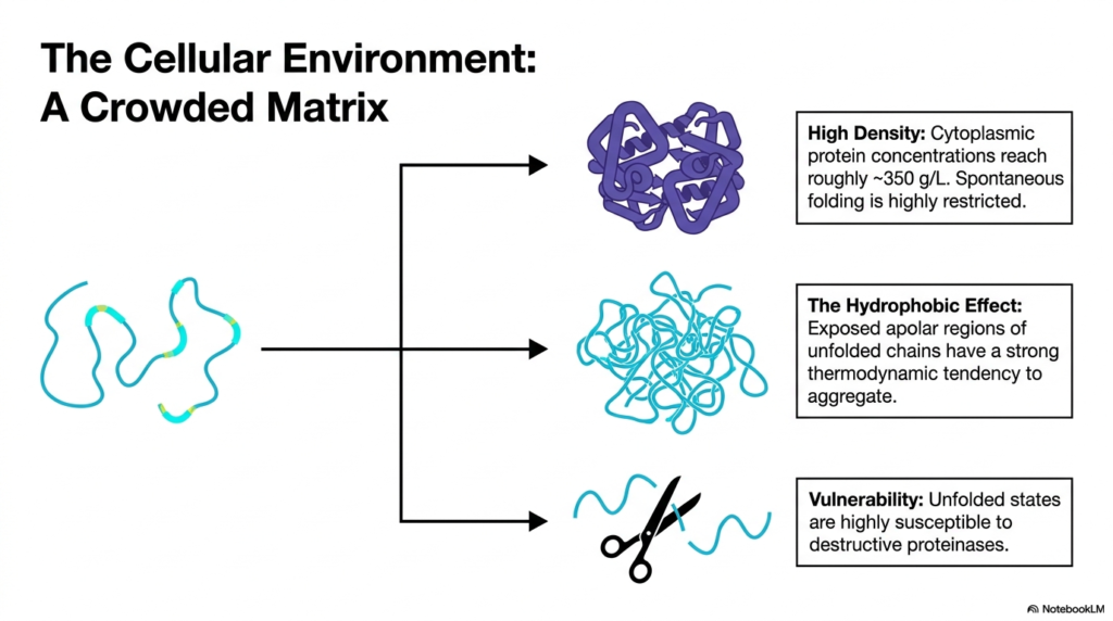
The cellular environment is far from a quiet, empty space; it is a highly crowded matrix that poses significant risks to early Protein Maturation. Cytoplasmic concentrations reach an astonishingly high density of roughly 350 grams per liter. In this packed space, molecules constantly collide with one another. Because of this extreme density, spontaneous, unassisted folding is highly restricted. An emerging linear chain faces intense physical interference from neighboring molecules, making the search for a stable three-dimensional conformation an incredibly treacherous process.
One of the most dangerous factors during Protein Maturation is the hydrophobic effect. When a chain unfolds, its apolar, water-fearing regions remain completely exposed to the aqueous cytoplasm. These exposed hydrophobic patches exhibit a strong thermodynamic tendency to associate with one another. Instead of folding correctly, neighboring unfolding chains can easily clump together, forming massive, non-functional aggregates. This aggregation is not merely an inconvenience; it represents a toxic event that can severely disrupt cellular metabolism and trigger widespread damage.
Furthermore, the vulnerable state of the unfolded chain makes Protein Maturation a race against time. Unfolded biological states are highly susceptible to destructive proteinases. These ubiquitous enzymes act as molecular scissors, prowling the cytoplasm to degrade damaged or unstructured biological material. If a newly synthesized chain remains unfolded for too long, proteinases will recognize it as biological waste and rapidly degrade it before it ever attains its native function.
The combination of extremely high density, dangerous hydrophobic interactions, and aggressive degradative enzymes creates a hostile environment for new molecules. The biological system must deploy robust protective strategies to shield these fragile structures immediately upon synthesis. Recognizing the harsh realities of this crowded cellular matrix is essential for understanding why complex protective mechanisms and auxiliary helper systems evolved to guarantee survival. Without these adaptations, the cell would collapse under the weight of misfolded debris.
Slide 4: Early Stabilization and Protein Maturation in the Endoplasmic Reticulum
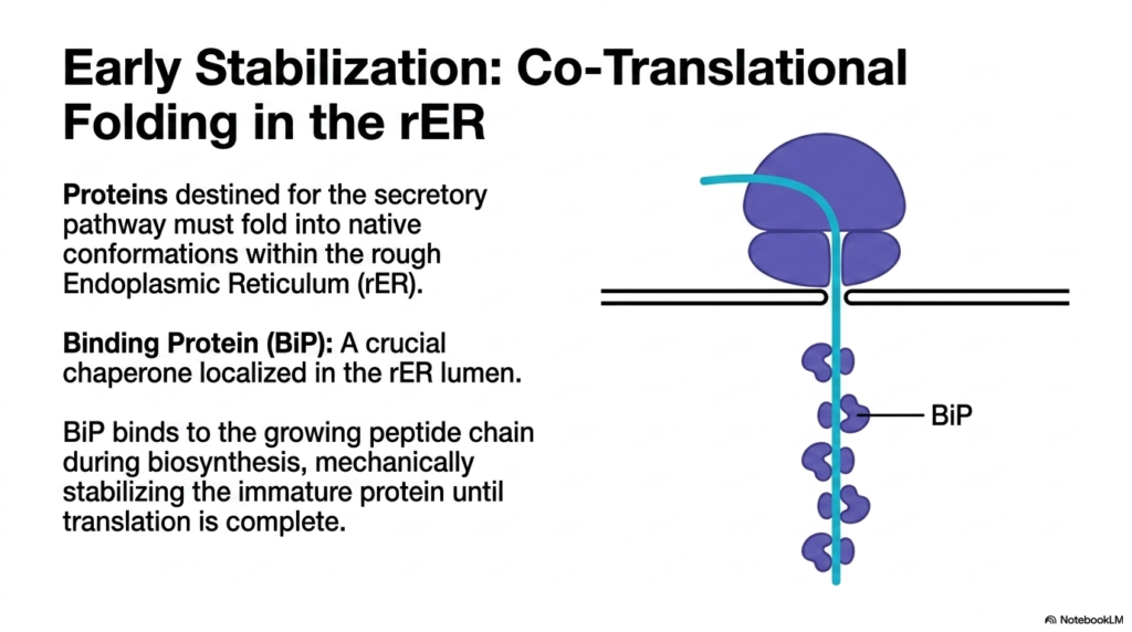
For many critical molecules, Protein Maturation begins with early stabilization during co-translational folding in the rough endoplasmic reticulum. Proteins destined for the secretory pathway, such as hormones or membrane receptors, cannot mature in the general cytoplasm. Instead, they must fold into their native conformations within the specialized lumen of this organelle. As the ribosome continues translation on the membrane surface, the growing peptide chain threads directly into the interior space, ensuring an immediate separation from the chaotic external environment.
Within this specialized compartment, Protein Maturation relies heavily on a crucial chaperone known as Binding Protein. This essential guardian localizes specifically within the lumen of the endoplasmic reticulum. It lies in wait, ready to interact with the nascent polypeptide the moment it emerges from the translocation channel. By binding directly to the growing chain during active biosynthesis, this chaperone provides immediate, localized protection against the harsh thermodynamics of the interior space.
The primary function of Binding Protein during Protein Maturation is to mechanically stabilize the immature structure. Since the entire sequence has not yet left the ribosome, the partial chain cannot fold properly. The attached chaperone acts as a physical placeholder, shielding vulnerable segments and preventing premature folding attempts. It holds the unfinished molecule in a safe, intermediate state, ensuring that the structural elements remain untangled and ready for proper assembly once the full sequence becomes available.
This co-translational strategy represents a brilliant evolutionary adaptation for handling complex structural requirements. By stabilizing the chain while it is still being manufactured, the biological system prevents disastrous early missteps. Once translation is complete, the localized chaperones release the intact chain, allowing it to proceed to the next necessary biochemical modifications. This early intervention is the foundational safeguard for all subsequent developmental steps in the secretory pathway, ensuring the ultimate viability of the secreted products.
Slide 5: Disulfide Bond Formation and Structural Bottlenecks in Protein Maturation
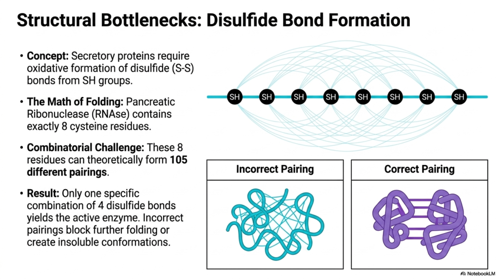
As molecules progress through the secretory pathway, Protein Maturation encounters major structural bottlenecks, most notably the oxidative formation of disulfide bonds. Secretory proteins exist in harsh extracellular environments and require robust covalent linkages between sulfur-containing groups to maintain their shape. These disulfide bonds act as permanent molecular safety pins, locking the folded architecture into place. However, forming these specific covalent connections requires precision. The specific pairing of cysteine residues is a delicate, complex task that can easily go wrong.
The classic example of Pancreatic Ribonuclease perfectly illustrates the severe mathematical challenges inherent to Protein Maturation. This specific enzyme contains exactly eight cysteine residues along its length. Because any one residue can potentially link with any other, the sequence faces an enormous combinatorial challenge. According to the fundamental math of folding, these eight residues can theoretically form one hundred and five completely different pairing combinations. The statistical probability of spontaneously achieving the correct architecture is remarkably low.
Despite the vast number of possibilities, a successful Protein Maturation event demands absolute accuracy. Out of the one hundred and five theoretical pairings, only one specific combination of four disulfide bonds yields the active, functional enzyme. If the molecule forms an incorrect covalent linkage, the entire developmental process halts. These incorrect pairings create rigid dead ends, completely blocking any further folding progress and trapping the biological material in an inactive, tangled state.
The consequence of incorrect pairing is the creation of insoluble, unstable conformations that the biological system cannot use. This profound structural bottleneck highlights the inherent risk of reliance on covalent stabilization. Because the stakes are so incredibly high, the cellular environment cannot rely on random chance to sort out the correct connections. It must utilize specialized molecular tools to monitor, break, and reform these bonds until the perfect, lowest-energy native state emerges. This guarantees the final product operates exactly as intended.
Slide 6: Error Correction Mechanisms During Protein Maturation
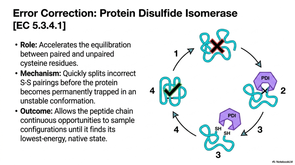
To overcome the massive combinatorial challenges of disulfide bonding, Protein Maturation requires dynamic error correction. This crucial task is carried out by Protein Disulfide Isomerase, a specialized enzyme that acts as a molecular proofreader. Its primary role is to accelerate the equilibration between paired and unpaired cysteine residues within the growing chain. When an incorrect covalent bond forms, the structure becomes trapped. This remarkable isomerase actively surveys the molecule, seeking out these rigid mistakes and intervening before the structural damage becomes an irreversible cellular crisis.
The catalytic mechanism of this enzyme is a fascinating aspect of Protein Maturation. The isomerase quickly splits the incorrect sulfur pairings, essentially cutting the flawed covalent ties. By severing these restrictive bonds, the enzyme rescues the molecule from being permanently trapped in an unstable, inactive conformation. This dynamic breaking and reforming action provides the trapped chain with a necessary second chance. It allows the tangled structure to relax, unfold slightly, and attempt to fold once again.
The ultimate outcome of this continuous error correction during Protein Maturation is structural perfection. The isomerase grants the peptide chain continuous, iterative opportunities to sample different configurations. By rapidly shuffling the chemical bonds, the enzyme allows the molecule to explore its energy landscape until it finally discovers its lowest-energy, native state. Once the correct, most stable structural arrangement is achieved, the isomerase disengages, leaving the functional molecule intact and ready for biological action.
This vital proofreading mechanism prevents the catastrophic accumulation of misfolded debris inside the endoplasmic reticulum. Without such dedicated and precise error correction, critical secretory pathways would quickly clog with useless, tangled polymers, leading to cellular toxicity. The action of this specific isomerase demonstrates how biological systems have evolved sophisticated quality-control networks to manage the sheer thermodynamic complexity of molecular assembly, ensuring that life processes continue smoothly.
Slide 7: Navigating X-Proline Isomerism in Protein Maturation
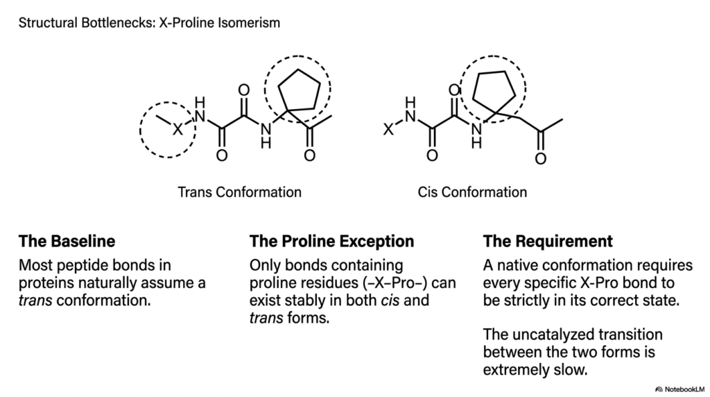
Beyond covalent linkages, Protein Maturation must also overcome significant structural bottlenecks related to peptide bond geometry, specifically X-Proline isomerism. As a baseline rule in biochemistry, the vast majority of peptide bonds naturally and comfortably assume a trans conformation. This arrangement minimizes physical crowding between adjacent amino acid side chains, allowing the backbone to remain stable. However, the unique, ring-like structure of the proline residue creates a distinct exception to this standard geometric rule.
The proline exception is a major hurdle during Protein Maturation. Unlike other amino acids, bonds containing a proline residue can exist stably in both the cis and the trans forms. The cyclic nature of the proline side chain alters the standard energy landscape, making the cis geometry relatively stable. Consequently, a newly synthesized chain will often emerge with a random mixture of both configurations scattered throughout its sequence, creating a chaotic and unpredictable starting point for structural assembly.
For successful Protein Maturation, however, there is a strict biological requirement. A functional, native conformation demands that every specific proline bond exist strictly in its designated state, whether cis or trans. The molecule cannot function if even a single proline hinge is bent in the wrong direction. Unfortunately, the uncatalyzed transition, or flipping, between these two distinct geometric forms is an extremely slow chemical process.
This slow conformational flip acts as a severe kinetic barrier. If left to its own devices, a complex molecule might take hours or even days to spontaneously arrange its proline hinges correctly. Such a delay is completely incompatible with the fast-paced demands of cellular life. Therefore, the biological system must find a way to dramatically accelerate this specific geometric transition to ensure molecular assembly occurs on a biologically relevant timescale, thereby preventing dangerous structural delays.
Slide 8: Overcoming Kinetic Barriers for Swift Protein Maturation
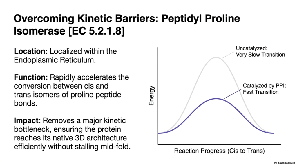
To solve the severe delays caused by complex bond geometry, Protein Maturation employs Peptidyl Proline Isomerase. This specialized biological catalyst is strategically localized within the endoplasmic reticulum, exactly where secretory molecules undergo their most critical developmental stages. Its sole function is to manipulate the stubborn proline hinges that otherwise cause major traffic jams in the folding pipeline. By targeting these specific regions, the enzyme provides a highly targeted solution to a widespread structural problem. This localized intervention guarantees that the structural assembly operates smoothly without fatal interruptions.
The primary mechanism of this isomerase during Protein Maturation involves a dramatic reduction in activation energy. As shown in the energy diagram, the uncatalyzed transition between the cis and trans isomers requires surmounting a steep energy barrier, resulting in a very slow transition. The catalyzed reaction, however, features a significantly lowered activation barrier. The isomerase rapidly binds to the proline bond and physically forces rotation, allowing a fast transition between the two stable geometric states.
The biological impact of this rapid acceleration on Protein Maturation is profound. The isomerase effectively removes a major kinetic bottleneck that would otherwise stall the entire developmental process. By ensuring that every proline hinge snaps quickly into its required orientation, the enzyme allows the overall three-dimensional architecture to assemble efficiently. The peptide chain can swiftly navigate its energy landscape without stalling mid-fold, avoiding prolonged exposure to dangerous degradative elements in the surrounding cellular matrix.
Without the dedicated action of this specific catalyst, the production of vital biological materials would grind to a halt. The graceful, rapid folding we observe in healthy biological systems is only possible because such kinetic barriers are actively managed and dismantled. This perfectly illustrates how the internal machinery prioritizes not just the accuracy of the final structure, but also the crucial speed of the overall developmental timeline necessary for survival.
Slide 9: Auxiliary Protectors in Cytoplasmic Protein Maturation
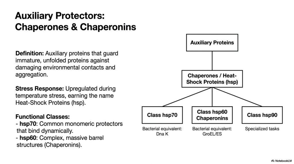
While the secretory pathway utilizes specialized enzymes, cytoplasmic Protein Maturation relies heavily on a distinct class of auxiliary protectors known as molecular chaperones and chaperonins. By definition, these auxiliary guardians are dedicated to shielding immature, unfolded chains against damaging environmental contacts. The bustling cytoplasm is rife with inappropriate interactive surfaces, and these protectors act as physical barriers. They surround the vulnerable sequences, preventing non-specific binding and halting the catastrophic aggregation that plagues densely packed biological environments, ensuring safe passage for newly minted structures.
A fascinating aspect of Protein Maturation is how these protectors respond to environmental danger. Many of these crucial guardians are rapidly upregulated during periods of severe temperature stress. When the cellular environment overheats, existing molecules begin to lose their structural integrity and unfold. To combat this crisis, the biological system triggers an emergency response, flooding the area with these specific helpers, earning them the famous title of Heat-Shock Proteins. Their sudden abundance prevents widespread thermal damage.
Cytoplasmic Protein Maturation relies on several distinct functional classes of these guardians, each tailored to specific developmental needs. The common monomeric protectors belong to the Hsp70 class and bind dynamically to early-unfolding chains. For more complex structural challenges, the system deploys class Hsp60 chaperonins, massive barrel-shaped structures that provide isolated sanctuaries. In bacterial models, these essential classes are known as Dna K and the GroEL complex, respectively, highlighting their ancient evolutionary origins.
Specialized tasks are also handled by the larger class of HSP90 proteins, which manage sophisticated signaling molecules. The collaborative effort among these various classes forms a comprehensive defensive network. Whether functioning as simple, dynamic shields or massive, sequestered chambers, these auxiliary proteins ensure that delicate chains survive the perilous journey from biosynthesis to functional independence, maintaining structural harmony and strict quality control throughout the biological landscape, regardless of external stressors.
Slide 10: The Dynamic Mechanism of hsp70 in Protein Maturation
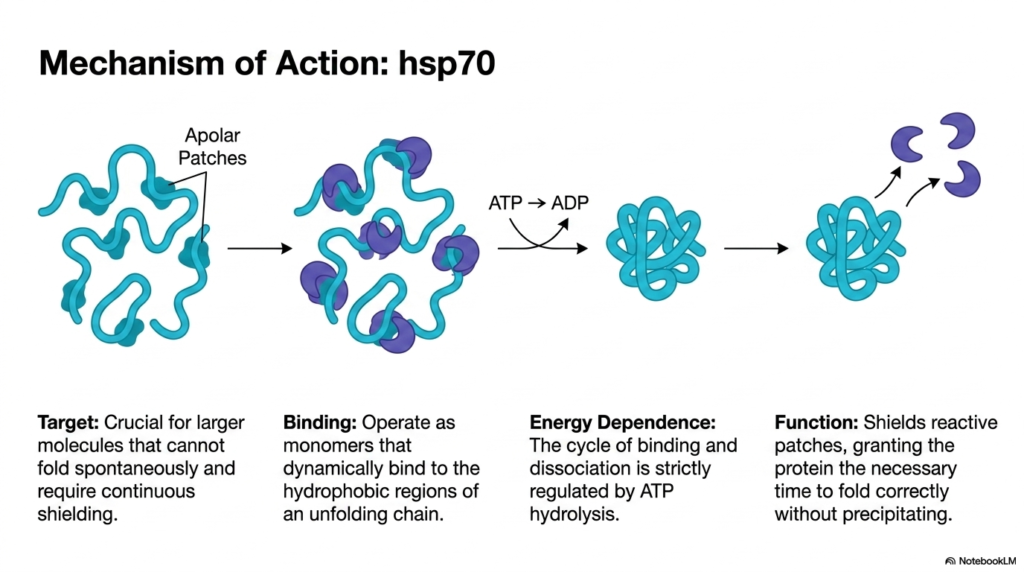
The precise mechanism of action for hsp70 is a critical component of dynamic Protein Maturation. This specific class of chaperone is absolutely crucial for larger molecules that simply cannot fold spontaneously. These massive chains take significant time to synthesize and organize, requiring continuous shielding throughout their lengthy development. The hsp70 molecules operate as vigilant monomers, actively scanning the cellular matrix to locate and protect these vulnerable, extended targets before they can form dangerous, inappropriate structural connections.
The binding process during Protein Maturation is highly selective and targeted. The hsp70 monomers dynamically bind directly to the exposed apolar, hydrophobic patches of an unfolding chain. These specific regions are the most reactive and dangerous parts of the molecule, eager to stick to any nearby surface. By covering these sticky patches like physical caps, the chaperone neutralizes the immediate threat of aggregation, keeping the linear string soluble and safely suspended in the busy cellular fluid.
This continuous shielding during Protein Maturation is strictly energy-dependent. The entire cycle of targeted binding and subsequent dissociation is driven by ATP hydrolysis. When the chaperone attaches to the reactive patch, it utilizes cellular energy to lock down tightly. Once the danger passes or the chain is ready to progress, the conversion of ATP to ADP alters the chaperone’s shape, forcing it to release its grip and detach from the target.
The ultimate function of this dynamic, energy-driven cycle is to grant the immature structure the necessary time to fold correctly without precipitating out of solution. By repeatedly binding, shielding, and releasing the reactive segments, hsp70 prevents dead-end interactions. This allows the complex architecture to slowly and safely find its ideal energetic state, proving that strategic, temporary interventions are essential for managing structural complexity and maintaining a healthy, functional biochemical environment.
Slide 11: The Isolated Folding Chamber for Advanced Protein Maturation
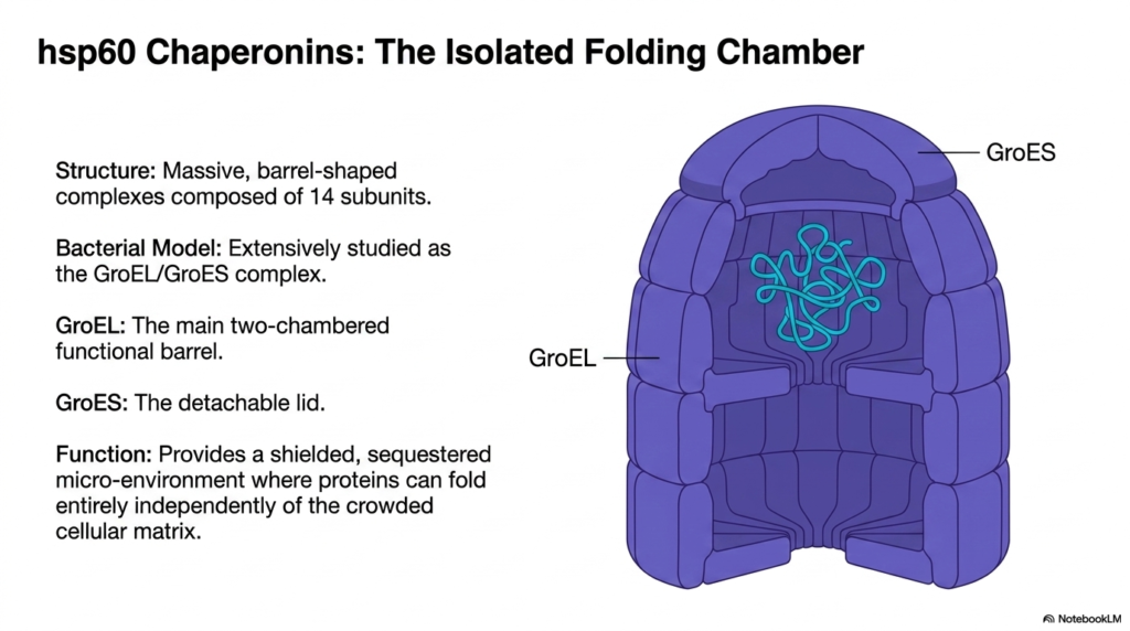
When dynamic shielding is not enough, Protein Maturation requires the specialized intervention of hsp60 chaperonins. These remarkable molecular machines function as isolated folding chambers, designed for complex cases that demand complete physical segregation from the cellular matrix. Structurally, these complexes are massive, barrel-shaped marvels composed of exactly fourteen identical subunits arranged in a highly organized double ring. This vast architectural scale allows them to encapsulate entire biological molecules, completely isolating them from external interference during the critical phases of development.
The bacterial model of this chamber is the GroEL-GroES complex, which has been extensively studied and provides profound insights into complex Protein Maturation. The primary architecture consists of GroEL, which forms the main two-chambered functional barrel. This sturdy cylinder provides the hollow interior space necessary for structural manipulation. Paired with this is the GroES component, which functions as a distinct, detachable lid. Together, these pieces form a secure, temporary biological vault for struggling peptide chains.
The ultimate function of this enormous complex in Protein Maturation is environmental control. It provides a heavily shielded, sequestered microenvironment where sensitive proteins can fold entirely independently of the crowded, dangerous cellular matrix outside. Inside this hollow sanctuary, the chaotic variables of high concentration and roaming proteinases are completely eliminated. The trapped chain is finally free to explore its energy landscape and arrange its complex architecture in absolute peace, guided only by its internal chemistry.
This physical isolation strategy represents the pinnacle of structural quality control. By physically enclosing the vulnerable target, the chaperonin ensures that even the most difficult, aggregation-prone sequences have a safe space to achieve their functional geometry. This sophisticated barrel mechanism highlights the extreme, energy-intensive measures the biological system will deploy to prevent toxic cellular buildup and ensure the successful deployment of large, complex biological machines required for survival.
Slide 12: The Alternating Chamber Mechanism in Protein Maturation
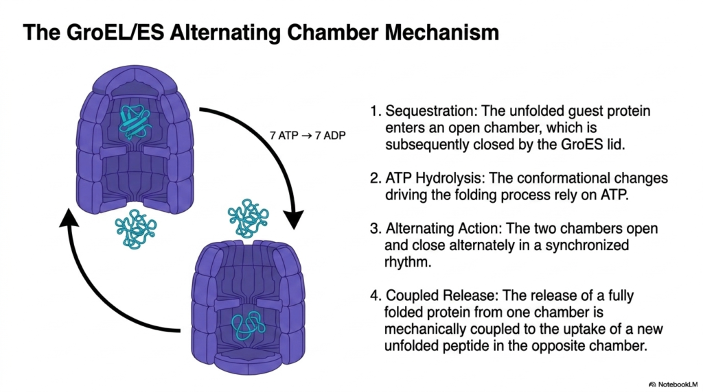
The operational cycle of the GroEL and GroES complex reveals a fascinating mechanical rhythm at the heart of advanced Protein Maturation. The process begins with the critical step of sequestration. An unfolded, highly vulnerable guest protein wanders into an open, empty chamber of the main barrel. Once inside, the biological vault is rapidly secured by attaching the GroES lid. This immediate closure completely traps the struggling sequence, locking it away from the external chaos.
Once sealed inside, Protein Maturation within the chamber relies heavily on active energy expenditure. The massive conformational changes that drive internal folding rely entirely on the continuous hydrolysis of ATP. The inner walls of the chamber physically shift and expand, changing their chemical properties to actively force the trapped sequence into a more compact, stable shape. This is an aggressive, mechanically driven operation that directly manipulates the structural geometry of the encapsulated target.
The sheer efficiency of this Protein Maturation engine is driven by its remarkable alternating action. The massive complex is divided into two distinct chambers that open and close alternately in a synchronized, biological rhythm. While one half of the barrel is sealed tight and actively folding a trapped molecule, the opposite half remains open, waiting to accept a new target. This alternating engine ensures a continuous, non-stop pipeline of structural processing, maximizing cellular efficiency.
Finally, the operation concludes with a brilliant coupled release. The ejection of a fully folded, functional molecule from the top chamber is mechanically coupled to the simultaneous uptake of a new, unfolded peptide in the opposite bottom chamber. This synchronized exchange guarantees that the complex is never idle. This precise, energy-driven machinery showcases the incredible mechanical sophistication required to force stubborn sequences into their vital, life-sustaining configurations, ensuring the cellular factory operates at peak capacity.
Slide 13: Comparative Analysis of Protectors in Protein Maturation
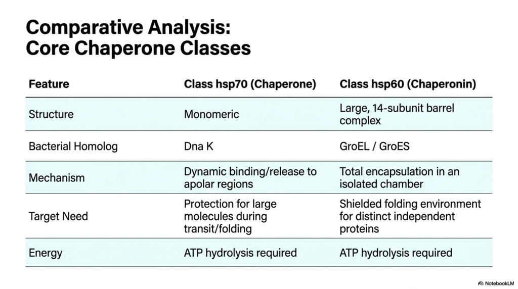
A thorough comparative analysis of the core chaperone classes highlights the versatile strategies employed during Protein Maturation. The biological system utilizes distinct structural approaches depending on the specific threat level. The class hsp70 chaperone functions as a simple, highly mobile monomeric unit, akin to a rapid-response team. Conversely, the class hsp60 chaperonin relies on a massive, stationary 14-subunit barrel complex. Their bacterial homologs, Dna K and the GroEL/GroES complex, respectively, confirm that this structural dichotomy is an ancient, universally conserved biological strategy.
The fundamental difference in their mechanisms reflects their distinct roles in Protein Maturation. The smaller hsp70 utilizes a strategy of dynamic binding and rapid release. It temporarily sticks directly to the dangerous apolar regions of a target, providing localized coverage. In stark contrast, the massive hsp60 barrel completely swallows the target. It utilizes total encapsulation, placing the entire vulnerable sequence within a completely isolated interior chamber, physically separating the molecule from the surrounding chaotic environment.
Because of these mechanical differences, they target entirely different needs during Protein Maturation. The dynamic hsp70 provides essential protection for large molecules actively in transit or during the early stages of folding, preventing catastrophic tangles. The giant hsp60 barrel, however, provides a shielded, highly controlled folding environment dedicated to distinctly independent molecules that have entirely failed to fold on their own. One prevents the fire, while the other acts as an intensive care unit.
Despite these dramatic differences in structure and application, both defensive classes share one critical limitation: energy dependence. Both mechanisms strictly require continuous ATP hydrolysis to function. Whether dynamically clamping onto a localized patch or driving the massive structural shifts of an entire barrel chamber, securing correct biological geometry demands a massive investment of cellular resources. This shared energy requirement underscores the immense thermodynamic cost of maintaining cellular order.
Slide 14: The Mitochondrial Import Challenge in Protein Maturation
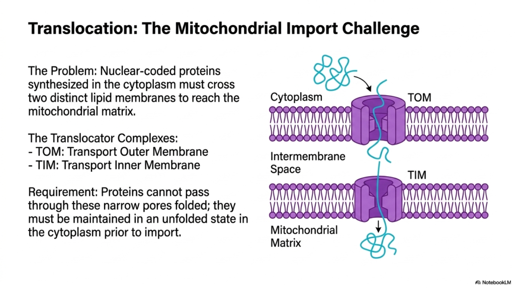
Protein Maturation often requires crossing severe spatial boundaries, presenting major logistical problems, such as the mitochondrial import challenge. Many specialized cellular machines are nuclear-coded, meaning they are fully synthesized out in the general cytoplasm. However, their ultimate functional destination is deep inside the mitochondrial matrix. To reach this isolated workspace, these chains must physically cross two distinct, highly impermeable lipid membranes. This complex geographical relocation adds a formidable layer of difficulty to the overall developmental pipeline, requiring specialized navigation tools.
Navigating these strict lipid barriers during Protein Maturation relies on dedicated molecular gates known as translocator complexes. The outer lipid boundary is guarded by the TOM complex, which stands for Translocase of the Outer Membrane. Directly beneath it, the inner lipid boundary is managed by the TIM complex, or Translocase of the Inner Membrane. These specialized pores must align perfectly to create a continuous, navigable channel straight from the busy cytoplasm into the deep interior.
The most severe physical requirement for this spatial Protein Maturation step concerns biological geometry. The translocator pores within the TOM and TIM complexes are incredibly narrow. Fully folded, three-dimensional structures are simply too wide to pass through these restrictive gates. Therefore, the molecules must be meticulously maintained in a completely unfolded, linear state in the cytoplasm prior to import. If the chain accidentally folds before transport, it becomes permanently locked out of its required destination.
This physical constraint forces the biological system to delay the final folding stages until the molecule has successfully reached the matrix. This requires a highly coordinated effort from the cytoplasmic chaperones to actively prevent premature organization. The intricate relationship between spatial location and structural timing illustrates how precisely the cellular machinery must choreograph its actions to ensure complex functional components reach their correct specialized organelles, avoiding massive logistical failures.
Slide 15: Step-by-Step Translocation in Protein Maturation
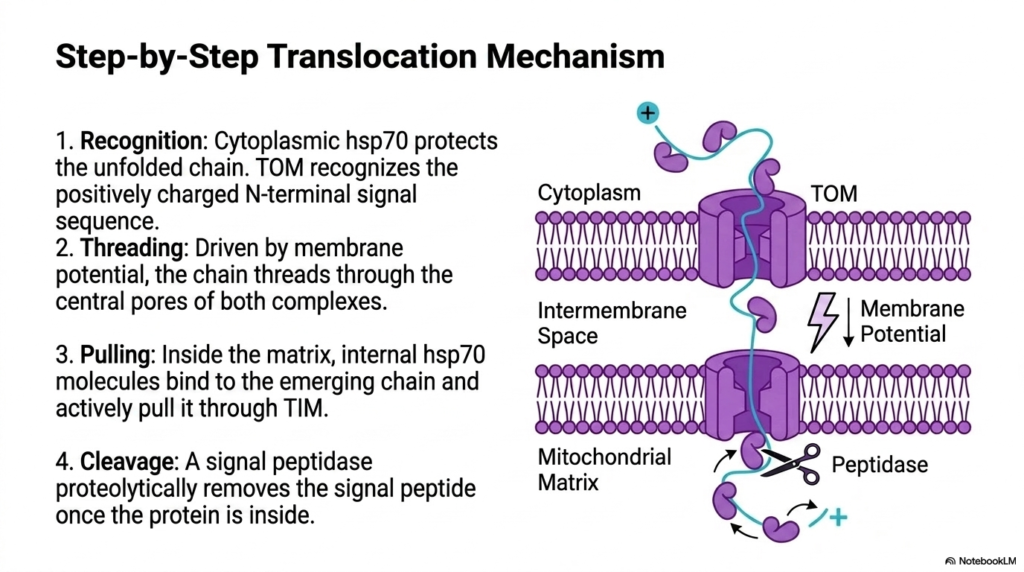
The final step-by-step translocation mechanism perfectly demonstrates the sheer complexity of spatial Protein Maturation. The operation begins with precise biochemical recognition. In the cytoplasm, dedicated hsp70 chaperones tightly coat the unfolded chain, actively protecting it from premature collapse. The target molecule possesses a positively charged N-terminal signal sequence acting as a chemical address label. The outer TOM complex specifically recognizes this unique biological barcode, ensuring that only the correct, authorized materials gain access to the interior.
Following initial recognition, Protein Maturation progresses into the active threading phase. Driven by an underlying electrochemical membrane potential, the linear chain begins to snake its way downward. It threads sequentially through the central pores of both the outer TOM and inner TIM complexes. This delicate movement requires the temporary stripping away of the protective cytoplasmic chaperones, leaving the exposed chain incredibly vulnerable as it squeezes through the tight translocator channels toward the deep interior space.
As the sequence emerges inside the matrix, the final pulling phase of Protein Maturation initiates. Internal hsp70 molecules, localized strictly within the mitochondria, immediately bind to the emerging linear chain. Functioning like a powerful molecular ratchet, these internal guardians actively and forcefully pull the rest of the molecule through the TIM complex. This energy-driven pulling mechanism prevents the chain from sliding backward, guaranteeing unidirectional transport across the double lipid boundary into the deep matrix.
The entire journey concludes with a definitive, irreversible cleavage event. Once the full length of the sequence successfully enters the matrix, a specialized signal peptidase acts like a biological scissor. It proteolytically removes the initial signal peptide, discarding the temporary address label. With the tag removed, the molecule is finally free to undergo its final folding steps within the matrix, completing its long structural evolution and becoming a fully active, essential metabolic machine ready to sustain cellular life.
Please read our Content Disclaimer Statement.
Check out our social media channels: