84. Eukaryotic Transcription: Biochemical Mechanisms and Regulation
Imagine having a vast library of blueprints, but no contractors to build the structures. That is a cell without gene expression. The core purpose of this slide deck is to demystify eukaryotic transcription, the critical mechanism cells use to read genetic blueprints and synthesize functional molecules. By analyzing these slides, students will grasp the biochemical machinery, regulatory networks, and structural dynamics that dictate cellular identity and metabolic function.
Slide 1: Eukaryotic Transcription Mechanisms: How Cells Read the Genetic Code
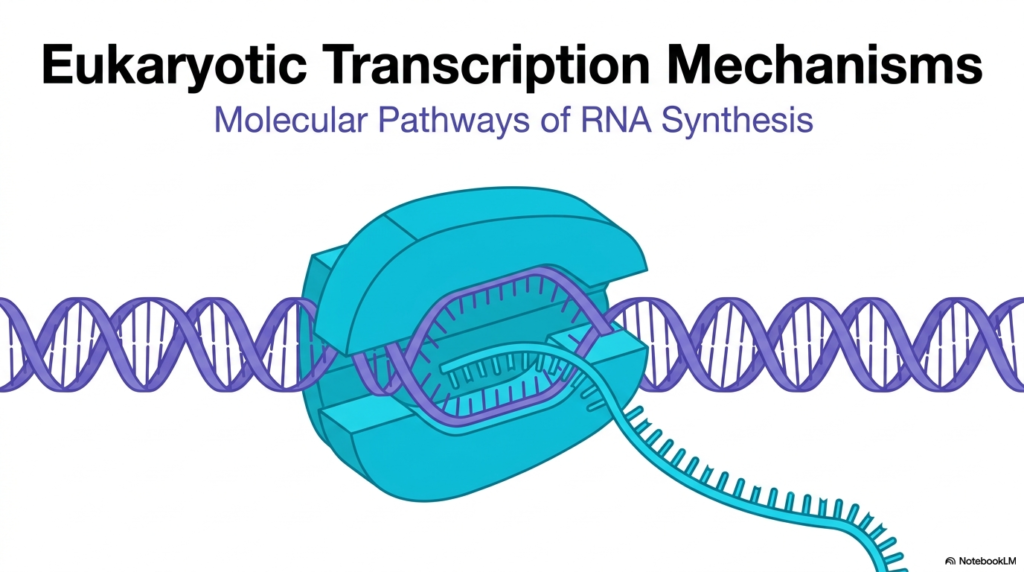
Eukaryotic transcription is the foundational process of life, acting as the bridge between static genetic information and dynamic cellular function. The visual provided illustrates the core molecular pathways of RNA synthesis, highlighting the massive enzymatic machinery required to read the DNA double helix. In eukaryotic transcription, the DNA remains safely housed within the nucleus while RNA polymerase transcribes the sequence into a temporary RNA message. This fundamental process ensures that the permanent genetic code is preserved while still allowing the cell to produce necessary proteins.
Understanding eukaryotic transcription requires an appreciation for the structural complexity of the enzyme shown. The light blue complex represents the RNA polymerase enzyme, specifically designed to clamp onto the purple DNA double helix. It pries the two strands apart, creating a localized transcription bubble. As the enzyme moves along the DNA, it reads the template and synthesizes the bright teal RNA strand. This visual perfectly captures the mechanical nature of eukaryotic transcription, where molecular machines perform precise physical tasks to read biological information.
While the slide offers a simplified representation, it is essential to remember that this process is highly regulated and intricate in biological systems. Standard biochemical knowledge beyond the immediate source material notes that this polymerase does not act alone; it requires dozens of accessory proteins to function efficiently. The newly synthesized RNA strand, shown exiting the enzyme, will undergo extensive modifications before it can be utilized. Ultimately, mastering the fundamentals of eukaryotic transcription prepares students to dive into the nuanced regulatory mechanisms explored in the following slides.
Students must recognize that the molecular pathways of RNA synthesis depicted here form the basis of all cellular responses. Every time a cell adapts to a new environment, it relies on eukaryotic transcription to produce the specific tools needed for survival. The subsequent slides will break down this overarching view into specific biochemical steps, revealing the elegant logic that governs how and when genes are activated.
Slide 2: The Role of Eukaryotic Transcription: Rewriting the Genetic Blueprint
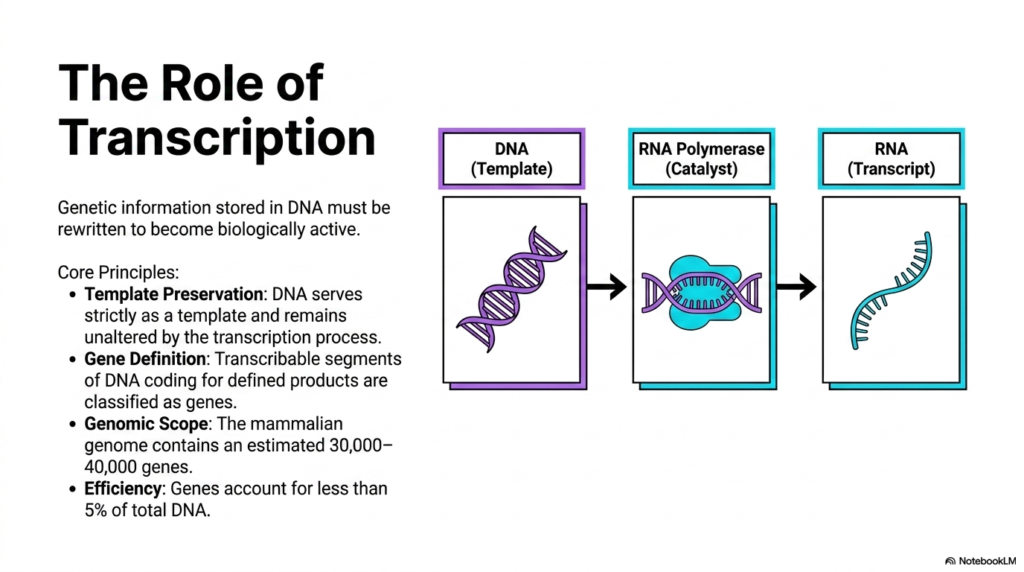
The primary role of eukaryotic transcription is to rewrite genetic information stored in DNA so it becomes biologically active. The slide breaks down this concept through a clear flowchart: DNA acts as the template, RNA polymerase serves as the catalyst, and the final transcript is RNA. A core principle of eukaryotic transcription is template preservation. The DNA strictly serves as a template and remains entirely unaltered by the transcription process. This ensures the genomic archive is protected and can be used repeatedly without degradation across the lifespan of the cell.
Another crucial concept is gene definition. Within the vast landscape of the genome, only specific transcribable segments coding for defined products are classified as genes. Eukaryotic transcription selectively targets these regions rather than reading the entire DNA sequence indiscriminately. The genomic scope highlighted in the source reveals that the mammalian genome contains an estimated thirty to forty thousand genes. This massive library of information requires a highly organized system of eukaryotic transcription to ensure the correct genes are transcribed at the precise moment they are needed by the cell.
The efficiency of this biological system is remarkable. The source notes that genes account for less than five percent of total DNA. This means the vast majority of the genome consists of non-coding regions. Eukaryotic transcription is a highly specific process that must navigate this enormous genetic sea to find the exact five percent that codes for functional products. Students should appreciate that while the slide outlines these core principles, standard external biochemical texts emphasize that the remaining ninety-five percent of DNA still plays vital structural and regulatory roles.
Understanding these core principles establishes a framework for studying molecular biology. The flow from DNA to RNA represents the first critical step of the central dogma. By maintaining template preservation and targeting specific genes, eukaryotic transcription acts as the master control switch for cellular activity. The machinery that executes this targeted synthesis will be the focus of the next deep dive into specific polymerases.
Slide 3: The Machinery of Eukaryotic Transcription: DNA-Dependent RNA Polymerases
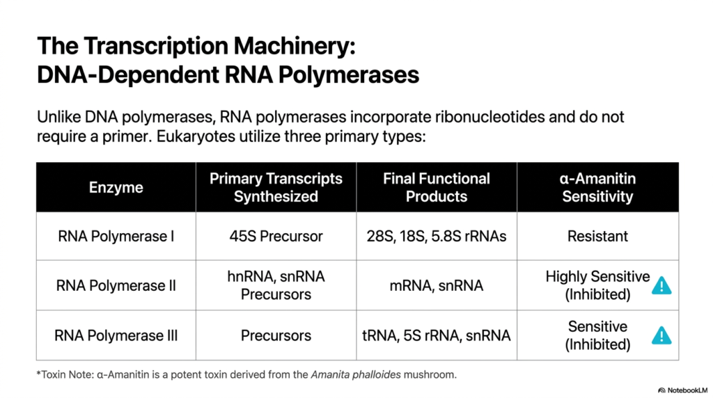
The enzymatic engines driving eukaryotic transcription are DNA-dependent RNA polymerases. Unlike DNA polymerases used during replication, RNA polymerases incorporate ribonucleotides and uniquely do not require a primer to initiate synthesis. The slide outlines the three primary types of polymerases utilized by eukaryotes, each tasked with synthesizing different classes of RNA. This division of labor is a hallmark of eukaryotic transcription, allowing the cell to independently regulate the production of messenger RNA, ribosomal RNA, and transfer RNA based on immediate metabolic demands and cellular conditions.
The table details the specific functions of each enzyme class. RNA Polymerase I synthesizes the 45S precursor, which eventually becomes the 28S, 18S, and 5.8S ribosomal RNAs. RNA Polymerase II is responsible for synthesizing heterogeneous nuclear RNA (hnRNA) and small nuclear RNA precursors, which mature into functional mRNA and snRNA. Finally, RNA Polymerase III creates precursors that yield transfer RNA, 5S rRNA, and other snRNAs. Eukaryotic transcription relies heavily on RNA Polymerase II for gene expression, as it produces the mRNA templates that will ultimately be translated into functional proteins.
A fascinating clinical and biochemical aspect of eukaryotic transcription is its vulnerability to specific toxins. The slide highlights alpha-amanitin, a potent toxin derived from the Amanita phalloides mushroom. The three polymerases exhibit distinct sensitivities to this compound. RNA Polymerase I is completely resistant, while RNA Polymerase III is sensitive and can be inhibited. Crucially, RNA Polymerase II is highly sensitive to alpha-amanitin. When this toxin enters a cell, it rapidly halts the eukaryotic transcription of mRNA, leading to devastating cellular failure and often fatal liver toxicity in humans.
Students should memorize these distinct polymerases and their products. The specific inhibition of RNA Polymerase II by alpha-amanitin is a classic biochemical principle frequently tested in medical board examinations. It perfectly illustrates how targeting specific components of eukaryotic transcription can have profound, systemic biological consequences.
Slide 4: Gene Structure in Eukaryotic Transcription: The PEP-CK Model
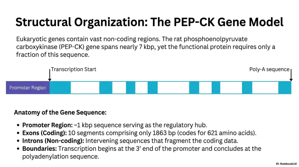
Eukaryotic genes are remarkably complex and contain vast non-coding regions. To illustrate this, the slide uses the rat phosphoenolpyruvate carboxykinase, or PEP-CK, gene as a structural model. This gene spans nearly seven kilobase pairs, yet the functional protein requires only a fraction of this sequence. During eukaryotic transcription, the entire span from the transcription start site to the polyadenylation sequence is read, creating a large primary transcript. This highlights a fundamental inefficiency built into eukaryotic transcription, where massive segments of genetic material must be processed down to a usable size.
The anatomy of the gene sequence features several critical landmarks. The promoter region is an approximately one kilobase pair sequence that serves as the regulatory hub, determining when and how vigorously eukaryotic transcription occurs. Following the promoter are ten distinct coding segments known as exons. These exons comprise only 1863 base pairs, which ultimately code for a 621 amino acid protein. The true complexity of eukaryotic transcription lies in the intervening non-coding sequences, or introns, that fragment the coding data and expand the overall physical size of the gene footprint.
The boundaries of the transcribable gene are strictly defined. Eukaryotic transcription begins precisely at the three-prime end of the promoter region and concludes at the specific polyadenylation sequence. Understanding this spatial arrangement is crucial for medical students, as mutations in either the promoter or the splice sites between exons and introns can lead to severe disease. Standard clinical knowledge notes that errors in processing these fragmented sequences frequently disrupt normal eukaryotic transcription outcomes, causing conditions like beta-thalassemia.
The PEP-CK gene serves as an excellent, typical example of genomic architecture. By studying this specific layout, students can visualize how regulatory hubs control the vast stretches of alternating coding and non-coding sequences. This structural complexity necessitates the elaborate RNA maturation processes that must immediately follow eukaryotic transcription.
Slide 5: Maturation Post-Eukaryotic Transcription: Crafting the Messenger RNA
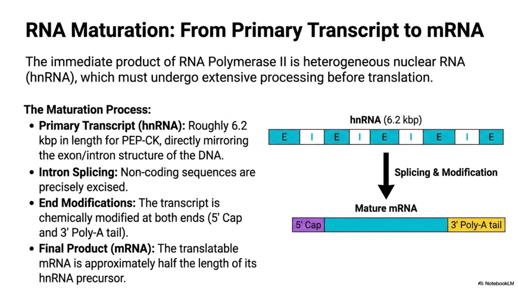
The immediate product generated by RNA Polymerase II during eukaryotic transcription is not ready for translation. This initial molecule is called heterogeneous nuclear RNA, or hnRNA, and it directly mirrors the alternating exon and intron structure of the genomic DNA. For the PEP-CK model, this primary transcript is roughly 6.2 kilobase pairs in length. A hallmark of eukaryotic transcription is that this bulky precursor must undergo extensive chemical processing and physical modification within the nucleus before it can ever be utilized by the cellular ribosomes.
The maturation process involves three major modifications. First, intron splicing must occur, where all the non-coding intervening sequences are precisely excised, and the exons are joined together. Second, the transcript undergoes crucial end modifications to ensure stability. A protective five-prime cap is added to the beginning, and a three-prime poly-A tail is added to the end. These modifications, which occur closely linked to eukaryotic transcription, are vital for protecting the delicate RNA molecule from rapid enzymatic degradation as it travels through the cellular cytoplasm.
The final product of this intensive maturation is the translatable messenger RNA. Due to the removal of massive intronic regions, this mature mRNA is approximately half the length of its original hnRNA precursor. Standard biochemical understanding, reaching beyond the slide, dictates that these post-processing steps are deeply integrated with eukaryotic transcription itself, often occurring co-transcriptionally while the polymerase is still moving. This ensures maximum efficiency and protects the transcript the moment it emerges from the enzyme channel.
Students must grasp that RNA maturation is as critical as the initial synthesis. Without precise splicing and protective capping, the products of eukaryotic transcription would be rapidly destroyed or translated into non-functional, potentially toxic proteins. This maturation phase represents a major quality control checkpoint in the flow of genetic information.
Slide 6: Initiating Eukaryotic Transcription: Remodeling Chromatin
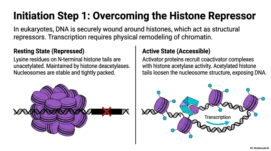
In order to begin eukaryotic transcription, the cellular machinery must first gain physical access to the genetic code. However, in eukaryotes, DNA is securely wound around octamers of histone proteins to form nucleosomes. These histones act as structural repressors. In the resting or repressed state, the lysine residues on the N-terminal histone tails remain unacetylated, a condition maintained by histone deacetylase enzymes. This causes the nucleosomes to be highly stable and tightly packed together, completely preventing the initiation of eukaryotic transcription by physically blocking the promoter regions from incoming enzymes.
To transition to an active state, eukaryotic transcription requires the physical remodeling of chromatin. Activator proteins recognize specific signals and recruit powerful coactivator complexes that possess histone acetylase activity. These enzymes add acetyl groups to the lysine residues on the histone tails. The addition of these negatively charged groups neutralizes the positive charge of the lysines, weakening their grip on the negatively charged DNA backbone. This chemical modification loosens the overall nucleosome structure, forcing the chromatin to uncoil and actively exposing the underlying DNA sequences.
This crucial first step acts as a massive regulatory gatekeeper for eukaryotic transcription. By default, the genome is locked away and silenced. Standard epigenetic knowledge demonstrates that this dynamic shifting between acetylated and unacetylated states is a primary mechanism cells use to globally regulate metabolic shifts and differentiation. Without successfully overcoming the histone repressor barrier, all subsequent steps of eukaryotic transcription simply cannot occur, regardless of how many transcription factors are present in the nuclear environment.
Recognizing chromatin remodeling is essential for medical students, as many modern cancer therapeutics specifically target histone deacetylase enzymes. By chemically manipulating how tightly DNA is wound, clinicians can artificially reactivate tumor suppressor genes, proving the immense biological significance of this initial phase of eukaryotic transcription.
Slide 7: Assembling the Core Machinery for Eukaryotic Transcription
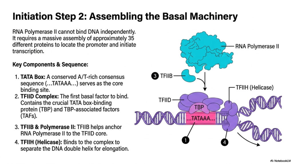
Once the chromatin is remodeled, the next hurdle in eukaryotic transcription is assembling the necessary enzymatic complexes. RNA Polymerase II is a massive enzyme, yet it cannot locate the promoter or bind to the DNA independently. It requires the coordinated assembly of approximately thirty-five different proteins to form the basal machinery. The assembly process begins at a specific landmark called the TATA box, a highly conserved sequence rich in adenine and thymine bases that serves as the primary core binding site for eukaryotic transcription.
The sequence of assembly is strictly ordered. The very first basal factor to bind the DNA is the TFIID complex. TFIID contains the crucial TATA box-binding protein, known as TBP, along with several TBP-associated factors. Once TFIID is firmly anchored to the sequence, it acts as a landing pad for the rest of the machinery. Next, TFIIB joins the complex; this factor specifically helps anchor the massive RNA Polymerase II to the TFIID core, ensuring the enzyme is precisely positioned over the start site to begin eukaryotic transcription.
The final major piece highlighted in the slide is TFIIH. This complex is vital because it possesses helicase activity. TFIIH binds to the growing structure and utilizes ATP to separate the DNA double helix, creating the initial transcription bubble required for elongation. It is important to note, drawing on broad biochemical principles, that this massive basal transcription complex represents the absolute minimum requirement for eukaryotic transcription to occur at a baseline level, serving as the foundation upon which all other regulatory signals build.
The intricate choreography of these transcription factors demonstrates the high level of control the cell maintains over gene expression. By requiring such a massive, multi-protein assembly just to position the polymerase, eukaryotic transcription ensures that accidental or premature RNA synthesis is exceedingly rare, conserving valuable cellular energy.
Slide 8: The Elongation Trigger in Eukaryotic Transcription
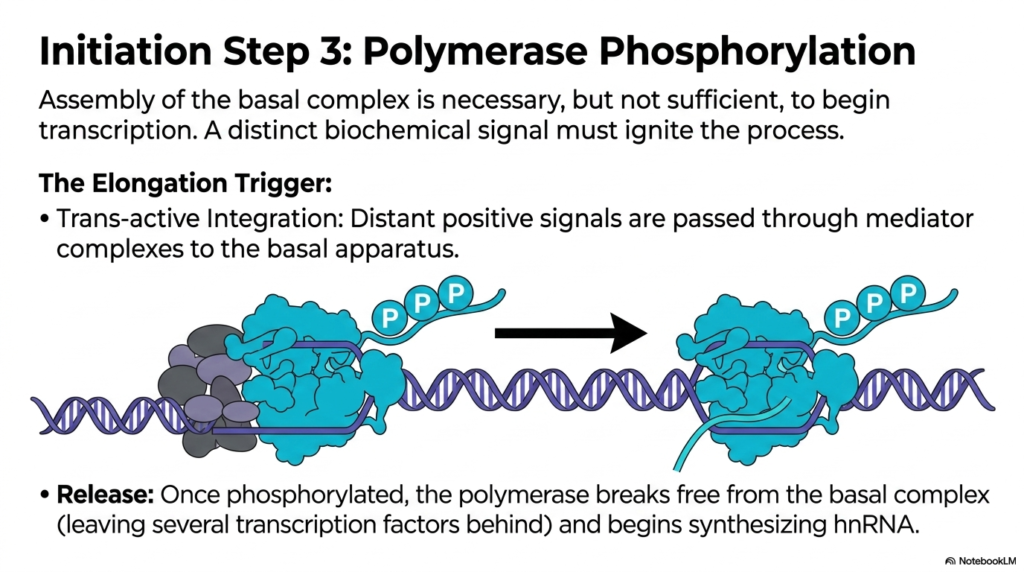
Even after the massive basal complex is fully assembled over the TATA box, the process is stalled. The physical assembly of these proteins is strictly necessary, but it is entirely insufficient to actually begin eukaryotic transcription. A distinct, powerful biochemical signal must be provided to ignite the polymerase engine and drive it forward. This elongation trigger involves trans-active integration, where distant positive biological signals are passed through massive mediator complexes down directly to the basal apparatus to initiate eukaryotic transcription.
The critical biochemical event that serves as the trigger is the phosphorylation of the polymerase. The source image clearly illustrates multiple phosphate groups being attached to the extended tail of RNA Polymerase II. Standard biochemical texts specify that it is actually the TFIIH complex, acting as a kinase, that rapidly adds these phosphates to the polymerase tail. This influx of negative charge dramatically alters the structural conformation of the enzyme, fundamentally changing its affinity for the initiation factors during eukaryotic transcription.
Once heavily phosphorylated, the polymerase violently breaks free from the restrictive basal complex. It leaves several of the general transcription factors, like TFIID, behind on the promoter while it propels itself forward along the DNA track. This physical release marks the official transition from the initiation phase to the elongation phase of eukaryotic transcription, allowing the newly liberated enzyme to begin rapidly synthesizing the heterogeneous nuclear RNA strand.
This phosphorylation checkpoint is a brilliant evolutionary adaptation. It ensures that eukaryotic transcription only proceeds when the cell has fully integrated all metabolic signals and actively committed to producing the transcript. Medical students will recognize that disrupting this specific kinase activity halts gene expression immediately, a mechanism often exploited by several natural cellular inhibitors to freeze cell growth. Understanding this trigger is fundamental to mastering the dynamics of eukaryotic transcription.
Slide 9: The Elongation Phase of Eukaryotic Transcription
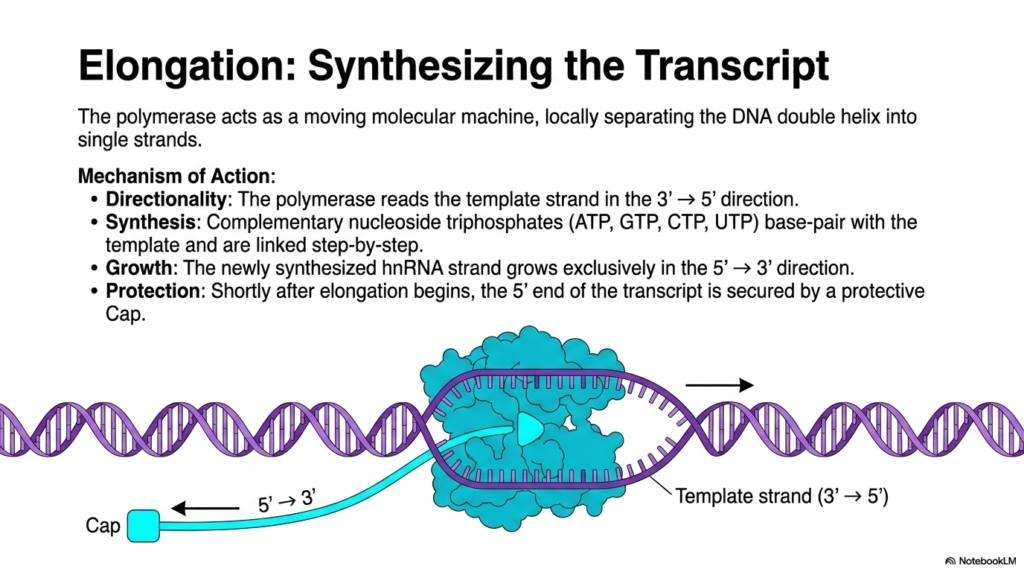
During the elongation phase, RNA Polymerase II transforms into an incredibly rapid, moving molecular machine. It physically travels along the template, locally separating the DNA double helix into single strands just ahead of its active site and rewinding them immediately behind it. A strict rule of directionality governs eukaryotic transcription: the polymerase must read the exposed DNA template strand exclusively in the three-prime to five-prime direction. This unidirectional movement ensures the genetic code is interpreted correctly and consistently every single time eukaryotic transcription occurs.
The actual mechanism of synthesis relies on complementary base pairing. Free-floating nucleoside triphosphates, specifically ATP, GTP, CTP, and UTP, enter the enzyme and base-pair with the exposed DNA template. The polymerase then catalyzes the formation of phosphodiester bonds, linking these nucleotides step-by-step. Because the template is read backward, the newly synthesized hnRNA strand grows exclusively in the five-prime to three-prime direction. This fundamental biochemical antiparallel mechanism is an absolute universal constant not just in eukaryotic transcription, but in virtually all nucleic acid synthesis.
To ensure the survival of the new genetic message, protection mechanisms are deployed almost instantly. The slide highlights that shortly after elongation begins and the new RNA strand emerges from the polymerase, the five-prime end of the transcript is secured by a specialized protective cap. Standard biochemistry confirms this is a heavily modified guanine nucleotide. This immediate capping is a defining feature of eukaryotic transcription, safeguarding the fragile transcript from ubiquitous cellular exonucleases that would otherwise destroy the bare RNA sequence.
The speed and precision of this elongation phase are truly staggering. As the massive enzyme slides continuously along the DNA track, it constantly proofreads its work, ensuring the high fidelity of eukaryotic transcription. This molecular manufacturing process highlights the elegant mechanical principles that underpin all living systems, seamlessly translating static genetic code into dynamic, functional chemical messengers ready for cellular use.
Slide 10: Concluding Eukaryotic Transcription: The Termination Phase
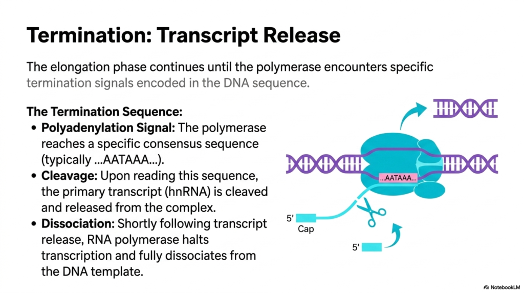
The high-speed elongation phase cannot continue indefinitely; it must be precisely halted to create a transcript of the exact correct size. Eukaryotic transcription continues unabated until the speeding polymerase enzyme encounters specific, hard-coded termination signals physically embedded directly within the DNA template sequence. The visual identifies the primary termination sequence as the polyadenylation signal. When the polymerase reaches this highly conserved consensus sequence, which typically reads AATAAA, it serves as an uncompromising molecular stop sign for the entire eukaryotic transcription machinery.
Encountering this signal triggers a dramatic sequence of events. Upon reading the AATAAA sequence, the primary hnRNA transcript is rapidly cleaved. Specific endonucleases cut the RNA strand, physically detaching the completed genetic message from the polymerase complex. This cleavage event marks the definitive end of the primary synthesis phase of eukaryotic transcription. It also simultaneously creates the perfect substrate for the subsequent addition of the protective three-prime poly-A tail, a modification essential for the RNA’s eventual journey to the ribosomes.
Following the cleavage and release of the transcript, the RNA polymerase is left transcribing a useless, uncapped tail of RNA. Shortly following the primary transcript release, the RNA polymerase completely halts its activity. It undergoes structural changes that cause it to fully dissociate from the DNA template. Standard knowledge beyond the slide indicates that specialized proteins actively dismantle the remaining polymerase complex, allowing the enzyme to be recycled for another complete round of eukaryotic transcription at a different promoter site.
Understanding this termination phase is vital for appreciating how eukaryotic cells prevent the wasteful, dangerous over-transcription of the genome. By utilizing specific sequence landmarks to dictate strict end boundaries, eukaryotic transcription ensures that cellular energy and raw nucleotide resources are meticulously conserved. The successfully released transcript is now ready to finalize its maturation processes, while the dissociated polymerase awaits its next biological assignment.
Slide 11: Regulating Eukaryotic Transcription: Defining Cellular Identity
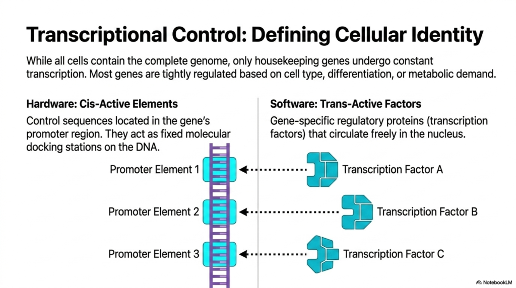
One of the most profound biological realities is that while all cells contain the exact same complete genome, they exhibit radically different forms and functions. This differentiation is entirely governed by the strict control of eukaryotic transcription. Only basic housekeeping genes undergo constant, unregulated transcription to maintain baseline cellular survival. The vast majority of genes are tightly regulated based on specific cell types, stage of differentiation, or immediate metabolic demand. This selective eukaryotic transcription is what allows a neuron to behave differently than a liver cell despite possessing identical DNA.
The slide elegantly divides this regulatory system into hardware and software. The hardware consists of cis-active elements. These are specific control sequences located directly within the gene’s own promoter region. Because they are physically built into the DNA strand, they act as permanent, fixed molecular docking stations. Without these localized cis-elements, eukaryotic transcription would lack the target sites necessary to organize regulatory complexes. They form the static infrastructure that allows specific genes to be recognized by the dynamic cellular environment.
The software of this system comprises trans-active factors. These are gene-specific regulatory proteins, commonly called transcription factors, that circulate freely within the nucleoplasm. Unlike the fixed DNA elements, these proteins are mobile and act as the dynamic decision-makers for eukaryotic transcription. Transcription Factor A, B, or C will recognize and bind to their corresponding Promoter Elements. External biochemical logic dictates that the specific combination and concentration of these freely floating factors at any given moment determines whether eukaryotic transcription will be heavily activated or completely suppressed.
The interaction between the permanent DNA hardware and the mobile protein software creates an incredibly versatile regulatory network. This precise mechanism of eukaryotic transcription allows the organism to rapidly adapt to environmental changes, utilizing a single genomic library to execute thousands of distinct, cell-specific biological programs throughout its lifespan.
Slide 12: Metabolic Networking in Eukaryotic Transcription: The PEP-CK Promoter
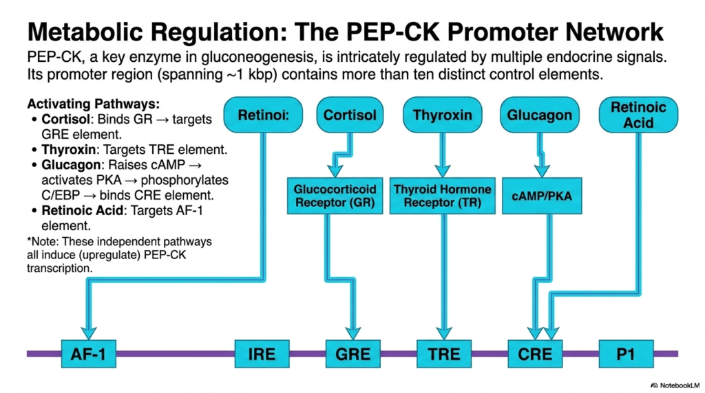
To understand the sheer complexity of eukaryotic transcription, the structural analysis returns to the PEP-CK model. PEP-CK is a key enzyme driving gluconeogenesis, the creation of new glucose. Because glucose homeostasis is critical for survival, this gene is intricately regulated by multiple, simultaneous endocrine signals. The slide illustrates a massive promoter network spanning approximately one kilobase, containing more than ten distinct control elements. This demonstrates that eukaryotic transcription is not controlled by a simple on-off switch, but rather by a highly sophisticated biological rheostat that measures diverse systemic inputs.
The visual maps out five major activating pathways that converge on this promoter to upregulate eukaryotic transcription. Hormones like Retinol and Retinoic Acid target the AF-1 elements. Cortisol binds directly to the Glucocorticoid Receptor, which then docks at the GRE element. Thyroxin utilizes the Thyroid Hormone Receptor to target the TRE element. Standard endocrinology pathways explain that Glucagon acts indirectly; it raises cellular cAMP levels, which activates PKA, leading to the phosphorylation of C/EBP proteins that ultimately bind the CRE element. All these independent pathways act to powerfully induce eukaryotic transcription of PEP-CK.
It is essential to recognize the physical layout of these cis-active elements on the DNA string. The AF-1, IRE, GRE, TRE, and CRE sites are arranged sequentially along the promoter. This spatial organization allows multiple different transcription factors to bind simultaneously without physically interfering with one another. This multi-receptor binding capability is a hallmark of complex eukaryotic transcription, enabling the cell to require multiple activating signals before committing to the energetic expense of synthesizing metabolic enzymes.
Students should view this promoter not just as a static sequence, but as a sophisticated biological sensor. By requiring varied inputs like cortisol and glucagon to initiate eukaryotic transcription, the body ensures that gluconeogenesis only occurs during systemic fasting or severe stress, perfectly matching gene expression to whole-body physiological needs.
Slide 13: Signal Integration in Eukaryotic Transcription: The CBP/p300 Complex
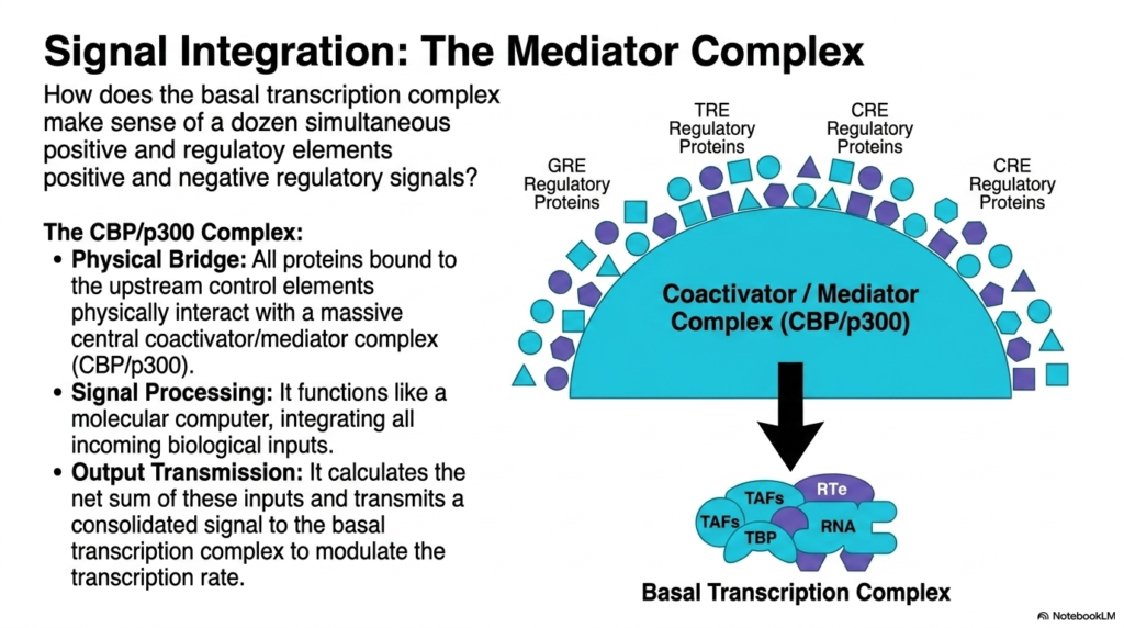
A fundamental question in molecular biology is how the basal machinery makes sense of a dozen simultaneous positive and negative regulatory signals. The answer lies in massive signal integration structures central to eukaryotic transcription. The slide highlights the CBP/p300 complex, which acts as the ultimate intermediary between the dozens of trans-active regulatory proteins bound upstream and the basal transcription complex sitting on the TATA box. Without this massive integrating structure, eukaryotic transcription would be chaotic, unable to prioritize conflicting metabolic demands.
The CBP/p300 complex functions first as a massive physical bridge. All the diverse regulatory proteins bound to the upstream control elements, such as the GRE and CRE binding factors, physically reach out and interact with this central coactivator. This physical contact is absolutely essential for signal transduction during eukaryotic transcription. Secondly, the complex acts as a biological signal processor. It functions much like a molecular computer, structurally shifting and modifying itself to integrate all the incoming biochemical inputs from the various hormonal pathways.
Finally, the mediator complex is responsible for output transmission. It essentially calculates the net sum of all positive and negative inputs and transmits a single consolidated signal directly to the basal transcription complex. External literature confirms that CBP/p300 possesses intrinsic histone acetylase activity, further aiding in chromatin uncoiling. By delivering this unified command, the mediator complex directly dictates the final firing rate of the RNA polymerase, perfectly modulating the speed and frequency of eukaryotic transcription to match the precise biochemical consensus of the cell.
The concept of the mediator complex is incredibly crucial for advanced medical biochemistry. It elegantly explains how a human cell can experience competing signals, such as simultaneous starvation and stress hormones, and still produce a perfectly graded, biochemically calibrated response. By utilizing massive integrating complexes like CBP/p300, the process of eukaryotic transcription achieves a profound level of computational logic that rivals the processing power of modern synthetic microprocessors.
Slide 14: Repressing Eukaryotic Transcription: Insulin’s Inhibitory Mechanism
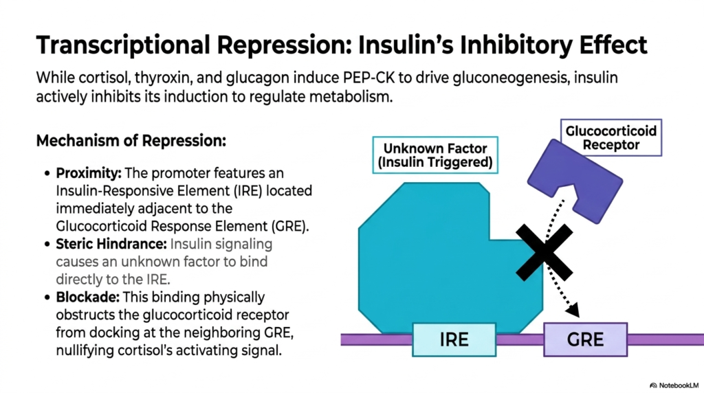
While multiple pathways activate the PEP-CK gene to drive gluconeogenesis, there must be a mechanism to turn this process off after a meal. This highlights the vital role of active repression in eukaryotic transcription. Insulin aggressively counteracts the effects of cortisol, thyroxin, and glucagon, actively inhibiting the induction of PEP-CK to regulate overall metabolism. This dynamic interplay showcases how eukaryotic transcription can be rapidly shut down to prevent the dangerous overproduction of glucose when the body is already in a fed, high-energy state.
The mechanism of this profound repression relies entirely on the physical proximity of the regulatory elements. The PEP-CK promoter features an Insulin-Responsive Element, or IRE, located immediately adjacent to the Glucocorticoid Response Element, or GRE. When insulin signaling pathways are triggered, they cause an unknown, massive regulatory factor to bind directly to this IRE site. In the realm of eukaryotic transcription, physical location is everything, and the strategic placement of the IRE site is a deliberate evolutionary design meant to facilitate direct protein competition.
The binding of this insulin-triggered factor creates severe steric hindrance. The massive physical bulk of the protein bound at the IRE structurally obstructs the adjacent GRE site. Because of this localized blockade, the glucocorticoid receptor is physically prevented from docking at its neighboring element, completely nullifying cortisol’s strong activating signal. Standard biochemistry texts use this exact model to teach how localized physical blockades act as incredibly dominant off-switches in eukaryotic transcription, proving that repressive factors can easily override multiple activating pathways through sheer structural interference.
Understanding this specific mechanism is fundamental for students studying diabetes and metabolic syndrome. The failure of insulin to properly exert this steric hindrance results in runaway gluconeogenesis, a hallmark of diabetic pathology. It perfectly demonstrates how a microscopic physical blockade in eukaryotic transcription can dictate macro-level human disease states.
Slide 15: Synthesizing the Mechanisms of Eukaryotic Transcription
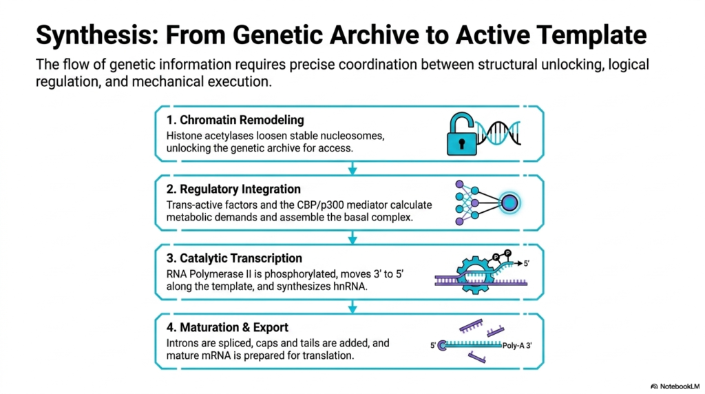
The flow of genetic information requires the precise coordination of multiple complex biochemical phases. This final slide provides a masterful synthesis of the entire process, demonstrating that eukaryotic transcription relies upon structural unlocking, logical regulation, and mechanical execution. The first mandatory step is chromatin remodeling. Here, histone acetylases chemically loosen stable nucleosomes, effectively unlocking the genetic archive so the underlying DNA sequences can be physically accessed. Without this initial structural breach, the entire system of eukaryotic transcription remains permanently paralyzed in a repressed state.
Once the DNA is exposed, the second phase involves deep regulatory integration. Trans-active factors bind to the promoter and utilize the massive CBP/p300 mediator complex to calculate complex metabolic demands. This computational phase ensures that eukaryotic transcription only occurs when absolutely physiologically necessary, ultimately resulting in the assembly of the basal complex over the TATA box. Following assembly and phosphorylation, the third phase initiates: catalytic transcription. The massive RNA Polymerase II engine moves three-prime to five-prime along the template, mechanically synthesizing the raw hnRNA strand.
Finally, the raw genetic output must be refined through maturation and export. The bulky hnRNA undergoes extensive processing where non-coding introns are rapidly spliced out. Protective chemical caps and poly-A tails are added to the extremes of the molecule, and the resulting mature mRNA is carefully prepared for translation. Standard biochemical frameworks dictate that this final modification phase is deeply coupled with the mechanical action of eukaryotic transcription itself, ensuring a seamless pipeline from static DNA code to dynamic, export-ready molecular messenger.
By mastering these four integrated steps, students gain a comprehensive understanding of cellular biology. Eukaryotic transcription is not merely a single chemical reaction, but an elegant, multi-stage industrial process that carefully translates the inert genetic archive into the vital proteins that sustain all complex life.
Please read our Content Disclaimer Statement.
Check out our social media channels: