91. Principles of DNA Cloning: A Biochemical Overview
The ability to rewrite genetic instructions represents a monumental leap in biological science. The core purpose of this slide deck is to demystify these complex biochemical mechanisms for emerging medical professionals and scientists. By analyzing each step, students will master the structural and functional principles of DNA cloning. Through this exploration of DNA cloning, the intricate dance of microscopic molecules becomes clear. Mastering DNA cloning opens the door to creating life-saving therapeutics and driving modern genetic innovation.
Slide 1: Principles of DNA Cloning: Decoding the Blueprint of Life
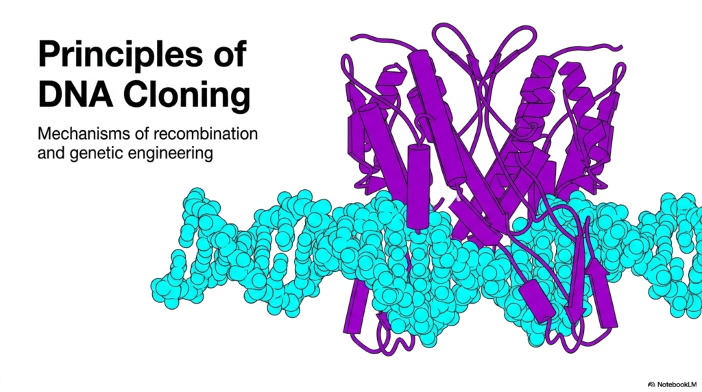
The visual foundation of this slide illustrates a precise physical interaction between a protein and a nucleic acid. A vivid purple protein structure, representing an active enzyme, intimately interacts with a bright cyan double-stranded nucleic acid molecule. This imagery captures the exact physical binding required to alter genetic codes. The entire process relies on these biological machines successfully locating and firmly binding to specific target sequences along the helix.
Understanding this physical interaction is crucial for mastering genetic engineering. The visual depicts a specialized protein folding perfectly around the double helix. The alpha helices and beta sheets of the protein create a unique structural shape that fits directly into the structural grooves of the genetic material. This precise structural alignment ensures that the enzyme acts only at the correct location, avoiding random alterations to the genetic code.
This foundational principle sets the stage for everything that follows in the laboratory. By grasping how these biological macromolecules interact on a physical level, researchers can design complex experiments that selectively isolate individual genes. The image serves as a powerful visual reminder that all advanced biotechnology begins with the elegant, physical reality of molecular binding and specific enzymatic action across the genome.
The implications of these structural interactions extend far beyond basic research. When the enzyme locks onto its target sequence, it triggers a precise biochemical cascade that scientists harness for targeted modifications. Recognizing this specific interaction paves the way for advanced methodologies in gene therapy and diagnostics. A solid grasp of this initial recognition event is the first necessary step in understanding the entire molecular workflow.
Slide 2: The Three-Pillar Methodology of DNA Cloning: Reshaping Biological Potential
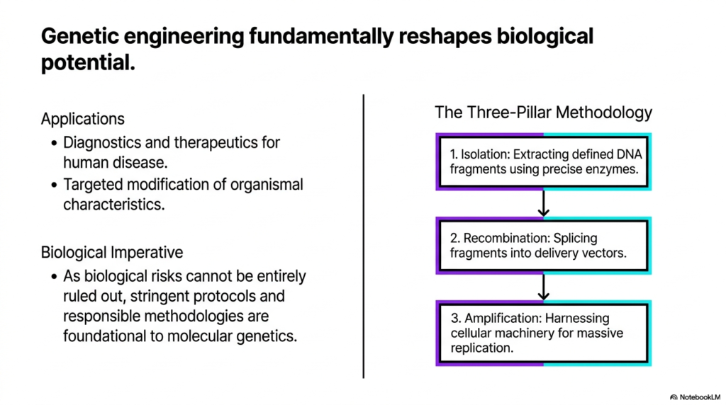
The quest to cure disease begins with understanding how to manipulate fundamental biological building blocks. The core purpose of this slide deck is to break down these exact methodologies for college and medical students. By exploring the overarching workflow of DNA cloning, learners will see how theoretical concepts translate into laboratory reality. Understanding DNA cloning involves mastering three distinct phases. DNA cloning empowers scientists to create targeted modifications that fundamentally reshape what biological organisms can achieve.
This slide outlines the overarching workflow, categorized into a three-pillar methodology that serves as an operational roadmap. The first essential pillar is isolation. During this phase, precise biological enzymes act to extract highly specific fragments of genetic material from a much larger, complex genome. This isolation step is crucial because researchers must target and secure exact genetic sequences without interference from surrounding irrelevant biological material.
Following isolation comes the critical recombination phase. In this subsequent step, the previously extracted genetic fragments are carefully spliced into specialized delivery vectors. These microscopic vectors function as biological transport vehicles, safely carrying the desired sequence into a new cellular environment. This seamless integration ensures the new host cell can read, interpret, and process the introduced genetic instructions as if they natively belonged to the organism itself.
The final operational pillar highlights amplification, the phase where biological scale is achieved. Once the engineered delivery vector successfully enters a host cell, the host’s natural cellular machinery takes over the heavy molecular lifting. The host cell begins massive, continuous replication of the inserted genetic material alongside its native genome. This relentless multiplication yields vast quantities of the targeted sequence for future laboratory use.
Together, these three pillars form the absolute backbone of modern genetic engineering. Moving from initial isolation through recombination to amplification provides a scalable, reliable method for producing necessary biological materials. This structured approach guarantees that researchers can produce enough of a specific gene or protein to conduct meaningful experiments, run diagnostic tests, or manufacture life-saving pharmaceutical treatments.
Slide 3: Restriction Endonucleases in DNA Cloning: Molecular Scalpels at Work
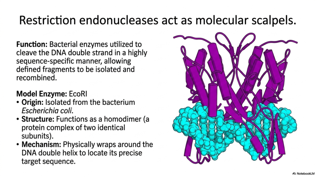
The precision of modern surgery is nothing compared to the exactness of a molecular scalpel. The core purpose of this slide deck is to elucidate the vital biochemical tools used in this exact science. By focusing on restriction endonucleases, students will learn how enzymes drive the mechanics of DNA cloning. The success of DNA cloning relies entirely on these microscopic tools. Without them, the targeted manipulation required in DNA cloning would be completely impossible.
This slide introduces restriction endonucleases, specifically focusing on the model enzyme EcoRI. These bacterial enzymes function to cleave the double strand in a highly sequence-specific manner. EcoRI originates from the common bacterium Escherichia coli, where it naturally serves as a defense mechanism against invading viruses. In the laboratory setting, scientists repurpose this natural defense system to isolate defined fragments for future genetic recombination.
Structurally, EcoRI operates as a homodimer, meaning it is a protein complex composed of two identical subunits working in perfect synchronization. The visual representation highlights this dimeric structure wrapping around the target molecule. This physical wrapping is not a passive event; it is an active scanning process. The homodimer physically interacts with the double helix, feeling along the chemical structure until it locates its precise target sequence.
The mechanism of action relies on an incredible level of molecular recognition. The two identical subunits of the homodimer ensure that both strands of the double helix are engaged simultaneously. When the enzyme locates its specific binding site, it undergoes a conformational change. This shape shift positions the catalytic centers of the enzyme perfectly over the chemical bonds that need to be severed, ensuring maximum precision.
Understanding the structure and function of these molecular scalpels is paramount for any biochemical student. By utilizing these enzymes, researchers can predictably and reliably cut genetic material at known locations. This predictability is fundamental to piecing different biological fragments together. The elegant structure of EcoRI demonstrates how form dictates function at the absolute smallest scales of biology.
Slide 4: EcoRI Target Sequences in DNA Cloning: Finding the Perfect Palindrome
Imagine reading a sentence that says the exact same thing forward and backward; nature uses this same trick for molecular precision. The core purpose of this slide deck is to highlight the specific signals that guide biochemical processes. By examining target sequences in DNA cloning, students will grasp the concept of genetic symmetry. The entire practice of DNA cloning depends on finding these sequences. In DNA cloning, symmetry provides the exact location for enzymatic action.
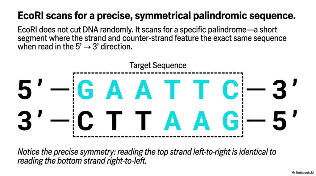
This slide details the specific target sequence that the EcoRI enzyme seeks out. EcoRI does not cut randomly along the genetic strand. Instead, it scans for a precise, symmetrical palindromic sequence. A biological palindrome occurs when a short segment of the strand and its counter-strand feature the exact same sequence when read in the standard five-prime to three-prime direction.
The visual breaks down the exact target sequence for EcoRI, which is GAATTC. When reading the top strand from left to right, the sequence is GAATTC. Remarkably, when reading the bottom strand from right to left, the sequence is identical. This precise symmetry is not a coincidence; it is a structural necessity that matches the symmetrical homodimer structure of the enzyme itself.
Because the enzyme is made of two identical halves, it requires a binding site that also presents an identical face on both sides. The identical subunits of EcoRI bind to the identical halves of the palindromic sequence. This matching of a symmetrical protein to a symmetrical nucleic acid ensures that the enzyme is perfectly centered over the target area before any cutting takes place.
Recognizing palindromic sequences is a foundational skill in molecular genetics. Researchers study these specific sequences to map out where a particular enzyme will make its cuts across an entire genome. By cataloging these palindromes, scientists can select the most appropriate molecular scalpel for their specific experimental needs, ensuring that they isolate the exact genetic fragment required for their unique work.
Slide 5: Enzymatic Cleavage in DNA Cloning: Breaking the Phosphodiester Bond
Severing the backbone of life requires incredible chemical force applied at a microscopic focal point. The core purpose of this slide deck is to explain the fundamental chemical reactions that make genetic manipulation possible. By studying enzymatic cleavage in DNA cloning, students will understand the actual chemical bonds being broken. DNA cloning hinges on this specific hydrolytic reaction. Every successful procedure in DNA cloning requires this exact bond separation.
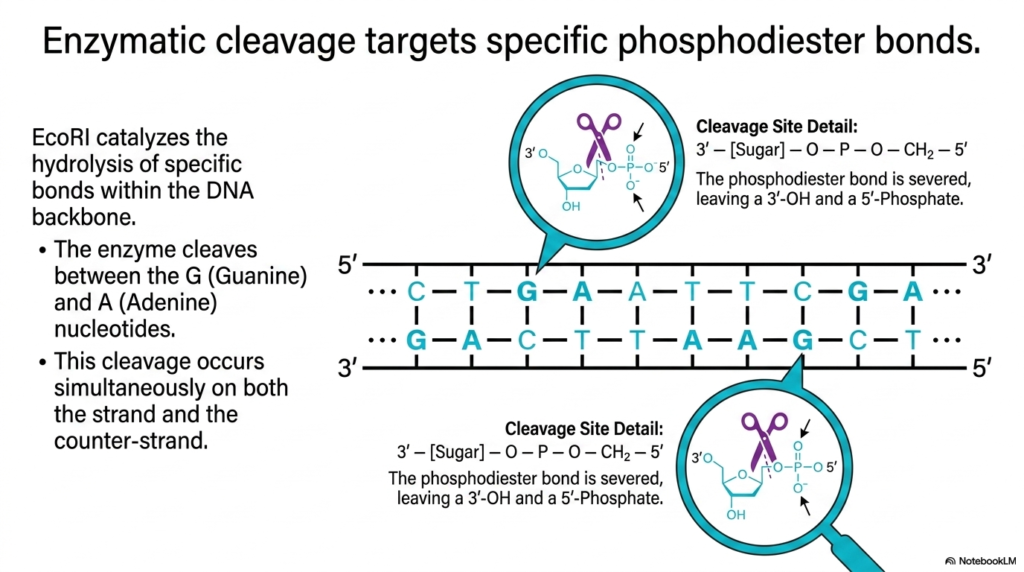
This slide focuses on the exact chemical event catalyzed by the EcoRI enzyme. The enzyme functions by targeting and breaking specific phosphodiester bonds within the structural backbone. The visual details how EcoRI catalyzes the hydrolysis of the bond situated directly between the Guanine and Adenine nucleotides. This cleavage does not happen on just one side; it occurs simultaneously on both the strand and the counter-strand.
The microscopic view provided on the slide highlights the anatomy of the cleavage site. A phosphodiester bond links the sugar of one nucleotide to the phosphate group of the next. The enzyme introduces a water molecule to sever this specific connection. Once the bond is successfully broken, the reaction leaves behind a distinct chemical signature: a three-prime hydroxyl group and a five-prime phosphate group.
Understanding the exact nature of the severed bond is essential for later steps in the process. The chemical state of the newly exposed ends dictates how they will interact with other molecules. Because the enzyme consistently leaves a specific hydroxyl and phosphate arrangement, researchers can predict exactly how these ends will behave when introduced to new genetic fragments. The chemistry is entirely standardized.
This standardized chemical reaction is a marvel of biological engineering. By executing this precise hydrolysis, the enzyme creates reliable, reproducible endpoints. Students must recognize that genetic engineering is not just moving letters around on a page; it involves concrete chemical reactions breaking robust covalent bonds. Mastering this biochemical reality provides a deeper appreciation for the mechanics of molecular biology.
Slide 6: Staggered Cuts and Sticky Ends in DNA Cloning: The Chemistry of Connection
Creating a puzzle piece is useless unless it has the perfect complementary shape to connect with another. The core purpose of this slide deck is to reveal how biological fragments are engineered to fit together perfectly. By examining staggered cuts in DNA cloning, medical students will understand the basis of molecular reassembly. DNA cloning utilizes these sticky ends to create custom constructs. Without these cohesive overhangs, DNA cloning would lack complete efficiency.
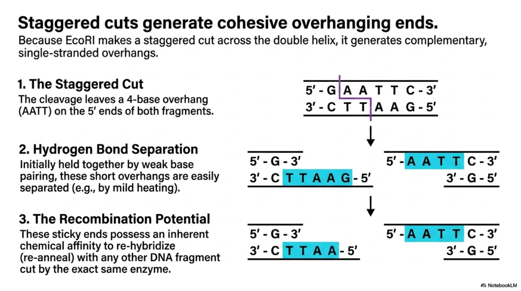
This slide explains the concept of staggered cuts and the resulting cohesive overhanging ends. Because the EcoRI enzyme makes a staggered cut diagonally across the double helix rather than a blunt cut straight through, it generates complementary, single-stranded overhangs. The visual demonstrates how the cleavage leaves a specific four-base overhang, specifically AATT, on the five-prime ends of both newly separated fragments.
Initially, the two strands are held together by weak hydrogen base pairing. Following the staggered cut, these short overhangs can be easily separated by applying mild heat. Once separated, these exposed single-stranded sequences are colloquially referred to as sticky ends. They are considered sticky because they possess an inherent chemical affinity to re-hybridize or re-anneal with complementary single-stranded sequences.
The recombination potential of these sticky ends is the driving force behind assembling new genetic combinations. Because they are driven by natural base-pairing rules, an AATT overhang will naturally seek out and bind to a TTAA overhang. This means that any genetic fragment cut by the exact same EcoRI enzyme will feature compatible sticky ends, allowing them to connect flawlessly.
This universal compatibility allows scientists to mix and match genetic material from entirely different organisms. As long as both sources are prepared with the same specific enzyme, their sticky ends will find one another and temporarily bond. Understanding this inherent chemical affinity explains how researchers can insert human genes into bacterial systems, bridging the gap between vastly different biological kingdoms.
Slide 7: Plasmids in DNA Cloning: The Ideal Delivery Vectors
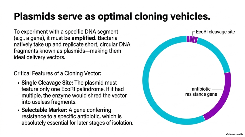
Delivering a fragile message across a hostile environment requires a robust and specialized vehicle. The core purpose of this slide deck is to demonstrate how scientists safely transport genetic material into new host cells. By focusing on plasmids in DNA cloning, students will learn the anatomy of effective biological vectors. DNA cloning relies heavily on these circular molecules. An optimized vector is the cornerstone of successful DNA cloning.
This slide introduces plasmids as the optimal cloning vehicles for genetic engineering. To experiment with a specific genetic segment, it must be successfully transported and amplified within a host. Bacteria natively take up and replicate short, circular genetic fragments known as plasmids. By hijacking this natural bacterial process, scientists utilize these circular structures as ideal delivery vectors for custom genetic payloads.
The visual outlines the critical features required for a plasmid to function as an effective cloning vector. The first vital feature is a single cleavage site. The plasmid must feature only one specific EcoRI palindrome. If the circular plasmid contained multiple target sites, the enzyme would shred the vector into multiple useless fragments. A single site ensures the circle simply opens to accept new material.
The second absolutely critical feature is the inclusion of a selectable marker. This is typically a gene that confers resistance to a specific antibiotic. When the plasmid is internalized by a bacterium, this marker allows the bacterium to survive exposure to that drug. This resistance mechanism is absolutely essential for later stages of the process, allowing researchers to isolate successful subjects.
Understanding plasmid architecture is fundamental to designing a successful biochemical experiment. Students must recognize that vectors are carefully engineered tools, not just random biological circles. The precise placement of the cleavage site and the selectable marker turns a simple bacterial anomaly into a powerful scientific instrument. This engineered simplicity makes mass genetic distribution entirely possible.
Slide 8: DNA Ligase in DNA Cloning: Sealing the Recombinant Construct
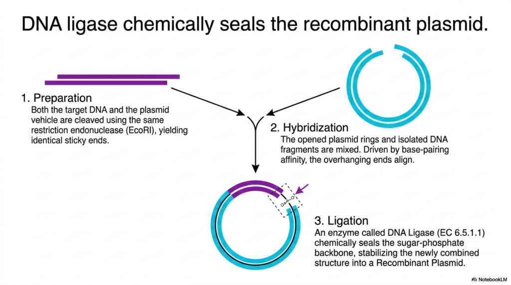
Even the most perfectly aligned puzzle pieces require a permanent chemical glue to stay together forever. The core purpose of this slide deck is to detail the exact enzymes responsible for finalizing genetic modifications. By studying DNA ligase within DNA cloning, students will grasp how transient connections become permanent structures. DNA cloning requires this final sealing step. Without DNA ligase, the constructs made during DNA cloning would completely fall apart.
This slide details the critical role of DNA ligase in chemically sealing the new recombinant plasmid. The process begins with preparation, where both the target sequence and the plasmid vehicle are cleaved using the identical restriction endonuclease. This preparation ensures that both components possess identical, complementary sticky ends, setting the stage for a seamless molecular connection between the two separate pieces.
The second phase is hybridization. The opened plasmid rings and the isolated genetic fragments are mixed together in a controlled environment. Driven by natural base-pairing affinity, the overhanging sticky ends align and form weak hydrogen bonds. While this alignment brings the pieces together perfectly, the connection remains structurally fragile. The sugar-phosphate backbone remains broken, requiring a final catalytic step for lasting stability.
The final phase is ligation. An enzyme called DNA Ligase chemically seals the breaks in the sugar-phosphate backbone. It catalyzes the formation of new phosphodiester bonds, bridging the gap between the adjacent nucleotides. This enzymatic action permanently locks the inserted fragment into the vector, stabilizing the newly combined structure into a robust, complete recombinant plasmid ready for cellular delivery.
Mastering the function of ligase completes the understanding of the initial engineering phase. While endonucleases act as molecular scalpels to cut material, ligase acts as the molecular glue. Students should appreciate that this enzyme requires cellular energy to forge these strong covalent bonds. The successful creation of a permanent, circular recombinant molecule marks the transition from chemical preparation to biological application.
Slide 9: Cellular Transformation in DNA Cloning: Internalizing the Blueprint
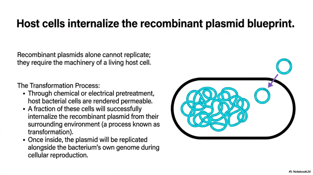
Creating a brilliant architectural blueprint means nothing if the construction crew never receives the plans. The core purpose of this slide deck is to explain how engineered genetic instructions are actually delivered to the living biological factories. By exploring transformation in DNA cloning, students will uncover how cells adopt foreign material. DNA cloning mandates this physical transfer of information. Transformation bridges the chemical and biological phases of DNA cloning.
This slide explains the critical transformation process where host cells internalize the recombinant plasmid blueprint. Recombinant plasmids are simply inert chemical constructs on their own; they cannot replicate without the complex machinery of a living host cell. Therefore, researchers must coax living bacteria into adopting these foreign circular vectors from their surrounding environment, a biological process formally known as transformation.
The visual highlights the mechanics of this cellular uptake. Bacterial cells naturally resist foreign material, so they must be rendered permeable through a pretreatment process. Scientists use chemical exposure or precise electrical shocks to temporarily weaken the bacterial membrane. These brief disturbances create microscopic pores, allowing a small fraction of the cells to successfully internalize the recombinant plasmids floating in the surrounding solution.
Once inside the host, the internal dynamics change entirely. The internalized plasmid is recognized by the bacterial machinery and is replicated alongside the bacterium’s own primary genome during regular cellular reproduction. The host cell essentially adopts the engineered blueprint as its own, dedicating its cellular resources to maintaining and expressing the newly introduced genetic instructions without distinguishing them from native material.
Understanding transformation highlights the delicate balance between chemistry and living biology. The inefficiency of the process dictates that massive amounts of cells and plasmids must be used to achieve success. Students must recognize that overcoming the cellular membrane barrier is one of the most significant challenges in genetic engineering. Successful internalization is the absolute prerequisite for subsequent biological manufacturing.
Slide 10: Antibiotic Selection in DNA Cloning: The Biological Filter
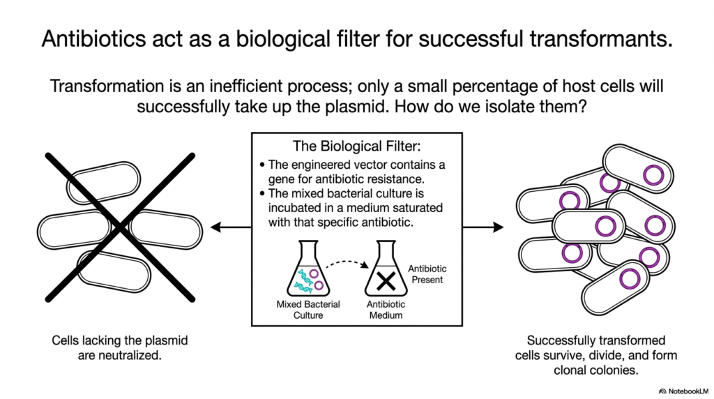
Finding a needle in a microscopic haystack requires a filter that simply burns away all the hay. The core purpose of this slide deck is to showcase the ingenious methods used to isolate microscopic success. By examining antibiotic selection in DNA cloning, students will learn how researchers leverage survival mechanics. DNA cloning relies on this harsh biological filter. Without selection, finding the correct cell in DNA cloning would be completely impossible.
This slide details how antibiotics act as a biological filter to identify successful transformants. Because transformation is an inherently inefficient process, only a minute percentage of the host cells will successfully take up the engineered plasmid. This creates a massive mixture of millions of empty cells alongside a few valuable modified cells. Researchers need a definitive method to isolate only the successful candidates.
The biological filter relies entirely on the selectable marker engineered into the plasmid. The vector contains a specific gene conferring antibiotic resistance. The entire mixed bacterial culture is transferred to an incubation medium saturated with that exact antibiotic. This creates a harsh, toxic environment for any standard, unmodified bacterium, applying massive selective pressure to the entire microscopic population simultaneously.
The visual clearly demonstrates the binary outcome of this exposure. The cells lacking the engineered plasmid are rapidly neutralized by the antibiotic drug, completely clearing them from the culture. In contrast, the successfully transformed cells utilize their newly acquired resistance gene to survive the toxic exposure. These surviving cells are free to divide and form visible, isolated clonal colonies.
This elegant application of selective pressure is a masterclass in biological engineering. By tying the desired genetic insert to a survival trait, scientists force the biology to sort itself out automatically. Students should grasp that this step is not about modifying cells, but strictly about identifying the rare successes. This harsh filtration guarantees that every surviving colony contains the necessary engineered blueprint.
Slide 11: Exponential Amplification in DNA Cloning: Mass Molecular Production
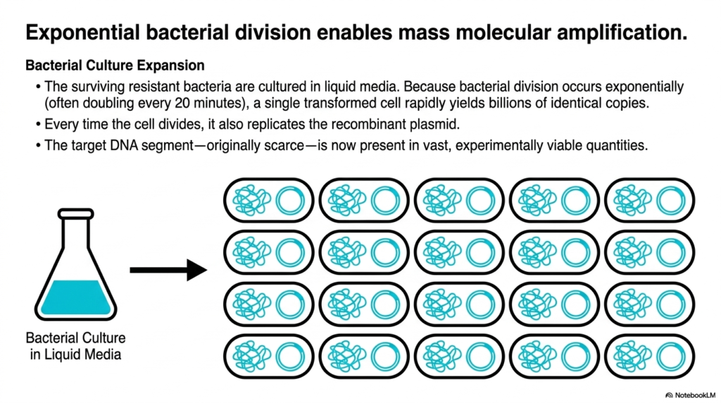
A single instruction manual is useless for a massive factory unless it can be rapidly copied for every worker. The core purpose of this slide deck is to explain how biological systems achieve massive scale. By observing bacterial amplification in DNA cloning, medical students will understand how microscopic alterations yield massive resources. DNA cloning harnesses natural exponential growth. This massive expansion is the ultimate goal of DNA cloning.
This slide illustrates how exponential bacterial division enables mass molecular amplification of the engineered construct. Once the surviving, resistant bacteria are successfully isolated, they are transferred into a nutrient-rich liquid culture. In this ideal environment, the bacteria begin to divide at an astonishing rate. Because bacterial division occurs exponentially, often doubling every twenty minutes, a single transformed cell rapidly yields billions of identical copies.
The visual depicts the rapid expansion of the bacterial culture inside the liquid media flask. With every single cellular division, the bacterium must copy all of its internal material, including the recombinant plasmid. Therefore, the replication of the host cell automatically drives the replication of the inserted genetic target. The host biology acts as a tireless, microscopic manufacturing plant.
The result of this exponential growth is a massive accumulation of the target material. The specific genetic segment, which was originally scarce and difficult to obtain, is now present in vast, experimentally viable quantities. By leveraging the natural reproductive speed of simple bacteria, scientists can generate enough raw genetic material to sequence, study, or utilize in widespread therapeutic applications.
Understanding this exponential biological scale is crucial for modern medicine. The production of life-saving treatments like synthetic insulin relies entirely on this exact amplification mechanism. Students must appreciate that the microscopic modifications made in earlier steps are ultimately magnified billions of times over. This step transforms a delicate molecular experiment into a robust industrial process capable of supporting global medical needs.
Slide 12: Plasmid Extraction and Excision in DNA Cloning: Retrieving the Product
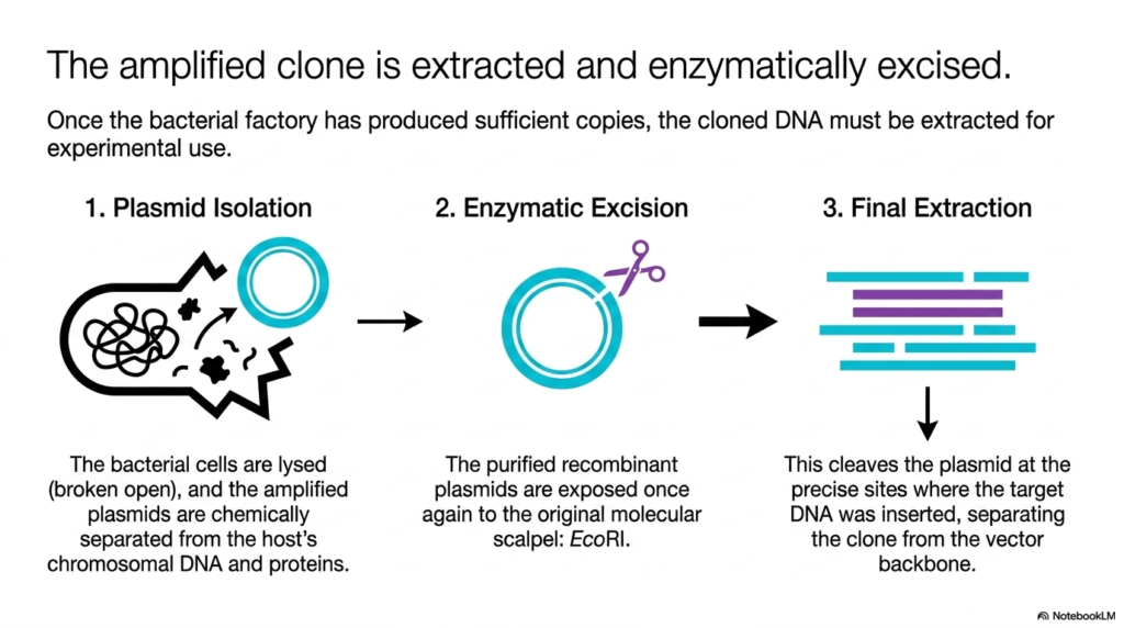
Manufacturing millions of products inside a factory means nothing if you cannot open the doors to ship them out. The core purpose of this slide deck is to reveal how the final genetic products are harvested from their biological hosts. By studying extraction techniques in DNA cloning, students will learn how to retrieve amplified materials. DNA cloning requires this destructive harvesting step. Excision finalizes the molecular phase of DNA cloning.
This slide details how the amplified genetic clone is extracted and enzymatically excised from the bacterial factory. Once the culture has produced sufficient copies through exponential division, the desired genetic material remains trapped inside billions of microscopic cellular walls. The first step to retrieve the product is plasmid isolation. The bacterial cells are chemically lysed, essentially broken open, releasing all internal contents.
Following cellular lysis, the amplified circular plasmids must be chemically separated from the host bacterium’s chromosomal debris and surrounding proteins. This purification yields a concentrated sample of just the recombinant vectors. The next phase, enzymatic excision, uses the same molecular scalpel as the first step: EcoRI. The purified plasmids are exposed to this enzyme to cut the circular structures open once again.
The final extraction occurs as the enzyme cleaves the plasmid at the precise flanking sites where the target sequence was originally inserted. This specific severing separates the amplified clone from the larger vector backbone. The result is a mixture containing the originally desired genetic fragment and the discarded biological vehicle, both present in massive quantities ready for physical separation.
Mastering this extraction process highlights the cyclical nature of genetic engineering. Students observe how the exact same enzymatic tools used to build the construct are deployed again to dismantle it. This controlled retrieval is essential for recovering the pure amplified gene. Understanding chemical lysis and enzymatic digestion equips future scientists with the practical knowledge required to harvest biological products successfully.
Slide 13: Gel Electrophoresis in DNA Cloning: Physical Separation by Size
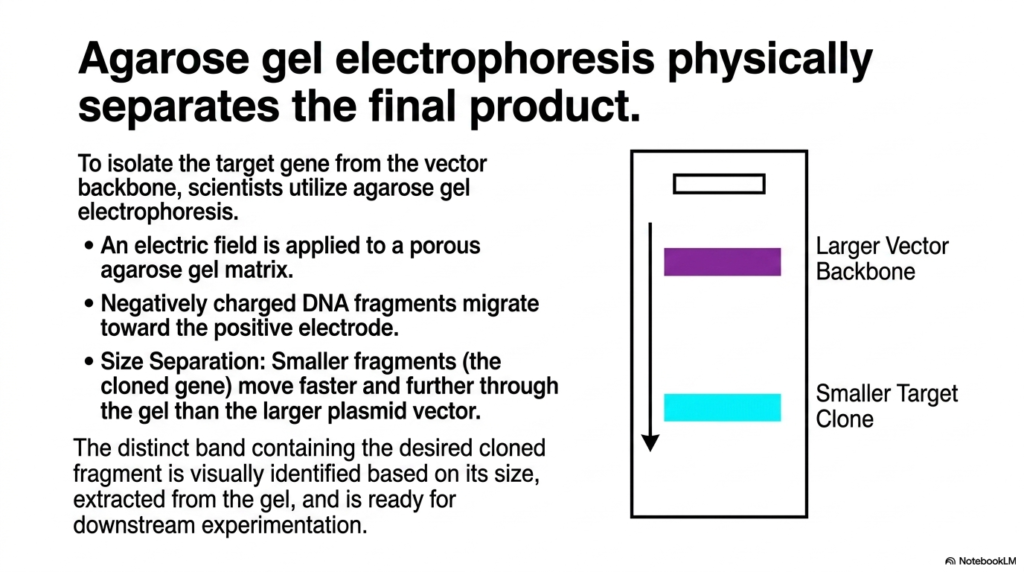
Sorting microscopic fragments mixed in a clear liquid requires a physical force that exploits their fundamental chemical properties. The core purpose of this slide deck is to explain the analytical techniques used to purify genetic material. By examining agarose gel electrophoresis in DNA cloning, students will grasp the mechanics of molecular sorting. DNA cloning relies on this electrical separation. Electrophoresis provides the final pure product in DNA cloning.
This slide demonstrates how agarose gel electrophoresis physically separates the final desired product from the molecular mixture. To isolate the target gene from the discarded vector backbone, scientists utilize this essential laboratory technique. An electric field is applied across a porous agarose gel matrix. Because the genetic backbone carries a uniform negative charge, the fragments are naturally propelled toward the positive electrode.
The visual highlights the principle of size separation as the fragments travel through the porous gel. The agarose acts like a microscopic sieve. The smaller fragments, which represent the isolated cloned gene, encounter less resistance and move much faster and further down the gel matrix. Conversely, the larger, bulky plasmid vector backbone gets tangled in the matrix and travels a much shorter distance.
This differential migration results in distinct physical bands forming within the gel. The distinct band containing the desired smaller fragment is easily identified visually based on its relative size and travel distance. Once identified, this specific area of the gel is physically cut out, and the pure amplified fragment is extracted, finally ready for complex downstream experimentation and therapeutic development.
Understanding electrophoresis is absolutely fundamental for any laboratory-based biochemical work. It provides a visual confirmation that the entire complex engineering process was successful. Students must realize that this physical sorting technique bridges the gap between invisible molecular chemistry and visible, workable physical samples. This elegant electrical separation guarantees the purity of the final biological product before clinical application.
Slide 14: The DNA Cloning Cycle: A Continuous Biochemical Workflow
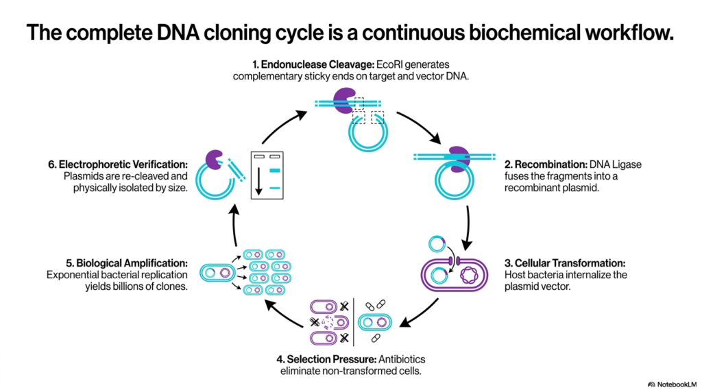
Every complex process in nature operates in cycles, and human-engineered biology is no different. The core purpose of this slide deck is to synthesize all individual mechanisms into one cohesive big picture. By reviewing the entire workflow of DNA cloning, students will appreciate the continuous, interdependent nature of these biochemical steps. DNA cloning is a seamless loop of innovation. The complete cycle of DNA cloning drives modern biotechnology forward.
This slide presents the complete cycle as a continuous, comprehensive biochemical workflow, summarizing the entire presentation. Step one shows endonuclease cleavage, where EcoRI generates complementary sticky ends on both the target and the vector. Step two highlights recombination, illustrating how DNA Ligase fuses the specific fragments together into a unified recombinant plasmid, structurally ready for the next biological phase.
Step three transitions into cellular biology through transformation, where living host bacteria internalize the engineered plasmid vector. Step four applies necessary selection pressure, utilizing antibiotics to rapidly eliminate any non-transformed cells, ensuring only the successes survive. Step five showcases biological amplification, where the surviving cells undergo exponential bacterial replication to yield billions of identical microscopic clones harboring the custom genetic instructions.
The final step, electrophoretic verification, represents the conclusion and extraction. The amplified plasmids are enzymatically re-cleaved and physically isolated by their specific size on a gel matrix. This cyclical visualization reinforces how every distinct chemical, biological, and physical step perfectly relies on the success of the preceding action, creating a flawless assembly line for molecular manufacturing.
Grasping this continuous workflow is the ultimate objective for students of molecular genetics. It demonstrates that advanced biotechnology is not magic, but a series of highly controlled, rational biochemical interventions. By mastering each step in this continuous cycle, future medical professionals will be equipped to harness these exact mechanisms to develop the next generation of genetic therapies and diagnostics.
Please read our Content Disclaimer Statement.
Check out our social media channels: