155. Biochemistry of Eicosanoids: Pathways, Functions, and Impact
Have you ever wondered how your body dials up a fever, triggers localized pain after an injury, or stops a minor cut from bleeding—all within seconds? The answer lies in eicosanoids, powerful lipid signals that control acute cellular communication. This slide deck explores the Biochemistry of Eicosanoids, detailing their synthesis from membrane phospholipids, their metabolic divergence into prostaglandins and leukotrienes, their physiological impact across body systems, and their clinical modulation by anti-inflammatory drugs like aspirin.
Slide 1: Biochemistry of Eicosanoids – Essential Mechanisms of Local Signaling
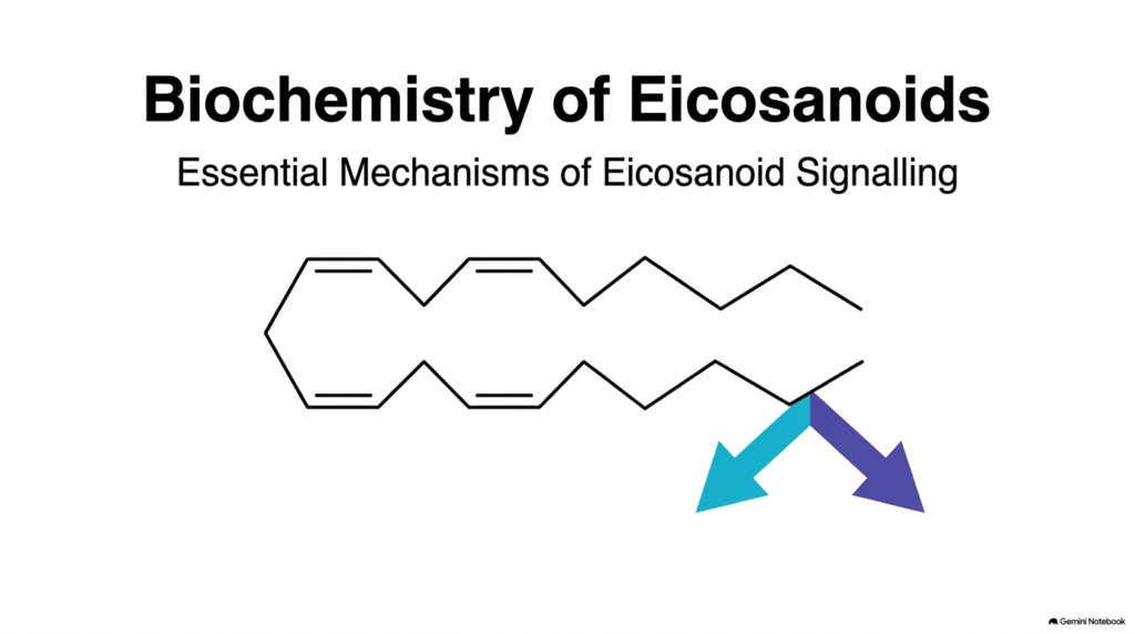
Eicosanoid biochemistry centers on a diverse group of oxygenated 20-carbon fatty acid derivatives that act as potent signaling molecules. Unlike classical hormones that travel through the bloodstream to distant organs, these bioactive lipids operate locally near their site of synthesis. Understanding eicosanoid biochemistry reveals how cells convert membrane phospholipids, specifically polyunsaturated fatty acids like arachidonate, into rapid chemical messengers. Arachidonate contains four cis double bonds, giving it a flexible shape that enzymes access easily during acute physiological responses.
When cells experience mechanical stress, cytokine stimulation, or chemical signals, specific enzymatic pathways quickly transform arachidonate into distinct functional families. The two primary metabolic pathways branching from arachidonate are the cyclooxygenase pathway and the lipoxygenase pathway. The cyclooxygenase pathway generates prostaglandins, prostacyclins, and thromboxanes, while the lipoxygenase pathway produces leukotrienes. Studying the Biochemistry of Eicosanoids helps medical students appreciate how these metabolic routes govern fundamental processes including vascular tone, platelet activation, bronchial constriction, and inflammatory cascades throughout the body.
Because these lipid mediators exert brief yet intense biological effects, tissues must tightly control their production and clearance. Each branch converts the parent polyunsaturated lipid into specialized ring structures or conjugated peptide complexes tailored for specific membrane receptors. By mastering these foundational metabolic pathways, students gain insight into how cells communicate locally and how targeted drugs manipulate inflammatory responses.
Slide 2: Biochemistry of Eicosanoids – Localized Autocrine and Paracrine Action
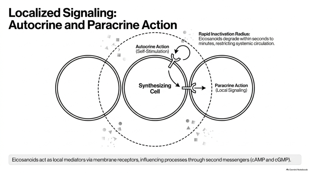
A core principle of eicosanoid biochemistry is their restricted, localized mechanism of action. Unlike endocrine hormones that circulate globally, these lipid mediators act primarily through autocrine and paracrine signaling. A synthesizing cell releases these active molecules into the immediate extracellular space, where they bind to specific G-protein-coupled receptors on either the producing cell itself or adjacent target cells. The Biochemistry of Eicosanoids relies on this spatial restriction to keep potent physiological responses confined to injured or inflamed tissues without causing systemic disruption.
To maintain this localized control, these signaling molecules possess an extremely short biological half-life. Rapid enzymatic inactivation breaks down these active lipids within seconds to minutes of secretion. This rapid degradation prevents the molecules from entering systemic circulation in active form, effectively creating a defined sphere of influence around the synthesizing cell. Receptor engagement triggers intracellular signaling cascades that primarily alter secondary messenger levels, such as cyclic adenosine monophosphate or cyclic guanosine monophosphate, to modify cellular behavior.
Exploring the Biochemistry of Eicosanoids helps future clinicians understand why inflammatory symptoms like localized swelling, redness, and pain remain localized to damaged tissue. By restricting signaling to immediate cellular neighbors, the body coordinates precise cellular defenses while protecting healthy distant organs from unnecessary exposure to inflammatory triggers.
Slide 3: Biochemistry of Eicosanoids – Membrane Cleavage and Arachidonate Release
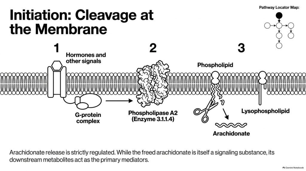
Eicosanoid biochemistry begins at the plasma membrane, where arachidonate remains esterified within membrane phospholipids. Because free arachidonate concentrations remain low inside resting cells, the initiation step requires precise enzymatic liberation. External stimuli, such as hormones, neurotransmitters, or physical trauma, activate membrane-bound G-protein complexes. This receptor activation triggers cytosolic phospholipase A2, officially classified as enzyme 3.1.1.4, to translocate directly to membrane bilayers. Understanding eicosanoid biochemistry requires recognizing phospholipase A2 as the primary rate-limiting checkpoint of the pathway.
Activated phospholipase A2 specifically hydrolyzes the sn-2 ester bond of membrane phospholipids, cleaving the molecule into free arachidonate and a lysophospholipid. While lysophospholipids participate in cellular remodeling, free arachidonate serves as a vital precursor fatty acid for subsequent enzymatic transformations. Although free arachidonate possesses minor intrinsic signaling capacity, its downstream oxygenated metabolites drive the major physiological actions associated with acute tissue responses.
Because phospholipase A2 controls the initial supply of precursor molecules, inhibiting this enzyme halts downstream mediator production. Anti-inflammatory steroids promote lipocortin synthesis, which inhibits phospholipase A2 activity and blocks precursor release. Through the Biochemistry of Eicosanoids, students can see how controlling membrane phospholipid cleavage offers a powerful medical target to suppress widespread inflammatory cascades before downstream metabolites form.
Slide 4: Biochemistry of Eicosanoids – The Branching Pathways: COX vs. LOX
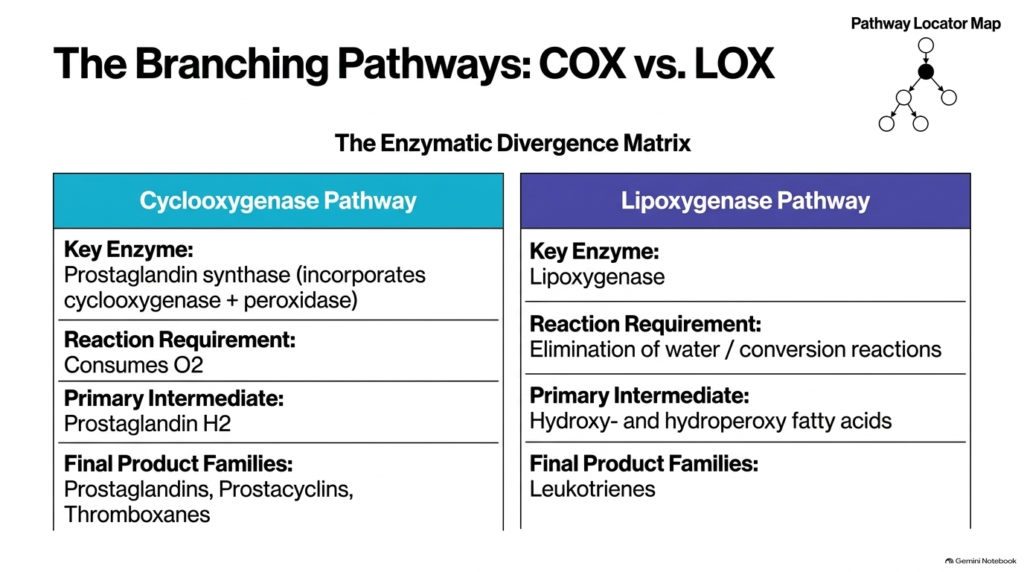
Once phospholipase A2 liberates arachidonate, the biochemistry of eicosanoids divides into two major enzymatic pathways: the cyclooxygenase pathway and the lipoxygenase pathway. The cyclooxygenase pathway depends on prostaglandin synthase, a bifunctional enzyme complex that combines cyclooxygenase and peroxidase activities. This oxygen-consuming pathway converts arachidonic acid into the central intermediate, prostaglandin H2. Prostaglandin H2 subsequently feeds into downstream synthases to generate prostaglandins, prostacyclins, and thromboxanes. Analyzing the Biochemistry of Eicosanoids highlights how enzyme availability determines which specific product family dominates within a given tissue.
In contrast, the lipoxygenase pathway utilizes lipoxygenase enzymes to oxygenate arachidonate without forming ring structures. This route involves water elimination and conversion reactions, creating hydroperoxy and hydroxy fatty acid intermediates that yield leukotrienes. While cyclooxygenase products primarily regulate vascular tone, pain, and fever, lipoxygenase products act as major mediators in respiratory dynamics and allergic inflammatory responses.
Comparing these branching routes within the Biochemistry of Eicosanoids demonstrates elegant metabolic coordination in human physiology. Individual cell types express specific enzyme profiles based on their specialized biological functions. For instance, blood platelets express thromboxane synthase to favor clotting, whereas vascular endothelial cells express prostacyclin synthase to maintain smooth blood flow and inhibit aggregation. Understanding this metabolic divergence helps students predict how selective pharmacological inhibitors shift the balance between pro-inflammatory and anti-inflammatory signals.
Slide 5: Biochemistry of Eicosanoids – Prostaglandin Synthase and PGH2 Synthesis
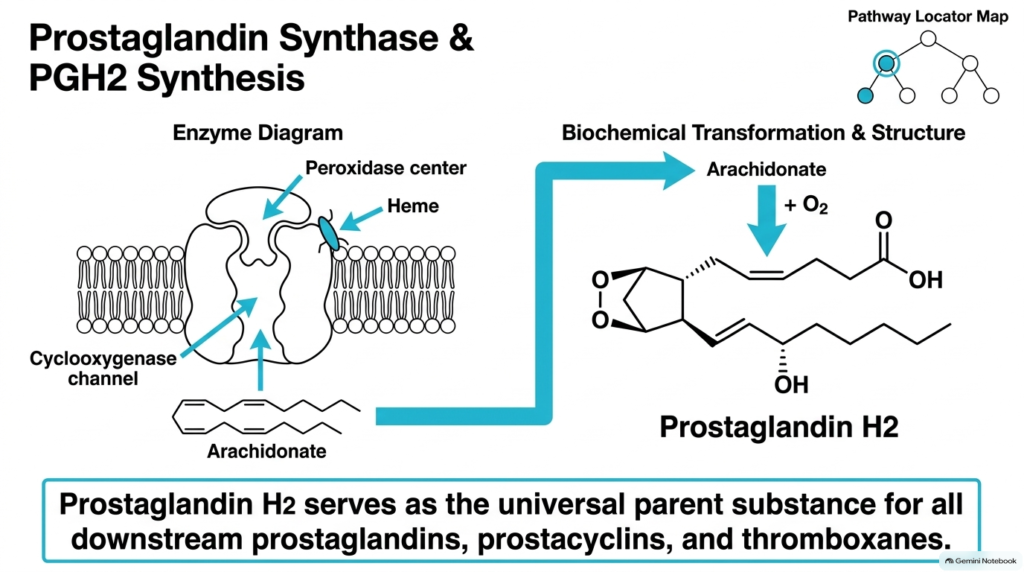
In eicosanoid biochemistry, prostaglandin synthase plays a pivotal role by carrying out two sequential catalytic reactions within a single membrane-bound protein complex. Free arachidonate enters the hydrophobic cyclooxygenase channel of the enzyme, where two molecules of oxygen are added to form the unstable endoperoxide intermediate, prostaglandin G2. Next, the enzyme’s heme-containing peroxidase center reduces this endoperoxide to produce prostaglandin H2. Mastering eicosanoid biochemistry requires understanding how prostaglandin H2 serves as the universal precursor for all downstream prostaglandins, prostacyclins, and thromboxanes.
The structural transformation from arachidonate to prostaglandin H2 introduces a characteristic five-membered cyclopentane ring flanked by two aliphatic side chains, along with an endoperoxide bridge across carbons nine and eleven. This fundamental structural arrangement stabilizes the carbon backbone while preparing it for rapid conversion by downstream terminal synthases. Because prostaglandin H2 is chemically unstable, cells rapidly process it into specialized final products before it degrades spontaneously.
Studying this double-step catalytic mechanism in the Biochemistry of Eicosanoids provides vital context for drug design. The cyclooxygenase channel contains specific amino acid residues that position arachidonate for precise oxygenation. Blocking this narrow channel prevents oxygen incorporation, effectively shutting down prostaglandin H2 synthesis and all derivative pro-inflammatory signaling molecules.
Slide 6: Biochemistry of Eicosanoids – Structural Profile of Prostaglandins
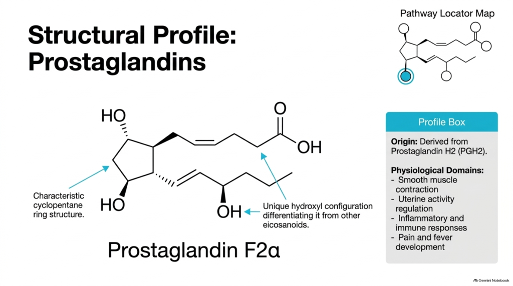
A defining feature of eicosanoid biochemistry is the precise molecular architecture that dictates biological activity. Prostaglandins, including prostaglandin F2alpha, retain the five-membered cyclopentane ring formed during initial precursor processing. Specific functional groups attached to this ring, such as unique hydroxyl configurations at key carbon positions, differentiate prostaglandins from other eicosanoid classes. In Biochemistry of Eicosanoids, students learn that small variations in stereochemistry and hydroxyl orientation change how these lipids fit into selective cell-surface receptors.
Derived directly from prostaglandin H2, classic prostaglandins mediate a wide range of physiological domains across diverse organ systems. In smooth muscle tissue, prostaglandin F2alpha stimulates potent contractions, playing a central role in uterine activity regulation during childbirth and vascular tone control. Prostaglandins also drive inflammatory and immune responses, working alongside bradykinin and histamine to promote pain, fever, and local vasodilation during acute injury.
Understanding these molecular structures in the Biochemistry of Eicosanoids explains why synthetic prostaglandin analogs are valuable clinical therapeutics. By modifying functional groups on the cyclopentane ring, pharmacologists design stable drugs that selectively target reproductive, ocular, or gastric receptors while minimizing off-target side effects.
Slide 7: Biochemistry of Eicosanoids – Structural Profile of Prostacyclins
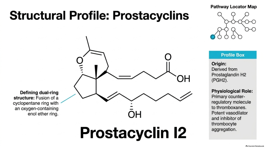
Examining eicosanoid biochemistry reveals how specialized enzymatic modifications alter basic core structures to produce distinct physiological functions. Prostacyclins, exemplified by prostacyclin I2, have a defining dual-ring structure formed by fusing a cyclopentane ring with an oxygen-containing enol ether ring. Prostacyclin synthase converts prostaglandin H2 into this bicyclic configuration within vascular endothelial cells. The Biochemistry of Eicosanoids shows how this double-ring arrangement provides the geometry needed to bind prostacyclin receptors on smooth muscle and platelet membranes.
Physiologically, prostacyclin I2 serves as the primary counter-regulatory molecule to thromboxanes in cardiovascular homeostasis. It is a potent vasodilator that relaxes vascular smooth muscle cells, decreasing peripheral resistance and lowering blood pressure. Furthermore, prostacyclin I2 strongly inhibits platelet aggregation, preventing clot formation on healthy vessel walls and maintaining unimpeded circulation throughout the cardiovascular network.
In the broader context of eicosanoid biochemistry, the balance between prostacyclin I2 and thromboxane A2 determines vascular health. Endothelial injury reduces local prostacyclin production, shifting the physiological equilibrium toward platelet activation and vessel constriction. Recognizing this structural and functional relationship helps medical students understand conditions like atherosclerosis and arterial thrombosis.
Slide 8: Biochemistry of Eicosanoids – Structural Profile of Thromboxanes
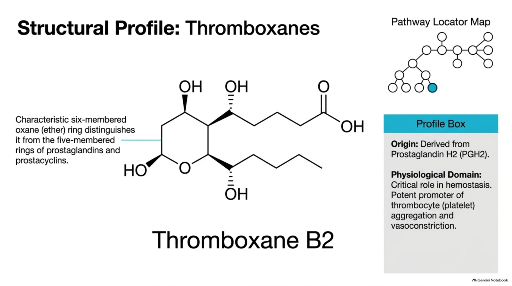
A fascinating structural transition in eicosanoid biochemistry occurs during thromboxane synthesis. Thromboxane synthase converts prostaglandin H2 into thromboxanes, which feature a six-membered oxane ether ring rather than the five-membered cyclopentane ring found in prostaglandins. Thromboxane B2 represents the stable, inactive metabolite of short-lived thromboxane A2. In the Biochemistry of Eicosanoids, this distinct oxane ring structure distinguishes thromboxanes from prostacyclins and cyclopentane prostaglandins, enabling selective binding to specific thromboxane receptors on blood platelets.
Thromboxanes play a critical physiological role in hemostasis and blood clotting dynamics. Produced abundantly by activated platelets, thromboxane A2 potently promotes thrombocyte aggregation, causing platelets to adhere to one another at sites of vascular injury. At the same time, thromboxane A2 induces powerful vasoconstriction, narrowing damaged blood vessels to reduce blood loss and stabilize initial clot formation during acute tissue trauma.
Connecting molecular shape to clinical outcomes in the Biochemistry of Eicosanoids clarifies why antiplatelet therapies target this pathway. Because platelets lack nuclei, they cannot synthesize new enzymes. Irreversibly inhibiting thromboxane synthesis permanently impairs platelet aggregation for the platelet’s entire lifespan, providing effective protection against ischemic cardiac events.
Slide 9: Biochemistry of Eicosanoids – The Lipoxygenase Pathway and Leukotrienes
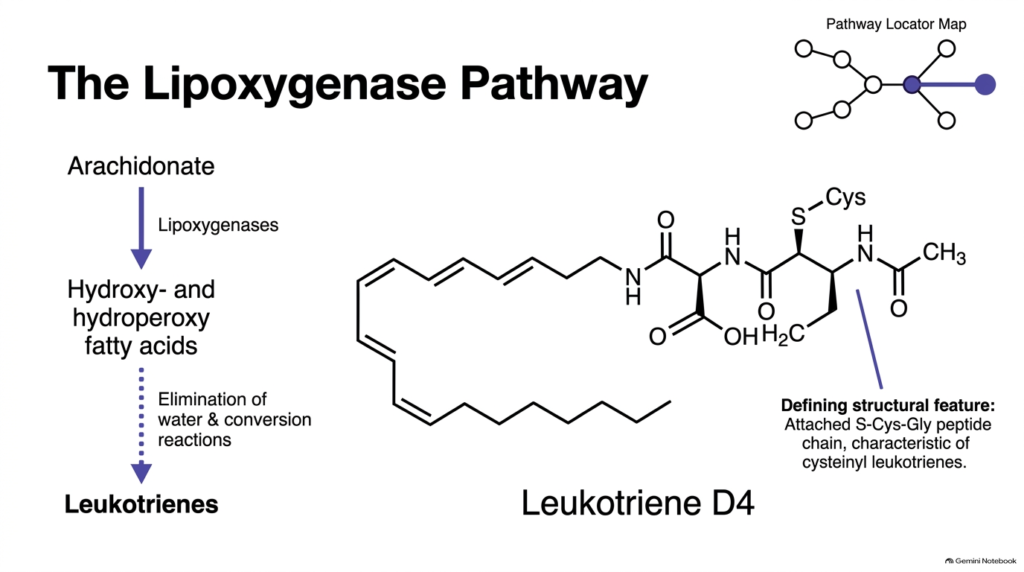
Beyond the cyclooxygenase route, eicosanoid biochemistry includes the lipoxygenase pathway, which generates linear oxygenated fatty acid derivatives called leukotrienes. Lipoxygenase enzymes insert molecular oxygen into arachidonic acid to form hydroperoxy and hydroxy fatty acid intermediates. Subsequent elimination of water and structural conversions convert these intermediates into active leukotrienes. Exploring the Biochemistry of Eicosanoids reveals that leukotrienes lack the cyclic ring features of prostaglandins, retaining an extended carbon chain with conjugated double-bond systems.
A defining structural feature of cysteinyl leukotrienes, such as leukotriene D4, is the covalent attachment of an S-cysteine-glycine peptide chain to the linear hydrocarbon backbone. Leukotriene synthases link these amino acid residues to produce potent biological mediators involved in hypersensitivity reactions. Leukotriene D4 is a potent bronchoconstrictor that increases vascular permeability, driving airway narrowing and tissue edema during acute asthma attacks.
Understanding the biochemistry of eicosanoids along the lipoxygenase branch highlights key therapeutic opportunities in respiratory medicine. Because leukotrienes drive severe allergic airway inflammation and mucus hypersecretion, pharmacological agents that block leukotriene receptors or inhibit 5-lipoxygenase provide vital maintenance therapy for asthma patients. By preventing leukotriene-mediated airway constriction, these drugs help restore normal pulmonary function and improve quality of life.
Slide 10: Biochemistry of Eicosanoids – Structural Comparison of Eicosanoid Families
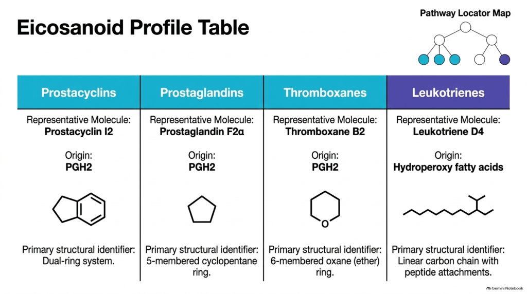
A comparative analysis of eicosanoid biochemistry categorizes these four major lipid families by precursor origin, representative molecules, and defining ring architectures. Prostacyclins, represented by prostacyclin I2, derive from prostaglandin H2 and feature a dual-ring system consisting of fused cyclopentane and enol ether rings. Prostaglandins, represented by prostaglandin F2alpha, also originate from prostaglandin H2 but feature a standalone five-membered cyclopentane ring. The biochemistry of eicosanoids shows how these structural differences allow target tissues to distinguish between closely related signals.
Thromboxanes, represented by thromboxane B2, also originate from prostaglandin H2 but have a six-membered oxane ether ring. In contrast, leukotrienes, represented by leukotriene D4, originate from hydroperoxy fatty acids generated by lipoxygenase rather than prostaglandin H2. Leukotrienes lack a ring structure entirely, displaying a linear carbon chain adorned with peptide attachments like cysteine and glycine residues.
Summarizing these structural profiles in the biochemistry of eicosanoids reinforces how subtle chemical modifications yield vastly different physiological outcomes. From smooth muscle relaxation to platelet aggregation and bronchial constriction, each family utilizes its unique carbon skeleton to bind selective receptor subtypes, orchestrating complex cellular responses with high specificity.
Slide 11: Biochemistry of Eicosanoids – Systems Impact and Physiological Integration
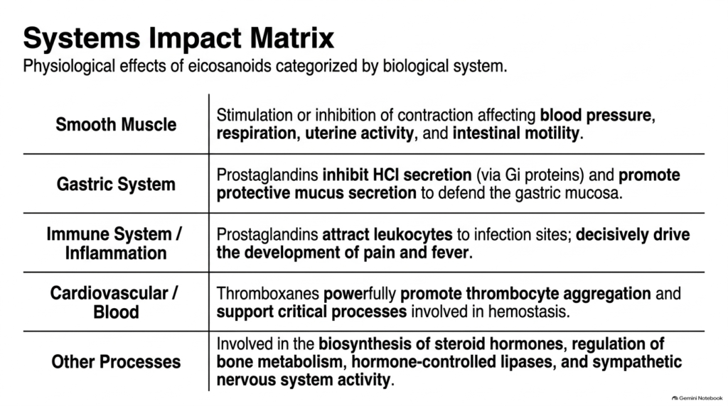
The broad physiological impact of eicosanoid biochemistry spans multiple organ systems throughout the human body. In smooth muscle, eicosanoids either stimulate or inhibit contraction, thereby directly influencing blood pressure, airway diameter, uterine activity during labor, and intestinal motility. In the gastric system, prostaglandins inhibit hydrochloric acid secretion through Gi-protein pathways while stimulating protective mucus secretion. Students analyzing the Biochemistry of Eicosanoids see how these lipid signals balance cellular functions to maintain mucosal integrity and tissue homeostasis.
In the immune system, prostaglandins recruit white blood cells to infection sites and drive localized pain and fever. In the cardiovascular system, thromboxanes promote platelet aggregation and vasoconstriction to support hemostasis, while prostacyclins exert opposing protective effects. Beyond these classic roles, eicosanoids participate in steroid hormone biosynthesis, regulate bone metabolism, influence hormone-sensitive lipase activity, and support sympathetic nervous system signaling.
Understanding eicosanoid biochemistry across these integrated body systems helps medical students see why systemic diseases often involve widespread lipid dysregulation. Because these mediators touch nearly every physiological domain, pharmacological interventions that alter eicosanoid levels produce broad systemic effects that require careful clinical monitoring.
Slide 12: Biochemistry of Eicosanoids – Pharmacological Inhibition and the Aspirin Blockade
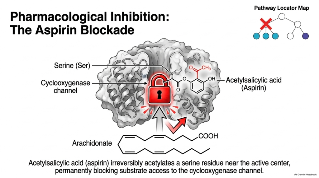
Pharmacological manipulation is a major clinical application in eicosanoid biochemistry. Acetylsalicylic acid, commonly known as aspirin, exerts its therapeutic effects by irreversibly inhibiting cyclooxygenase enzymes. Aspirin transfers an acetyl group directly to a specific serine residue located near the active site inside the cyclooxygenase channel. Studying the Biochemistry of eicosanoids shows that this covalent acetylation creates a permanent steric barrier, blocking substrate access and preventing arachidonate from entering the active channel.
Because aspirin attaches covalently to the serine residue, cyclooxygenase inactivation is irreversible. Cells must synthesize entirely new enzyme proteins to restore prostaglandin H2 production. In nucleated cells like vascular endothelial cells, protein synthesis eventually replenishes enzyme levels. However, blood platelets lack nuclei and cannot manufacture new proteins, leaving them permanently unable to produce thromboxanes for the remainder of their seven-to-ten-day circulatory lifespan.
This molecular blockade in the Biochemistry of Eicosanoids explains why low-dose aspirin serves as a foundation for cardiovascular prevention. By permanently disabling platelet cyclooxygenase without permanently suppressing endothelial prostacyclin synthesis, aspirin selectively reduces blood clot formation, reducing the risk of heart attacks and ischemic strokes in high-risk clinical populations.
Slide 13: Biochemistry of Eicosanoids – Clinical Consequences of Cyclooxygenase Inhibition
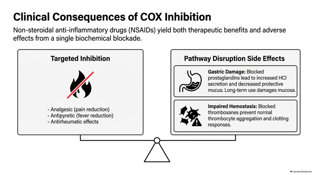
In clinical practice, the Biochemistry of Eicosanoids explains both the therapeutic benefits and adverse side effects associated with non-steroidal anti-inflammatory drugs. Inhibiting cyclooxygenase produces powerful targeted therapeutic outcomes, including pain reduction (analgesic effects), fever reduction (antipyretic effects), and decreased joint inflammation (antirheumatic effects). However, because cyclooxygenase enzymes produce protective baseline prostaglandins alongside inflammatory mediators, evaluating the Biochemistry of Eicosanoids demonstrates why enzyme blockade creates a double-edged physiological balance.
Pathway disruption side effects arise when protective prostaglandin synthesis is suppressed. Blocking stomach prostaglandins increases hydrochloric acid secretion while decreasing protective mucus production, leading to gastric mucosal erosion, ulceration, and potential gastrointestinal bleeding with prolonged drug use. Additionally, blocking thromboxane synthesis impairs hemostasis by preventing normal platelet aggregation and increasing bleeding tendencies in surgical and clinical settings.
Mastering eicosanoid biochemistry enables healthcare providers to balance therapeutic goals against potential toxicities. Recognizing these physiological trade-offs prompted the development of selective COX-2 inhibitors, which aim to suppress inflammatory prostaglandins at injury sites while sparing COX-1-mediated protective mechanisms in the gastric mucosa and vascular bed.
Slide 14: Biochemistry of Eicosanoids – Signal Termination and Rapid Enzymatic Degradation
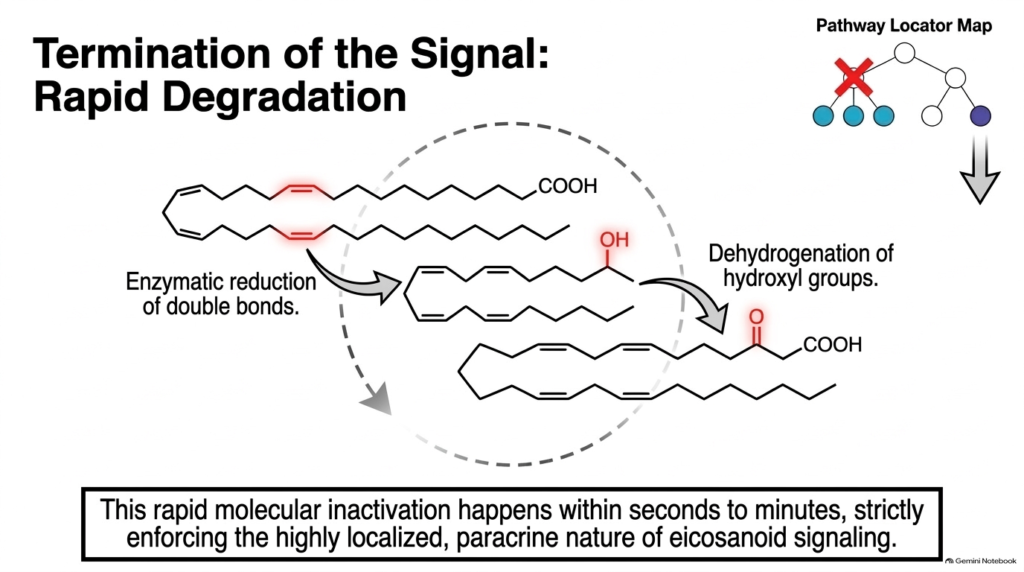
The final chapter in the Biochemistry of Eicosanoids focuses on how cells rapidly terminate lipid signals to prevent prolonged or exaggerated tissue responses. Once eicosanoids exert their localized effects, specialized catabolic enzymes quickly degrade these active molecules into inactive metabolites. Key enzymatic steps include reducing double bonds and dehydrogenating essential hydroxyl groups. Studying the Biochemistry of Eicosanoids shows that these chemical modifications destroy receptor-binding affinity, inactivating active signaling molecules within seconds to minutes.
This rapid molecular breakdown enforces the strictly localized, paracrine nature of lipid signaling. By rapidly clearing active mediators near their point of release, tissues prevent local inflammatory signals from diffusing into global circulation and causing unintended systemic reactions. Local catabolism keeps vascular tone changes, pain signals, and immune cell recruitment tightly controlled and self-limiting in healthy biological systems.
Through the Biochemistry of Eicosanoids, students appreciate that effective biological communication relies as much on swift signal clearance as on rapid signal generation. Understanding metabolic degradation pathways allows researchers to design synthetic analogs that resist enzymatic breakdown, prolonging therapeutic duration when long-acting local treatments are needed.
Please read our Content Disclaimer Statement.
Check out our social media channels: