153. Second Messengers: Biochemistry Signal Pathways
Have you ever wondered how a tiny hormone knocking on a cell’s surface can instantly change what happens deep inside? Cells rely on intricate communication networks to interpret external signals without letting those molecules cross the plasma membrane. This slide deck explores the foundational biochemistry of cellular signal transduction. Designed for college and medical school students, this visual guide breaks down how cells construct, regulate, and terminate molecular signals. By examining these pathways step by step, students can master the core mechanisms that drive cellular metabolism, physiological adaptation, and targeted medical therapies.
Slide 1: Introduction to Intracellular Second Messengers
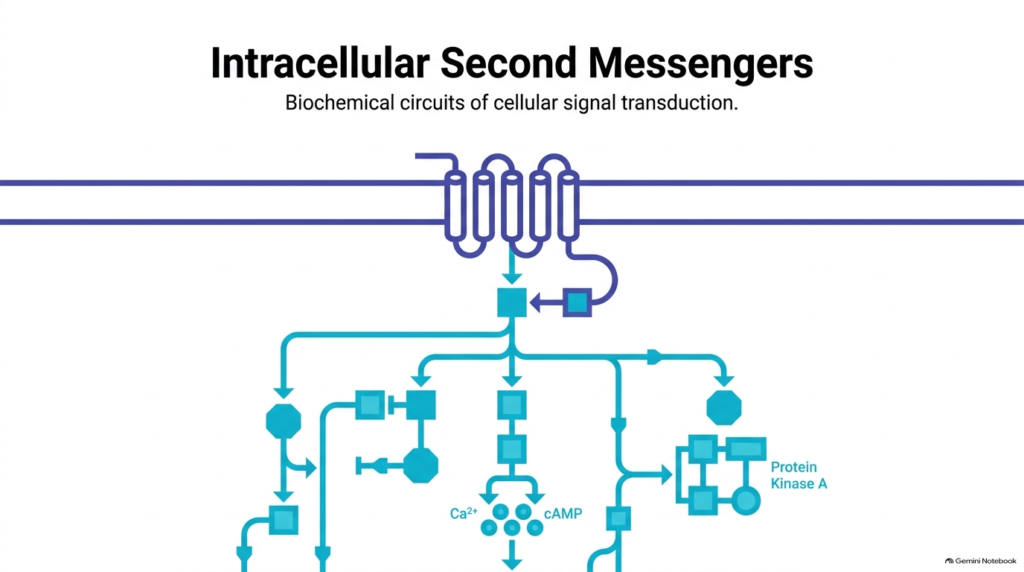
Cellular communication relies on sophisticated molecular relays to transmit extracellular information across the plasma membrane. When a primary ligand, such as a hormone or neurotransmitter, binds to a cell-surface receptor, it triggers a cascade of intracellular events. Rather than entering the cytoplasm directly, the primary signal triggers the rapid synthesis or release of internal signaling molecules. These internal molecules are known as second messengers. Second messengers act as biochemical relays that amplify the initial signal, allowing a single ligand-binding event to rapidly modify thousands of downstream enzymes. This dramatic amplification ensures that cells respond swiftly and efficiently to shifting physiological demands across various tissues.
The schematic illustrates how transmembrane receptors, such as seven-transmembrane G protein-coupled receptors, initiate complex signal networks within target cells. Activation of these receptors mobilizes membrane-bound G proteins, which in turn stimulate specific effector enzymes like adenylate cyclase. These enzymes generate abundant second messengers, including cyclic adenosine monophosphate and calcium ions, in the cytosol. Once formed, second messengers diffuse throughout specific cellular compartments to engage key target proteins, such as protein kinase A. Through these interconnected pathways, second messengers integrate diverse extracellular inputs into coordinated cellular outputs, governing vital metabolic processes ranging from glycogen breakdown to muscle contraction.
Slide 2: Defining the Intracellular Properties of Second Messengers
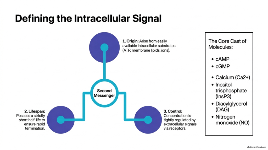
To understand cellular signal transduction, biochemists categorize signal molecules based on their functional characteristics and chemical origins. Small signaling molecules within the cell operate under three strict biochemical rules. First, second messengers arise from easily available intracellular substrates, such as adenosine triphosphate, membrane phospholipids, or inorganic ions. Second, second messengers possess a strictly short half-life to ensure rapid termination once the extracellular stimulus ceases. Third, extracellular signals acting through specific membrane receptors tightly regulate their intracellular concentration. These three properties ensure cellular signals remain fast, precise, and fully reversible during continuous metabolic regulation across tissues.
The core cast of second messengers includes several distinct chemical families that operate in different cellular locations. Key cyclic nucleotides include cyclic adenosine monophosphate and cyclic guanosine monophosphate, which act as primary intracellular messengers. Lipid-derived signaling molecules include inositol trisphosphate and diacylglycerol, both derived from the enzymatic cleavage of structural membrane phospholipids. Inorganic ions like calcium serve as powerful universal signals, while gaseous molecules such as nitrogen monoxide diffuse freely across lipid membranes. Together, these diverse second messengers allow cells to translate a wide variety of external stimuli into specific, highly tailored physiological responses, maintaining systemic physiological balance throughout the entire organism.
Slide 3: Cyclic AMP Synthesis and Second Messengers
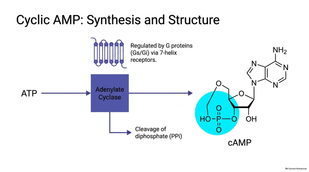
Cyclic adenosine monophosphate represents one of the most thoroughly studied second messengers in classical biochemistry. Adenylate cyclase catalyzes its synthesis; it is a multipass transmembrane enzyme located in the plasma membrane. Heterotrimeric G proteins tightly regulate adenylate cyclase activity, and seven-helix G protein-coupled receptors control them. Stimulatory G proteins activate adenylate cyclase to boost production, whereas inhibitory G proteins suppress its enzymatic activity. Upon activation, adenylate cyclase converts intracellular adenosine triphosphate into cyclic AMP through an intramolecular nucleophilic attack, releasing inorganic pyrophosphate as a high-energy byproduct into the cytoplasm. This regulated step guarantees that second messengers respond rapidly to extracellular stimuli.
The chemical structure of cyclic AMP reveals why second messengers can act as such effective signaling devices within living cells. Cleavage of high-energy diphosphate from ATP provides the thermodynamic driving force to form a cyclic phosphodiester bond between the ribose sugar ring’s three-prime and five-prime hydroxyl groups. This unique cyclic phosphodiester motif creates a distinct shape that downstream effector proteins recognize. Because second messengers like cyclic AMP are synthesized rapidly from abundant ATP pools, the cell can generate thousands of active signal molecules within milliseconds, creating an immediate metabolic shift in response to extracellular hormonal stimulation across various target tissues.
Slide 4: cAMP Activation Circuits and Second Messengers
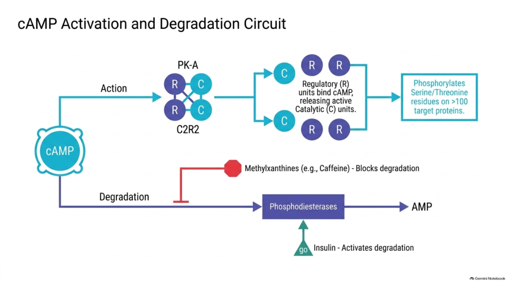
Once generated, cyclic AMP propagates signals by activating specific effector enzymes within the cytoplasm. The primary downstream target of cyclic AMP is protein kinase A, an inactive heterotetrameric complex consisting of two regulatory subunits and two catalytic subunits. As intracellular second-messenger concentrations rise, four cyclic AMP molecules bind cooperatively to the regulatory subunits. This binding induces a conformational change that liberates the active catalytic subunits. Once released, these catalytic enzymes phosphorylate specific serine and threonine residues on more than one hundred target proteins, dramatically altering cellular metabolic pathways and gene expression. Because second messengers act as metabolic switches, activating protein kinase A alters enzyme kinetics across the cell.
Signal termination is just as critical as signal activation to prevent uninhibited overstimulation. Phosphodiesterase enzymes break down cyclic AMP into inactive adenosine monophosphate, effectively resetting the cellular signaling system. Phosphodiesterase activity illustrates how pharmacological and physiological inputs regulate second messengers. Methylxanthines, such as caffeine, inhibit phosphodiesterase activity, blocking degradation and prolonging signal duration within target tissues. Conversely, the hormone insulin activates phosphodiesterase enzymes to accelerate second-messenger breakdown, showing how opposing hormonal inputs fine-tune intracellular signaling networks in real time to maintain metabolic equilibrium.
Slide 5: Phospholipid Cleavage Generating Second Messengers
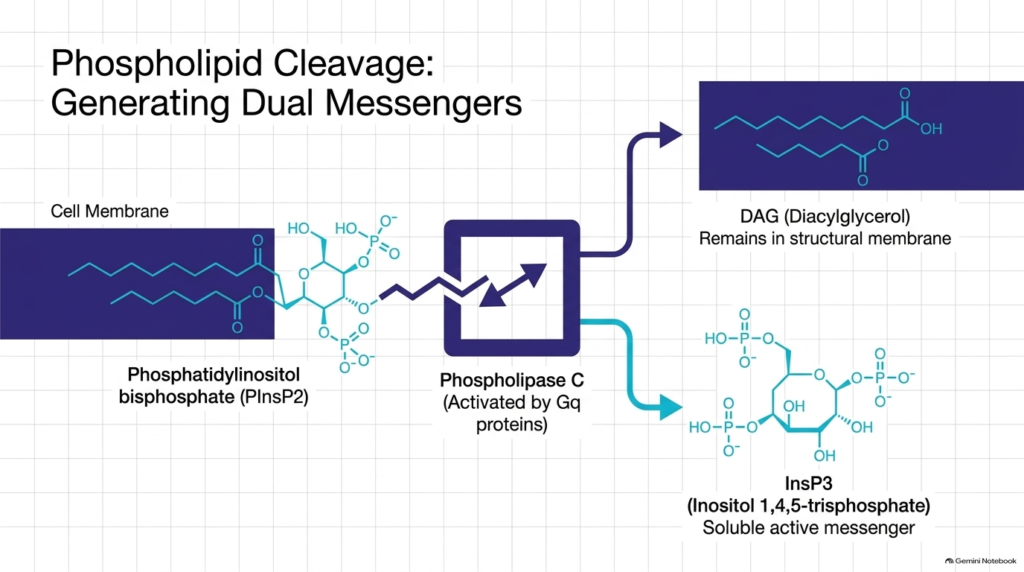
Cellular membranes serve not only as structural barriers but also as rich reservoirs for crucial signaling precursors. Phosphatidylinositol 4,5-bisphosphate is a minor phospholipid localized specifically in the inner leaflet of the plasma membrane. When cell-surface receptors engage stimulatory ligands, they activate Gq proteins, which then recruit the membrane-bound enzyme phospholipase C. Phospholipase C hydrolyzes phosphatidylinositol 4,5-bisphosphate, splitting this membrane lipid into two distinct second messengers. This cleavage shows how a single membrane lipid precursor can give rise to two complementary intracellular signaling pathways. Because second messengers transduce signals from membrane receptors, activating phospholipase C simultaneously initiates two distinct downstream pathways.
The two products generated by phospholipase C have dramatically different physical properties, enabling spatial bifurcation of signals. Diacylglycerol consists of two hydrophobic fatty acid tails attached to a glycerol backbone, so it remains embedded in the plasma membrane. In contrast, inositol 1,4,5-trisphosphate is a highly polar, water-soluble molecule that detaches from the membrane and enters the aqueous cytoplasm. Both of these second messengers carry out specialized signaling roles. By producing one membrane-bound signal and one cytosolic signal, second messengers efficiently coordinate simultaneous events at the cell boundary and within internal organelles, ensuring a comprehensive cellular response.
Slide 6: Spatial Divergence of Second Messengers
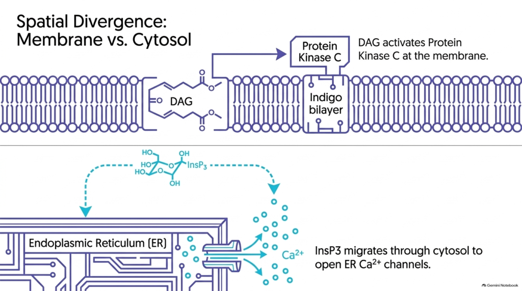
The spatial separation of diacylglycerol and inositol trisphosphate highlights the structural diversity among second messengers. Because diacylglycerol is nonpolar, it moves laterally within the lipid bilayer. There, it binds directly to protein kinase C, attracting the enzyme to the plasma membrane for full activation. Full activation of protein kinase C requires both lipid binding and calcium interaction. Through this localized mechanism, membrane-bound second messengers ensure that specific kinase cascades occur directly at the cell membrane, where key structural and transport proteins reside to alter membrane permeability and cell function. Because second messengers control membrane-associated enzymes, targeting protein kinase C to the lipid bilayer ensures rapid phosphorylation of adjacent target proteins.
Meanwhile, inositol trisphosphate acts as a mobile cytosolic messenger, diffusing rapidly from the plasma membrane to internal organelles. It targets ligand-gated calcium channels on the endoplasmic reticulum membrane. Inositol trisphosphate binding opens these channels, allowing stored calcium ions to flow down their concentration gradient into the cytoplasm. Through this mechanism, soluble second messengers bridge the physical gap between the cell membrane and intracellular storage depots. In this way, second messengers drive complex, multi-compartmental physiological responses from a single receptor activation event, mobilizing calcium stores throughout the cytoplasm.
Slide 7: Calcium Homeostasis and Second Messengers
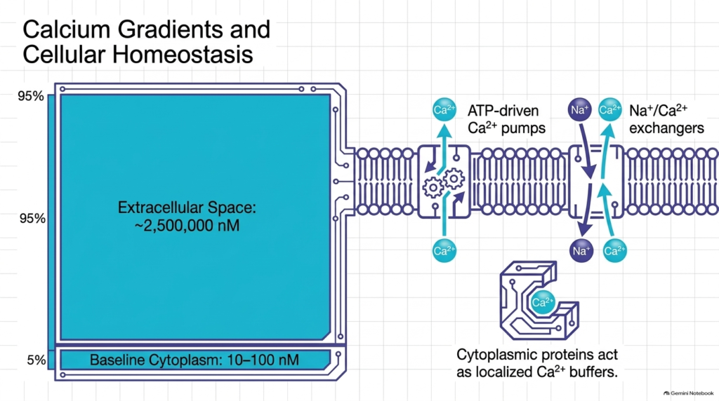
Calcium ions function as universal second messengers across virtually all eukaryotic cell types. To utilize calcium effectively as a signal, cells maintain an extraordinarily steep concentration gradient across the plasma membrane. Extracellular calcium concentration is approximately 2.5 millimolar, whereas baseline cytoplasmic calcium is kept extremely low, between 10 and 100 nanomolar. Maintaining this twenty-five-thousand-fold concentration gradient requires continuous metabolic energy, as resting cells must actively extrude calcium or sequester it within internal storage organelles like the endoplasmic reticulum. Because second messengers rely on low baseline levels, maintaining this steep calcium gradient enables rapid, high-contrast signaling whenever ion channels open.
Cells use specialized transport machinery to maintain low resting cytoplasmic calcium levels. Primary active transport relies on ATP-driven calcium pumps located in the plasma membrane and endoplasmic reticulum. Secondary active transport uses sodium-calcium exchangers that leverage the sodium gradient to export calcium ions from the cytosol. Additionally, specialized cytoplasmic proteins act as localized buffers to bind excess free calcium ions. When signaling events trigger a sudden influx, these transport mechanisms quickly restore baseline levels. Through these active transport systems, cells ensure that calcium ions function as reliable second messengers without disrupting resting cellular homeostasis.
Slide 8: Calcium Spikes, Cytotoxicity, and Second Messengers
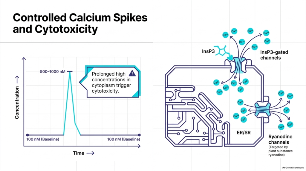
Cellular calcium signaling relies on controlled, transient spikes rather than sustained elevations. Upon receptor stimulation, cytoplasmic calcium levels rapidly rise from a baseline of 100 nanomolar to peak concentrations between 500 and 1,000 nanomolar. These transient calcium spikes activate specific downstream enzymes without damaging cellular structures. However, if cytoplasmic calcium remains elevated for prolonged periods, it triggers severe cytotoxicity, organelle damage, and apoptotic cell death. Therefore, cells strictly limit the duration and magnitude of calcium signals to protect cellular structural integrity. By keeping calcium signals brief, cells utilize these ionic second messengers for efficient signal transduction while avoiding catastrophic structural damage.
Calcium release into the cytoplasm occurs through distinct channel families embedded in the endoplasmic and sarcoplasmic reticulum membranes. Inositol trisphosphate-gated channels open in response to cytosolic inositol trisphosphate, releasing stored calcium into the cytoplasm. Similarly, ryanodine receptor channels facilitate calcium release in excitable tissues like muscle cells. Plant alkaloids like ryanodine specifically target these channels, illustrating their biological significance. Once released, these ionic second messengers rapidly activate target proteins before active pumps clear calcium back into internal stores. By regulating channel opening and closing, cells ensure second messengers act safely and predictably during every signaling event.
Slide 9: Calmodulin Sensing of Second Messengers
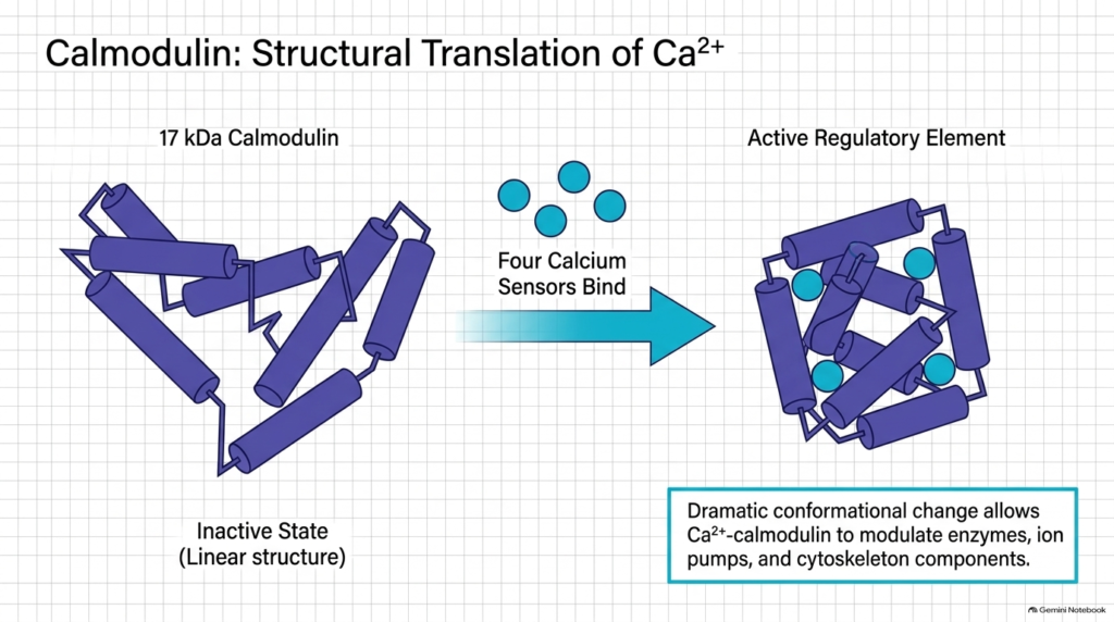
Because calcium ions cannot be chemically modified like organic molecules, cells rely on specialized sensor proteins to translate calcium signals into biochemical actions. Calmodulin is a highly conserved, acidic, seventeen-kilodalton protein that serves as the primary intracellular calcium sensor. In its inactive state, calmodulin adopts an extended linear conformation with low affinity for target enzymes. As calcium levels rise, calcium ions act as critical second messengers that bind to four specific EF-hand motifs within calmodulin, triggering a dramatic conformational change that alters protein function. Because calcium ions function as versatile second messengers, calmodulin provides the structural machinery required to sense small fluctuations in calcium ion concentration.
Binding of four calcium ions causes calmodulin to fold into a compact, globular active regulatory element. This conformational shift exposes hydrophobic surfaces that interact directly with downstream target enzymes, ion pumps, and cytoskeletal components. Activated calcium-calmodulin complexes regulate key enzymes, including myosin light-chain kinase and calmodulin-dependent protein kinases. Through this structural transition, cells convert simple ionic second messengers into complex adjustments in protein activity. This mechanism shows how inorganic second messengers regulate fundamental physiological processes, from smooth muscle contraction to long-term memory formation, by linking calcium binding to structural changes that fine-tune diverse enzymatic cascades.
Slide 10: Comparative Analysis of Second Messengers
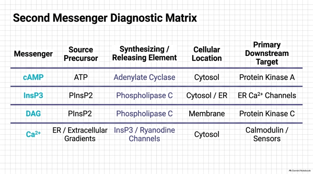
Understanding cell signaling requires comparing the distinct biochemical properties of major signaling molecules. The second messenger diagnostic matrix organizes these key molecules by precursor, synthesizing enzyme, cellular location, and primary downstream target. Cyclic AMP is produced from adenosine triphosphate by adenylate cyclase, acts in the cytosol, and primarily activates protein kinase A. Inositol trisphosphate is derived from membrane phospholipids by phospholipase C, diffuses through the cytosol, and targets endoplasmic reticulum calcium channels. Each pathway highlights how second messengers adapt specific chemical structures for targeted functional roles across diverse cell types. These pathways show how second messengers tailor their physical properties to produce distinct signaling outcomes.
Similarly, phospholipase C generates diacylglycerol alongside inositol trisphosphate, but diacylglycerol remains in the plasma membrane to activate protein kinase C. Calcium ions enter the cytoplasm from the extracellular space or endoplasmic reticulum stores through specialized channels, where they engage calmodulin and other sensors in the cytosol. Comparing these four core signaling pathways reveals common operational principles across diverse biological systems. All second messengers enable rapid signal amplification, spatial specificity, and precise temporal control. By integrating multiple regulatory loops, second messengers maintain essential metabolic functions and allow cells to adapt to constantly changing external environments.
Please read our Content Disclaimer Statement.
Check out our social media channels: