154. Signal Transduction Pathways: Guide to Cellular Signaling
Have you ever wondered how a single hormone molecule like insulin can instantly signal your body to store glucose, or how a tiny gas radical can relax blood vessels within seconds? Cells constantly receive chemical messages that dictate their survival, metabolism, and function. This slide deck explores the vital mechanisms of cellular communication. By comparing transmembrane receptors with gaseous diffusion, we break down how cells turn external cues into rapid physiological changes.
Slide 01: Introduction to Biochemical Cascades and Signal Transduction Pathways
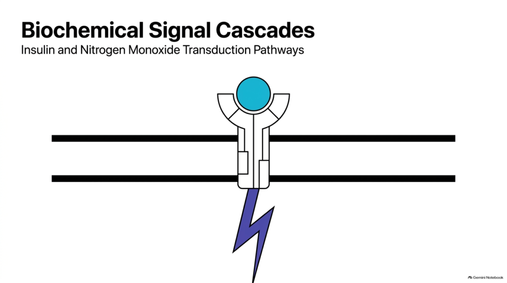
Cells rely on complex communication networks to sense their environment and maintain internal equilibrium. Biochemical signal cascades represent the primary mechanism by which extracellular signals trigger specific biological responses inside the cell. At the center of these communication networks are Signal Transduction Pathways, which convert physical or chemical stimuli into coordinated intracellular events.
When a ligand binds to a membrane receptor, it initiates a sequential chain reaction. This structural binding event alters the receptor conformation, allowing it to interact with downstream effector proteins. Signal Transduction Pathways amplify tiny initial signals into massive physiological outcomes, ensuring that a single molecular recognition event yields a robust cellular reaction.
These cascades govern essential processes including nutrient storage, gene transcription, and vascular tone. Understanding Signal Transduction Pathways provides crucial insight into human physiology and disease mechanisms. For example, metabolic disorders like type two diabetes arise when these intracellular relays fail to function properly.
This slide deck introduces two distinct signaling frameworks: endocrine signaling through transmembrane receptors and local paracrine signaling via dissolved gases. By examining insulin receptor activation and nitrogen monoxide diffusion side by side, students can appreciate the diversity of cellular communication architecture. Through this comparative approach, we will trace molecular interactions from initial membrane-binding events to nuclear gene expression and smooth muscle relaxation.
Slide 02: Endocrine and Paracrine Architectures in Signal Transduction Pathways
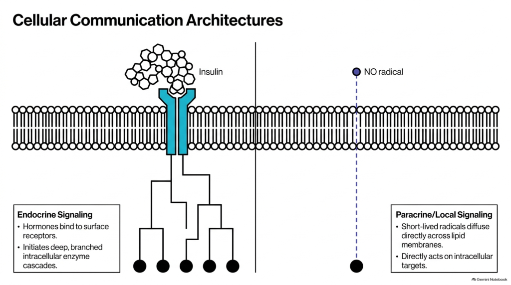
Cells employ distinct structural designs to send chemical signals over varying distances across human tissues. Endocrine communication relies on long-range hormones like insulin, which travel through the bloodstream to reach distant target tissues. These systemic Signal Transduction Pathways begin when the hormone binds to specific surface receptors, triggering branched intracellular enzyme cascades that spread throughout the cytoplasm.
In contrast, paracrine communication operates locally through short-lived molecules like the nitrogen monoxide radical. Because nitrogen monoxide is an uncharged gas radical, it diffuses directly across lipid cell membranes without needing a surface receptor interface. These localized Signal Transduction Pathways act on immediate neighboring cells to generate rapid, short-range physiological responses without systemic hormonal circulation.
Comparing these two paradigms highlights fundamental principles of biological molecular design. Endocrine networks generate sustained, multi-tissue responses through elaborate amplification cascades and protein phosphorylation networks. Local paracrine systems deliver instantaneous, highly targeted physiological adjustments by acting directly on intracellular target enzymes within target blood vessels.
Both architectural frameworks demonstrate how Signal Transduction Pathways maintain dynamic homeostasis across complex multicellular organisms. Whether through complex receptor-mediated cascades or direct gas diffusion, cells fine-tune their internal activity to match physiological demands. Recognizing these architectural differences helps explain why different organ systems utilize specialized chemical messengers for rapid or prolonged communication.
Slide 03: The Insulin Receptor Structure in Signal Transduction Pathways
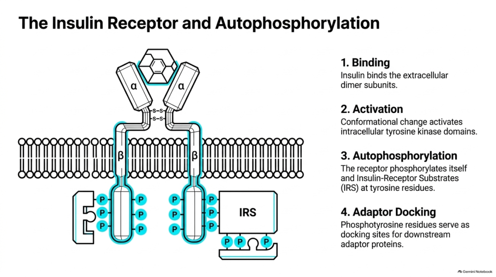
The insulin receptor serves as a classic example of a transmembrane tyrosine kinase involved in metabolic regulation. Structurally, the receptor exists as a preassembled dimer of two extracellular alpha subunits and two transmembrane beta subunits, linked by disulfide bonds. Insulin binding to the extracellular alpha subunits initiates one of the most vital Signal Transduction Pathways in human metabolism.
Ligand binding induces a critical conformational change that brings the intracellular beta subunits into close proximity. This structural shift activates the intrinsic tyrosine kinase domain located within each beta subunit. The receptor then undergoes autophosphorylation, adding phosphate groups to specific tyrosine residues on its own cytoplasmic tails and on insulin-receptor substrate proteins.
These phosphorylated tyrosine residues create high-affinity docking sites for downstream adaptor proteins containing specialized binding domains. By creating these docking sites, the insulin receptor establishes the foundation for multiple Signal Transduction Pathways that regulate glucose uptake and gene transcription. Without this initial autophosphorylation step, downstream signaling molecules cannot assemble at the inner membrane surface.
Proper insulin receptor function is essential for normal metabolic homeostasis throughout the body. Mutations or structural defects that disrupt autophosphorylation severely impair these Signal Transduction Pathways, leading to insulin resistance and metabolic pathology. Understanding this receptor architecture shows how cell-surface structural changes translate into powerful enzymatic activity in the cytoplasm.
Slide 04: The MAPK Kinase Cascade in Signal Transduction Pathways
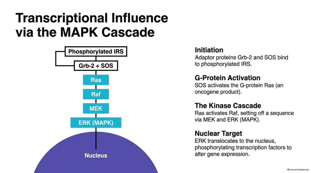
After the insulin receptor autophosphorylates, it can activate pathways that alter nuclear gene expression. Adaptor proteins Grb-2 and SOS bind directly to phosphorylated insulin-receptor substrates, initiating transcriptionally focused Signal Transduction Pathways. SOS functions as a guanine nucleotide exchange factor that converts the inactive G-protein Ras into its active GTP-bound form.
Active Ras recruits and activates the protein kinase Raf, launching a classical mitogen-activated protein kinase cascade. Raf phosphorylates and activates MEK, which subsequently phosphorylates ERK, also known as mitogen-activated protein kinase. These sequential phosphorylation steps in Signal Transduction Pathways serve as crucial molecular amplifiers, multiplying the original hormonal signal at each relay level.
Activated ERK detaches from cytoplasmic complexes and translocates across the nuclear envelope into the nucleoplasm. Inside the nucleus, ERK phosphorylates specific transcription factors, modulating their ability to bind promoter regions and stimulate target gene expression. This nuclear targeting alters protein synthesis patterns required for cell growth, division, and long-term metabolic adaptations.
The MAPK cascade shows how cell-surface signals reach nuclear genetic material with high specificity. Malfunctions in these Signal Transduction Pathways often lead to unregulated cell proliferation, making components like Ras and Raf prominent oncogene targets in cancer biology. Analyzing this pathway illustrates the seamless connection between membrane receptor activation and genomic transcription.
Slide 05: PI3K Activity and Lipid Signaling in Signal Transduction Pathways
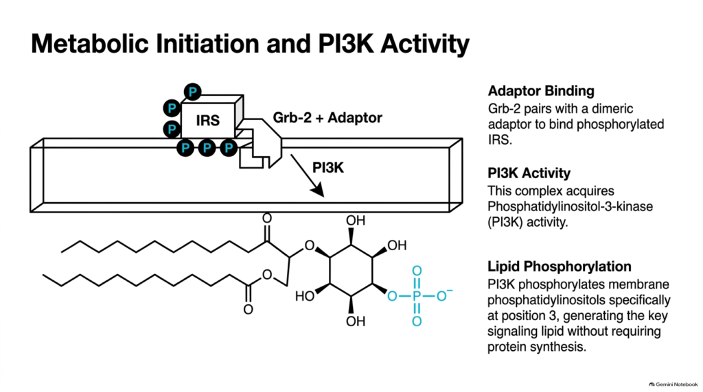
Beyond altering gene expression, insulin regulates immediate metabolic activity through lipid-modifying enzymes. Phosphorylated insulin-receptor substrate recruits adaptor complexes that activate phosphatidylinositol 3-kinase at the plasma membrane. This activation launches metabolic Signal Transduction Pathways that govern acute nutrient storage, cellular energy balance, and substrate utilization across peripheral tissues.
Active phosphatidylinositol 3-kinase catalyzes the addition of a phosphate group to the third position of the inositol ring of membrane phosphatidylinositols. This reaction generates phosphatidylinositol 3,4,5-trisphosphate directly within the inner leaflet of the lipid bilayer. Unlike protein synthesis pathways, these lipid-based Signal Transduction Pathways produce second messenger lipids within seconds to drive rapid physiological shifts.
The accumulation of phosphatidylinositol 3,4,5-trisphosphate alters the physical docking environment of the inner plasma membrane. Specialized signaling proteins containing pleckstrin homology domains recognize and bind tightly to these newly generated membrane lipids. This spatial recruitment concentrates key signaling enzymes right at the membrane interface where their substrates reside.
Lipid phosphorylation highlights the importance of non-protein second messengers in cellular signaling cascades. By generating membrane-bound lipid signals, these Signal Transduction Pathways bypass time-consuming transcriptional steps to execute instantaneous metabolic decisions. Understanding phosphatidylinositol 3-kinase activation reveals how cells modify their lipid membranes to coordinate complex biochemical cascades.
Slide 06: PK-B Activation and Glucose Transport in Signal Transduction Pathways
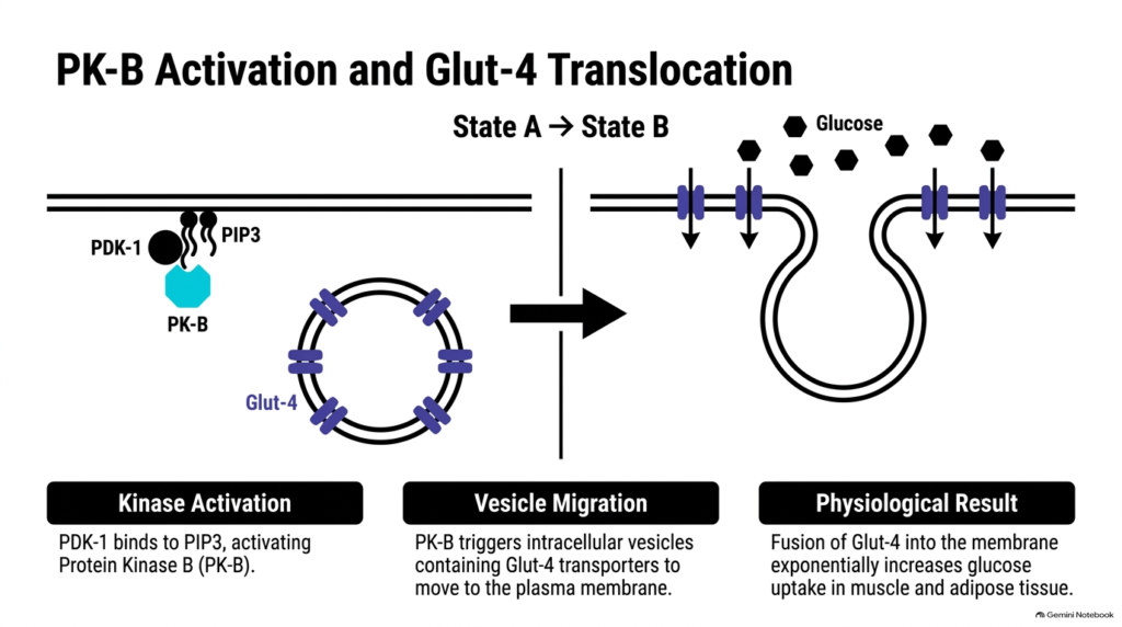
The lipid signals generated by phosphatidylinositol 3-kinase recruit two essential protein kinases to the plasma membrane. Phosphoinositide-dependent kinase-1 and protein kinase B both bind to membrane-bound phosphatidylinositol 3,4,5-trisphosphate. This co-localization allows PI3K to phosphorylate and fully activate Akt, driving central metabolic Signal Transduction Pathways.
Activated protein kinase B detaches from the plasma membrane and phosphorylates target proteins throughout the cytoplasm. One of its primary physiological targets is the intracellular vesicle pool carrying Glucose Transporter 4 proteins. Through these Signal Transduction Pathways, protein kinase B stimulates these specialized vesicles to translocate toward and fuse with the outer plasma membrane.
Vesicle fusion dramatically increases the concentration of functional glucose transporters embedded in the cell membrane. This structural insertion exponentially enhances glucose uptake from the bloodstream into skeletal muscle and adipose tissue cells. Consequently, these Signal Transduction Pathways rapidly lower blood glucose levels following a meal, restoring systemic metabolic balance.
This glucose transport mechanism illustrates how chemical signals translate into physical structural rearrangements inside target cells. Impairments at any point in these Signal Transduction Pathways reduce membrane glucose transporter expression, contributing to elevated blood sugar levels in diabetic patients. Mastering this pathway helps students connect enzymatic activation with whole-body tissue physiology.
Slide 07: Regulation of Glycogen Synthesis in Signal Transduction Pathways
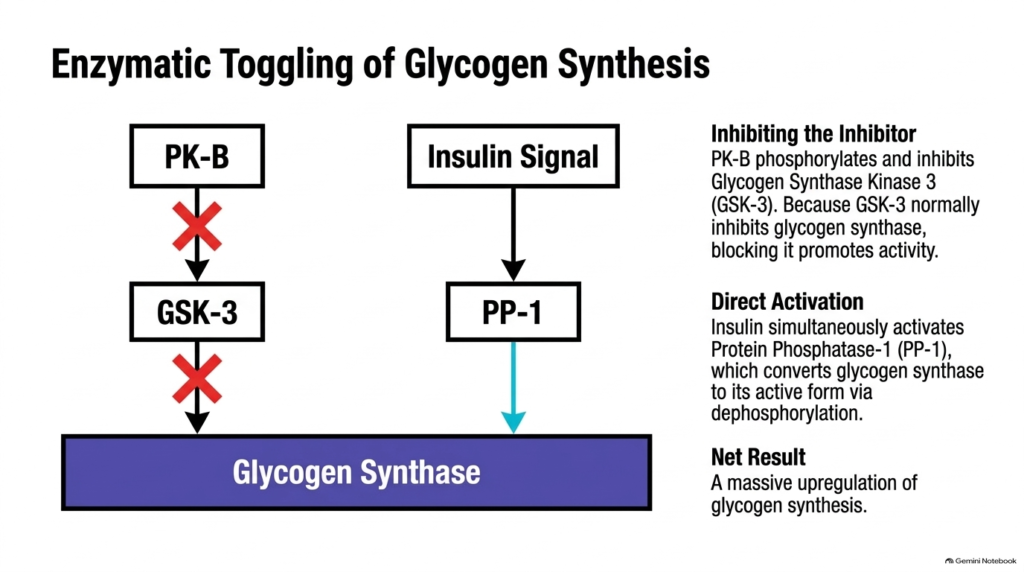
Insulin signaling also coordinates long-term glucose storage by regulating glycogen-synthesis enzymes in the liver. Protein kinase B phosphorylates glycogen synthase kinase-3, an enzyme that normally keeps glycogen synthase inactive by keeping it phosphorylated. In these Signal Transduction Pathways, phosphorylation of glycogen synthase kinase-3 inhibits its enzymatic activity, effectively removing its repressive effect on glycogen synthesis.
At the same time, insulin activates protein phosphatase-1 through complementary molecular mechanisms. Active protein phosphatase-1 removes inhibitory phosphate groups directly from glycogen synthase, converting it into its active, dephosphorylated form. By combining kinase inhibition with phosphatase activation, these Signal Transduction Pathways rapidly shift cellular machinery toward energetic storage and carbohydrate accumulation.
This dual regulatory mechanism represents a sophisticated biological logic known as double-negative control, or inhibiting the inhibitor. By inactivating glycogen synthase kinase-3 while activating protein phosphatase-1, insulin ensures maximal glycogen production without enzymatic competition. The net physiological result is a massive upregulation of glycogen synthesis in hepatic and muscle tissues.
Through these coordinated Signal Transduction Pathways, cells efficiently store excess dietary glucose as structured glycogen polymers. This precise enzymatic toggling prevents futile metabolic cycles where synthesis and breakdown occur simultaneously. This dual regulation shows how cells make decisive physiological shifts by controlling opposing metabolic enzymes.
Slide 08: Endothelial Synthesis of NO in Signal Transduction Pathways
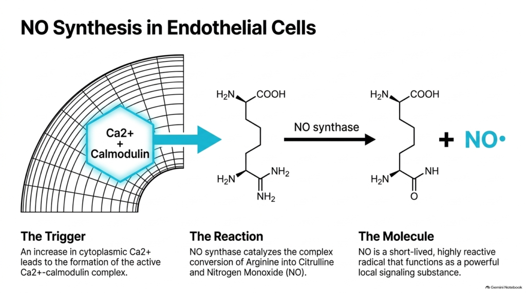
Turning to local paracrine communication, blood vessel dilation relies on endothelial synthesis of nitrogen monoxide. Vascular endothelial cells respond to chemical or mechanical stimuli by increasing intracellular calcium ion concentrations. Calcium ions bind to calmodulin, forming an active calcium-calmodulin complex that initiates crucial cardiovascular Signal Transduction Pathways.
The calcium-calmodulin complex binds to and activates the enzyme nitric oxide synthase within the endothelial cytoplasm. Nitric oxide synthase catalyzes a complex oxidation reaction that converts the amino acid arginine into citrulline and nitric oxide gas. Through these Signal Transduction Pathways, endothelial cells rapidly produce nitrogen monoxide radicals in response to heightened blood flow demands.
Nitric oxide is a tiny, uncharged radical molecule with an extremely short biological half-life. Because it is uncharged and gaseous, nitrogen monoxide diffuses out of endothelial cells instantly, without requiring transport proteins or membrane channels. These unique physical properties allow local Signal Transduction Pathways to operate over micro-distances surrounding blood vessels.
The enzymatic production of nitrogen monoxide represents a pivotal mechanism in circulatory physiology. Defects in endothelial nitrogen monoxide synthesis severely impair these Signal Transduction Pathways, contributing to hypertension and vascular disease. Examining arginine conversion provides insight into how simple chemical radicals regulate complex cardiovascular functions.
Slide 09: Paracrine Diffusion of NO in Signal Transduction Pathways
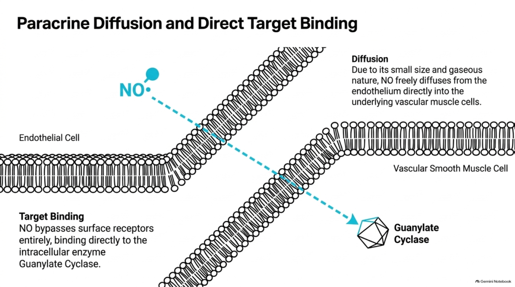
Once synthesized in endothelial cells, nitrogen monoxide diffuses across cellular boundaries to target adjacent tissues. Because of its small size and lipophilic, gaseous nature, nitrogen monoxide easily crosses the plasma membranes of both endothelial cells and underlying vascular smooth muscle cells. This direct diffusion bypasses the traditional cell-surface receptor-binding steps seen in hormonal Signal Transduction Pathways.
Inside smooth muscle cells, nitrogen monoxide binds directly to the iron atom in the heme group of soluble guanylate cyclase. This direct binding induces a conformational change that dramatically activates guanylate cyclase. Unlike membrane-bound hormone receptors, these intracellular signal transduction pathways use an internal soluble protein as their primary sensor for incoming gaseous signals.
This direct binding mechanism allows for extraordinarily rapid signal transmission between adjacent cell layers. Because nitrogen monoxide degrades within seconds, guanylate cyclase activation remains tightly localized to the site of gas release. Thus, these Signal Transduction Pathways restrict their physiological effects to local vascular beds without causing systemic blood pressure drops.
The paracrine diffusion model illustrates how physical properties like solubility and half-life dictate signaling behavior. By avoiding membrane receptors and circulating carriers, these gas-mediated Signal Transduction Pathways achieve targeted local communication. Studying nitrogen monoxide movement helps students appreciate how non-traditional messengers function in vascular control.
Slide 10: Smooth Muscle Relaxation and cGMP in Signal Transduction Pathways
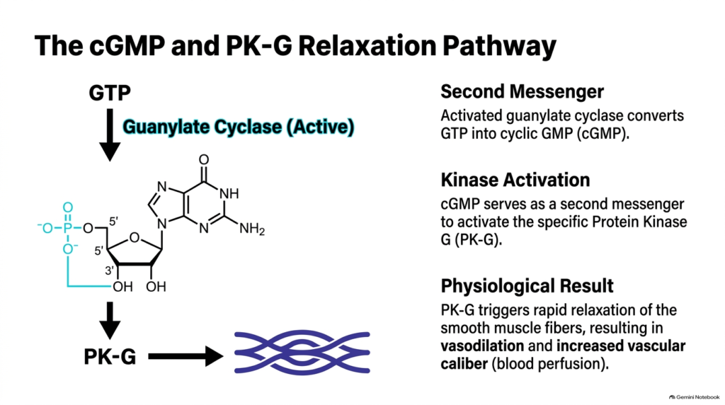
Activated guanylate cyclase inside vascular smooth muscle cells converts GTP into the key second messenger cyclic GMP. Accumulated cyclic GMP binds to and activates protein kinase G, launching intracellular Signal Transduction Pathways that control muscle contraction. Active protein kinase G phosphorylates specific ion channels and calcium pumps, lowering cytoplasmic calcium ion concentrations within the muscle cell.
Reduced intracellular calcium levels cause smooth muscle filaments to detach and relax around the vessel lumen. As smooth muscle cells relax, the internal diameter of blood vessels expands, a process called vasodilation. Through these cGMP-dependent Signal Transduction Pathways, blood flow increases significantly while local vascular resistance drops across active tissue beds.
Phosphodiesterase enzymes eventually terminate the cyclic GMP messenger signal by hydrolyzing cyclic GMP back into inactive GMP. By regulating phosphodiesterase activity, cells maintain precise temporal control over vessel diameter and blood perfusion. Inhibiting phosphodiesterase enzymes prolongs these relaxing Signal Transduction Pathways, providing a major therapeutic mechanism for cardiovascular conditions.
This pathway demonstrates how intracellular second messengers translate chemical signals into mechanical tissue relaxation. The generation of cyclic GMP and subsequent protein kinase G activation form the backbone of modern vascular pharmacology. Understanding this pathway explains how local blood flow dynamically adjusts to match metabolic needs.
Slide 11: Clinical Interventions and ANP in Signal Transduction Pathways
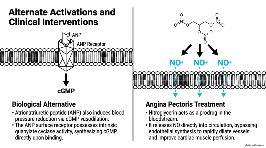
Pharmacological therapies often target cyclic GMP signaling to manage cardiovascular diseases such as angina pectoris. Nitroglycerin acts as a prodrug that breaks down in the bloodstream, releasing nitric oxide radicals directly into circulation. This pharmaceutical delivery bypasses damaged endothelial cells, activating smooth muscle Signal Transduction Pathways to dilate coronary arteries and relieve cardiac ischemia.
Endogenous peptide hormones can also trigger vasodilation through alternative receptor mechanisms. Atrial natriuretic peptide binds to transmembrane receptors with intrinsic guanylate cyclase activity in their intracellular domains. Ligand binding directly stimulates cyclic GMP synthesis without requiring nitrogen monoxide release, activating parallel Signal Transduction Pathways for blood pressure regulation.
Both nitroglycerin therapy and atrial natriuretic peptide signaling converge on cyclic GMP production inside vascular smooth muscle. Whether activated by exogenous gas donors or endogenous peptide hormones, these redundant Signal Transduction Pathways ensure effective regulation of vascular tone and fluid balance. These therapeutic models highlight how cardiovascular drugs exploit natural signaling machinery.
Understanding these parallel pathways allows medical students to connect molecular biology with clinical pharmacology. By targeting different entry points in these Signal Transduction Pathways, physicians can precisely control vasodilation in patients with heart disease. This clinical application demonstrates the practical importance of mastering signal transduction research.
Slide 12: Comparative Paradigms of Signal Transduction Pathways
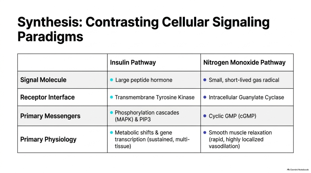
Comparing insulin and nitrogen monoxide signaling highlights two contrasting paradigms of cellular communication across biological systems. The insulin pathway utilizes a large peptide hormone that binds transmembrane tyrosine kinase receptors, triggering protein phosphorylation cascades and lipid modifications. These elaborate Signal Transduction Pathways produce sustained metabolic shifts and changes in gene expression across multiple organs.
Conversely, the nitrogen monoxide pathway relies on a small gas radical that diffuses across membranes to activate intracellular guanylate cyclase. This paracrine network generates cyclic GMP second messengers to induce rapid, highly localized smooth muscle relaxation. Comparing these Signal Transduction Pathways illustrates how signal molecule structure determines receptor location, signal speed, and overall physiological scope.
Both systems demonstrate the fundamental principles of biological signal amplification, specificity, and feedback control across tissues. While insulin coordinates systemic energy storage over hours, nitrogen monoxide adjusts local tissue perfusion within seconds. Integrating these two models gives students a complete conceptual framework for evaluating diverse Signal Transduction Pathways in medicine.
Ultimately, mastering these contrasting paradigms reveals how cells synthesize chemical inputs to maintain homeostasis. Whether managing blood glucose or regulating vascular diameter, cells employ tailored signaling machinery suited to their physiological demands. This comparative synthesis completes our detailed analysis of biochemical signal cascades.
Please read our Content Disclaimer Statement.
Check out our social media channels: