103. Hemoglobin Oxygen Transport: Mechanisms of Allosteric Regulation
Breathing is automatic, yet the microscopic journey of a single oxygen molecule is a marvel of biological engineering. Without a dedicated transport system, complex life would suffocate. This blog post explores the precise biochemical mechanisms that allow the body to sustain oxidative metabolism. The following slide deck breaks down the structures, allosteric regulation, and thermodynamic cycles that govern this vital process. Designed for medical and college students, this comprehensive guide reveals the chemical elegance keeping human tissues alive and thriving.
Slide 1: Introduction to Hemoglobin Oxygen Transport
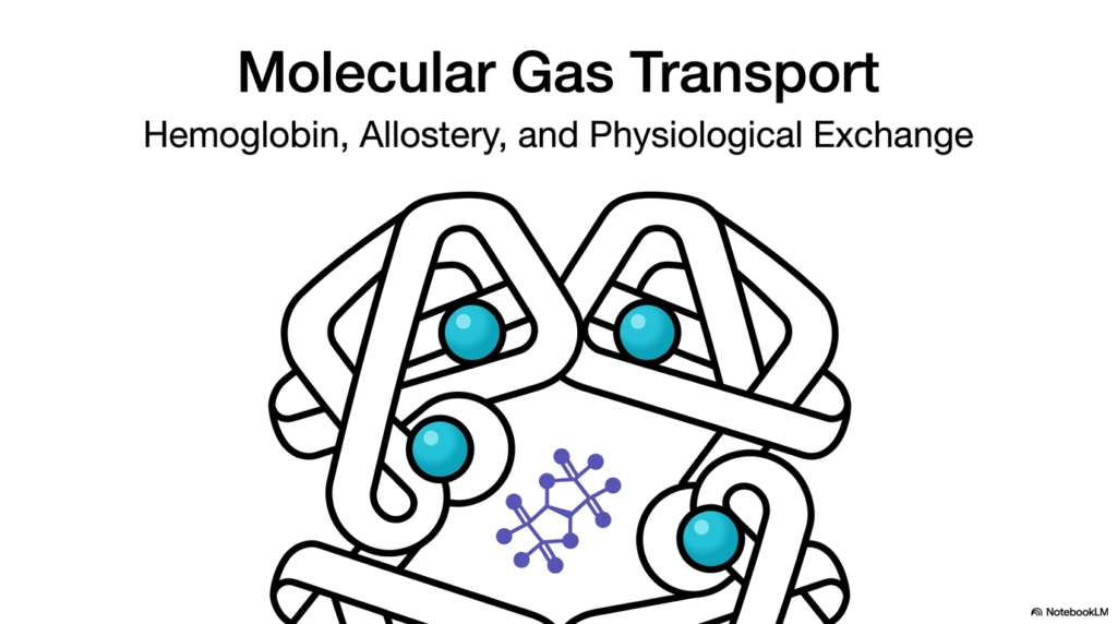
The process of Hemoglobin Oxygen Transport is an extraordinary feat of molecular biology that governs how complex life functions at the systemic level. This opening slide introduces the overarching theme of molecular gas exchange, focusing on the intricate metalloprotein that transports life-sustaining gases throughout the bloodstream. The visual prominently features the complex quaternary structure of this essential protein, highlighting its multiple subunits. By understanding Hemoglobin Oxygen Transport, medical students gain critical insight into the fundamental physiological process that sustains human tissue viability and function.
A core biochemical concept introduced here is allostery, which refers to structural alterations that occur when molecules bind to specific regulatory sites on a protein. This structural flexibility allows the transport protein to respond dynamically to the body’s shifting metabolic environments. Efficient Hemoglobin Oxygen Transport relies entirely on these shape-shifting capabilities to load gases in the lungs and unload them in peripheral tissues. Rather than acting as a simple, static container, the molecule functions as a highly responsive, finely tuned biological machine.
Mastering the mechanics of Hemoglobin Oxygen Transport provides the necessary foundation for understanding advanced respiratory physiology and pathology. The featured tetrameric structure sets the stage for exploring how four distinct protein subunits communicate and cooperate in structural harmony. This cooperative behavior ensures that the relentless cellular demand for oxidative energy is consistently met by a reliable fuel supply. The upcoming slides will dissect exactly how this complex, three-dimensional architecture governs physiological gas exchange at the microscopic level.
The intricate design of this protein demonstrates how form strictly dictates function in biochemistry. The spatial arrangement of the subunits creates specific binding environments that are highly sensitive to external chemical signals. Recognizing this structure-function relationship is crucial for any rigorous study of molecular gas transport. These concepts form the bedrock of understanding how the human body sustains continuous metabolic activity.
Slide 2: The Molecular Blueprint of Hemoglobin Oxygen Transport
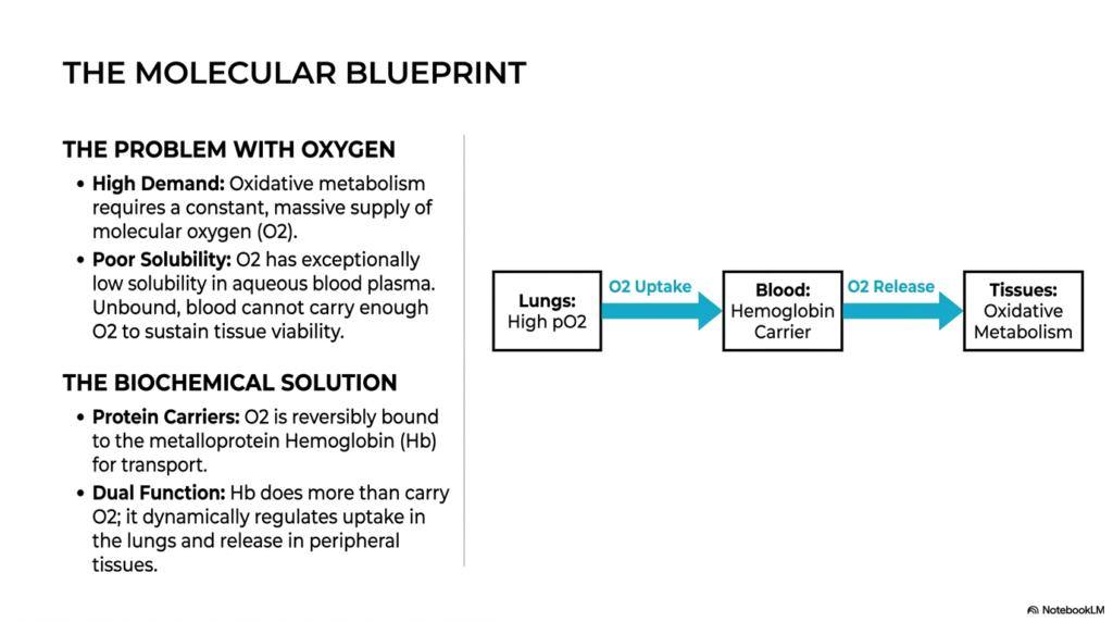
The necessity for Hemoglobin Oxygen Transport arises from a fundamental chemical limitation involving solubility and metabolic demand. Human tissues rely on oxidative metabolism, which requires a massive, uninterrupted supply of molecular oxygen. However, oxygen has an exceptionally low solubility in aqueous solutions such as blood plasma. Without a specialized mechanism like Hemoglobin Oxygen Transport, the bloodstream could never deliver enough dissolved gas to sustain even basic tissue viability. This biochemical puzzle forces the body to rely on specialized molecular machinery to prevent cellular asphyxiation.
To overcome this severe biological problem, nature developed a biochemical solution utilizing specialized protein carriers. The slide illustrates how oxygen reversibly binds to a metalloprotein to facilitate mass transit through the circulatory system. This mechanism highlights why Hemoglobin Oxygen Transport is far superior to simple diffusion. The protein carrier actively concentrates the gas, packing a massive payload into the red blood cells to meet the relentless demands of cellular respiration in peripheral tissues.
Furthermore, the slide emphasizes that this carrier performs a dual function, dynamically regulating both uptake and release. Effective Hemoglobin Oxygen Transport requires the molecule to bind gas tightly in the oxygen-rich environment of the lungs, yet release it easily in the oxygen-deprived environment of working muscles. This highly calibrated loading and unloading process ensures that vital tissues receive exactly the amount of fuel they need to maintain continuous oxidative metabolism.
The flowchart provided in the slide elegantly maps this continuous physiological cycle. It traces the journey from areas of high partial pressure in the lungs to areas of high metabolic consumption in the tissues. Understanding this directional flow is paramount for students studying the thermodynamic principles that drive biological gas exchange. By acting as an intermediary between the atmosphere and the cell, the protein carrier perfectly resolves the inherent limitations of aqueous blood plasma.
Slide 3: Myoglobin vs. Hemoglobin Oxygen Transport
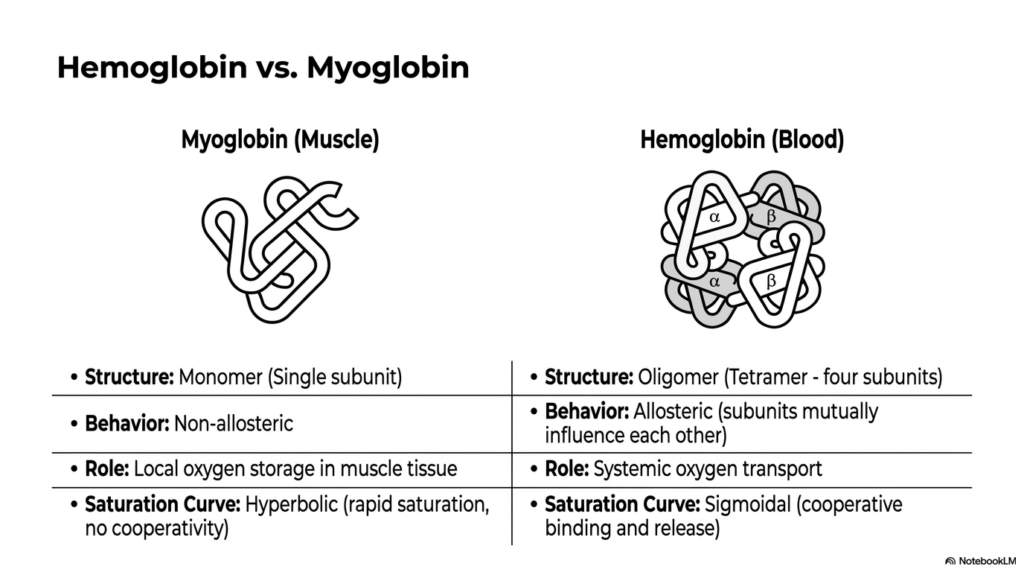
To truly appreciate the sophistication of Hemoglobin Oxygen Transport, one must compare it to the simpler mechanism of myoglobin. This slide presents a side-by-side structural and functional analysis of these two related, yet distinct, metalloproteins. Myoglobin functions as a monomer, meaning it consists of a single polypeptide chain designed strictly for local storage in muscle tissue. In contrast, the protein responsible for Hemoglobin Oxygen Transport is an oligomer, composed of four interacting subunits that make systemic delivery possible.
The structural differences between the two molecules dictate entirely different biochemical behaviors and saturation curves. Myoglobin exhibits a hyperbolic saturation curve, indicating that it binds gas rapidly and independently without any allosteric communication. This makes it an excellent storage unit but a poor delivery vehicle. Conversely, Hemoglobin Oxygen Transport relies on a sigmoidal saturation curve driven by cooperative binding. The four subunits of the tetramer influence one another, enabling the molecule to sense and respond to varying physiological environments.
This cooperative behavior is the defining feature of efficient Hemoglobin Oxygen Transport across the human body. Because the tetramer is an allosteric protein, it can retain its payload in the lungs and rapidly release it when it encounters the lower partial pressures in peripheral tissues. Myoglobin, lacking this cooperative release mechanism, would simply hold onto the gas, starving the tissues. Therefore, the oligomeric structure is an absolute biological necessity for dynamic, systemic gas transport.
The visual comparison between the single-subunit structure and the four-subunit complex perfectly illustrates this functional divergence. Students must recognize that while both proteins use similar metal-binding chemical groups, their overall quaternary structure dictates their physiological fate. The multi-subunit design transforms a simple storage molecule into a highly sophisticated, environment-sensing delivery system. This evolutionary adaptation is what allows large, multicellular organisms to sustain high metabolic rates across vast networks of circulatory vessels.
Slide 4: Allostery and Homotropic Effects in Hemoglobin Oxygen Transport
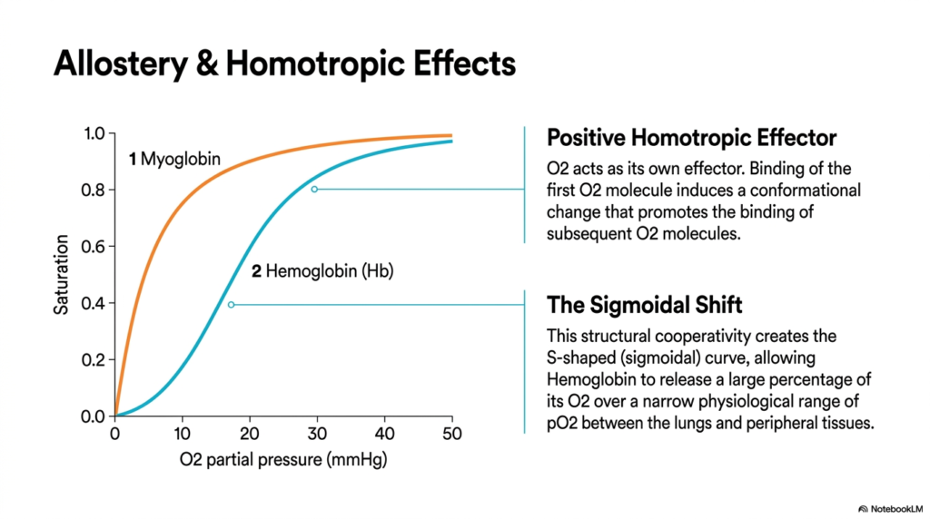
The sigmoidal curve central to Hemoglobin Oxygen Transport is physically generated by positive homotropic effects. This slide delves into the mathematics and structural mechanics of cooperative binding, illustrating how the ligand acts as its own effector. When the first molecule binds to a completely empty tetramer, the process is thermodynamically unfavorable. However, this initial binding event induces a crucial conformational shift that significantly increases the binding affinity of the remaining empty subunits for subsequent binding events.
This structural cooperativity is the molecular engine driving Hemoglobin Oxygen Transport. As each subsequent molecule attaches, the protein transitions from a tense state to a relaxed state, making it progressively easier to reach full saturation. The resulting S-shaped or sigmoidal curve allows the protein to release a massive percentage of its payload over a very narrow physiological range. Without this precise sigmoidal shift, Hemoglobin Oxygen Transport would be wildly inefficient, functioning poorly in both the lungs and the tissues.
The slide visually contrasts this sigmoidal behavior with the simple hyperbolic curve of a monomeric storage protein. The steep portion of the sigmoidal curve corresponds exactly to the partial pressures found in peripheral tissues. This means that a small drop in partial pressure triggers a massive release of the necessary gas. Understanding this homotropic regulation is vital for students, as it clearly demonstrates how protein conformation governs physiological efficiency in systemic circulation.
Biochemists refer to this phenomenon as allosteric regulation, where binding at one site influences the structural integrity of distant sites on the same macromolecule. The precise communication between the four subunits ensures that the molecule remains exquisitely sensitive to its immediate environment. This environment-sensing capability prevents premature release of the payload into the arteries while ensuring rapid offloading into the capillaries. Ultimately, this structural transition mechanism is the definitive hallmark of a sophisticated biological delivery system.
Slide 5: Metabolic Effectors and Hemoglobin Oxygen Transport
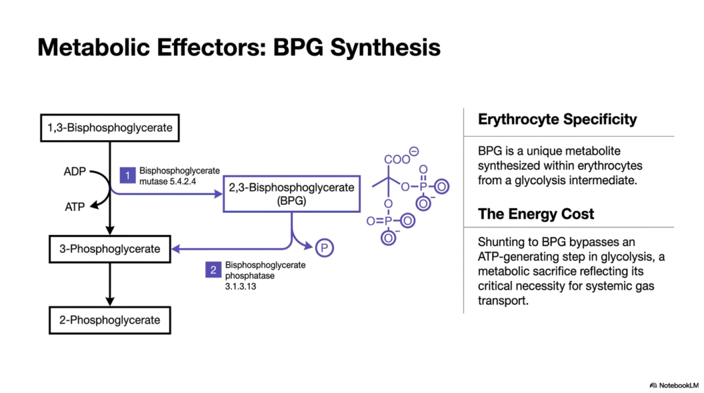
Beyond homotropic cooperativity, Hemoglobin Oxygen Transport is tightly regulated by specific metabolic effectors synthesized within red blood cells. This slide introduces 2,3-Bisphosphoglycerate (BPG), a critical heterotropic modulator that permanently alters the protein’s binding affinity. BPG is a unique metabolite produced by a side reaction of glycolysis, specifically catalyzed by bisphosphoglycerate mutase. The synthesis of this molecule is a specialized adaptation unique to erythrocytes, directly serving the complex needs of systemic gas delivery mechanisms.
The creation of BPG highlights the immense metabolic importance of efficient Hemoglobin Oxygen Transport. Shunting the glycolytic pathway to produce BPG actually bypasses a standard ATP-generating step in glycolysis. This represents a significant metabolic sacrifice for the erythrocyte, forfeiting potential cellular energy to ensure that the systemic delivery system functions properly. This deliberate energy cost underscores just how essential BPG regulation is to maintaining functional Hemoglobin Oxygen Transport under widely varying physiological demands.
Students must understand that this biochemical investment is non-negotiable for human survival. By heavily investing in BPG production, the red blood cell gains the ability to fine-tune the delivery of fuel to distant tissues. The enzymes involved in this shunt pathway are highly active in erythrocytes, constantly maintaining an optimal concentration of the effector molecule. This slide provides the necessary metabolic context, showing how cellular energy pathways are intimately linked to broader physiological requirements.
Without this specific metabolic diversion, the entire transport system would fail to unload sufficient amounts of the life-sustaining payload. The intricate balance between energy production and allosteric regulation demonstrates the beautiful economy of cellular biochemistry. The erythrocyte willingly sacrifices its own energy currency to guarantee that vital organs receive the chemical resources required for sustained oxidative respiration. This metabolic compromise is a cornerstone concept in understanding systemic hematology and cellular bioenergetics.
Slide 6: How BPG Shunts Hemoglobin Oxygen Transport
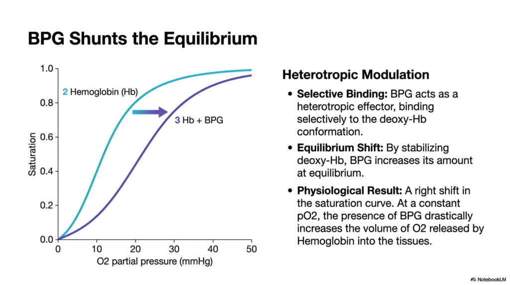
The physiological consequence of BPG synthesis is a profound shift in the equilibrium of Hemoglobin Oxygen Transport. This slide visualizes the mechanical impact of BPG through a clear graphical representation of heterotropic modulation. BPG acts by selectively binding to the central cavity of the deoxygenated protein conformation. By stabilizing this tense, deoxygenated state, the effector molecule shifts the structural equilibrium, making it considerably harder for the protein to hold onto its valuable payload.
This stabilization directly dictates the efficiency of Hemoglobin Oxygen Transport in the capillary beds. The graph illustrates a distinct rightward shift in the saturation curve when BPG is introduced to the system. At a constant partial pressure found in peripheral tissues, this rightward shift drastically increases the volume of gas released into the surrounding cells. Without BPG interacting with the tetramer, Hemoglobin Oxygen Transport would trap the gas in the blood, starving the tissues of necessary resources.
Therefore, BPG functions as a powerful biochemical lever, prying the protein apart to facilitate offloading where it matters most. The selective binding relies on precise electrostatic interactions between the negatively charged phosphate groups of BPG and the positively charged amino acids in the protein’s central cavity. Medical students must recognize this interaction as a perfect example of how small metabolites control massive physiological systems. The presence of this effector molecule guarantees that systemic delivery remains highly responsive.
This allosteric modulation serves as a crucial fail-safe, ensuring that the transport protein’s affinity never becomes too high. By shifting the saturation curve to the right, the biochemical environment of the erythrocyte actively promotes offloading. This shifting equilibrium is particularly important during states of high physiological stress or high altitude, where maximizing delivery efficiency becomes a matter of immediate survival. The elegant simplicity of this electrostatic interaction drives the complexity of human respiration.
Slide 7: The Bohr Effect in Hemoglobin Oxygen Transport
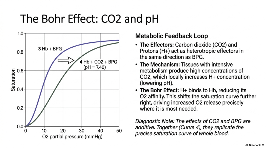
The chemical environment of active tissues introduces additional regulatory factors that optimize Hemoglobin Oxygen Transport. This slide explores the Bohr Effect, detailing how carbon dioxide and protons serve as potent heterotropic effectors. Tissues undergoing intensive oxidative metabolism rapidly produce high concentrations of carbon dioxide, which subsequently increases local proton concentration and lowers the ambient pH. This acidic environment acts as a chemical signal, biochemically alerting the red blood cells to areas of high metabolic demand.
The Bohr Effect powerfully enhances Hemoglobin Oxygen Transport precisely where tissues require the most support. Protons bind directly to specific amino acid residues on the protein structure, stabilizing the deoxygenated conformation and significantly reducing binding affinity. This creates an even further rightward shift in the saturation curve, driving increased release exactly where metabolism is highest. This feedback loop ensures that Hemoglobin Oxygen Transport dynamically adapts, delivering more fuel to tissues working the absolute hardest.
Importantly, the slide notes that the regulatory effects of protons, carbon dioxide, and BPG are completely additive. Together, these three heterotropic modulators replicate the exact, naturally occurring saturation curve of whole blood observed in living organisms. Understanding this cumulative regulation is essential for students analyzing respiratory mechanics. The combined biochemical forces of the Bohr Effect and metabolic effectors transform a simple carrier protein into a highly intelligent, environment-sensing delivery vehicle that perfectly matches physiological supply with cellular demand.
This intricate chemical interplay demonstrates the elegant thermodynamic principles governing systemic respiration. The waste products of cellular metabolism directly facilitate the acquisition of fresh energetic resources. By utilizing acidic byproducts as allosteric triggers, the biological system creates a perfectly self-regulating cycle. Medical professionals rely on this principle to understand pathological states involving acidosis or alkalosis, as these pH imbalances fundamentally disrupt the overall efficiency of the entire molecular gas transport mechanism.
Slide 8: Modes of CO2 in Hemoglobin Oxygen Transport
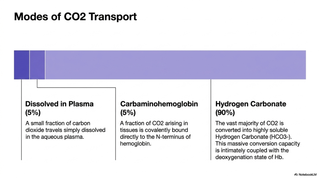
While delivering fuel is critical, managing metabolic waste is equally important for the overall cycle of Hemoglobin Oxygen Transport. This slide transitions to the mechanisms of carbon dioxide removal, outlining the three distinct modes of transport. Because of its low solubility, only about 5% of this waste gas travels dissolved in the aqueous plasma. Another five percent binds directly to the N-terminus of the protein subunits, forming a complex known as carbaminohemoglobin.
The vast majority of the waste, approximately ninety percent, undergoes a massive chemical conversion essential to Hemoglobin Oxygen Transport. It is rapidly converted into highly soluble hydrogen carbonate, more commonly known as bicarbonate. This extensive conversion capacity prevents the dangerous buildup of gas bubbles in the bloodstream and maintains strict acid-base balance. The process of Hemoglobin Oxygen Transport is closely coupled to this bicarbonate system, as the deoxygenation of the protein directly helps accommodate newly formed ions.
This detailed breakdown illustrates that the circulatory system requires sophisticated enzymatic assistance to handle cellular waste. The formation of carbamino compounds and bicarbonate ensures that tissues do not become dangerously acidic during intense physical exertion. Students evaluating these transport modes must recognize that clearing this waste is just as biochemically complex as delivering the initial fuel. The entire circulatory process is a delicate balancing act, heavily reliant on rapid, reversible chemical modifications to sustain continuous metabolic activity.
The exact distribution of these transport modes highlights the limitations of simple fluid dynamics in biological systems. Relying solely on plasma dissolution would lead to fatal toxicity within minutes. By utilizing the erythrocyte’s massive surface area and internal machinery, the body safely shuttles toxic byproducts back to the external environment. This multi-modal approach to waste management is a defining characteristic of advanced vertebrate physiology, perfectly complementing the primary respiratory delivery system.
Slide 9: Tissue Capillary Exchange in Hemoglobin Oxygen Transport
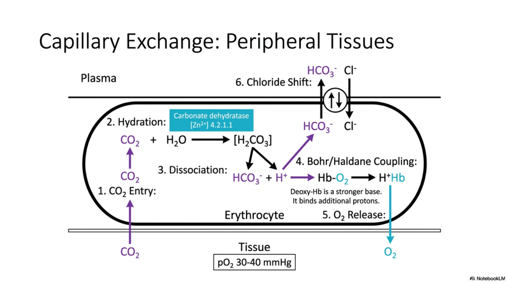
The physiological magic of Hemoglobin Oxygen Transport occurs at the microscopic interface between capillaries and peripheral tissues. This slide maps the complex enzymatic and ionic exchanges that occur within the erythrocyte as it navigates through deep tissue beds. Carbon dioxide freely diffuses into the red blood cell, where the enzyme carbonic anhydrase rapidly hydrates it into carbonic acid. This intermediate quickly dissociates into bicarbonate and a free proton, initiating a cascade of vital structural shifts.
This biochemical cascade is the physical execution of Hemoglobin Oxygen Transport at the cellular level. The newly generated proton binds directly to the allosteric protein, forcing it into its tense state and ejecting the molecular payload into the oxygen-starved tissue. This remarkable mechanism is known as the Bohr/Haldane coupling. It demonstrates how Hemoglobin Oxygen Transport utilizes the very waste products of the tissue to mechanically pry the protein open, forcing it to relinquish its precious cargo.
Simultaneously, the massive efflux of bicarbonate into the blood plasma requires a balancing mechanism known as the chloride shift. To maintain electrical neutrality across the erythrocyte membrane, chloride ions rush into the cell as bicarbonate exits. Students must study this diagram carefully, as it visualizes the simultaneous execution of gas delivery, waste processing, and ionic balancing. This synchronized capillary exchange represents the absolute pinnacle of biochemical efficiency, ensuring that the cellular environment remains chemically stable.
The role of carbonate dehydratase cannot be overstated, as it accelerates the hydration reaction by a factor of millions, matching the rapid speed of circulatory transit. Without this specific metalloenzyme, the entire coupling process would grind to a devastating halt. The erythrocyte acts as a microscopic chemical processing plant, seamlessly exchanging molecules across its lipid bilayer. This complex choreography of ions and protons guarantees the ongoing success of systemic respiration and tissue survival.
Slide 10: Lung Capillary Exchange in Hemoglobin Oxygen Transport
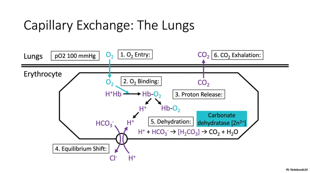
When venous blood returns to the pulmonary system, the process of Hemoglobin Oxygen Transport completely reverses the biochemical machinery involved. This slide illustrates capillary exchange in the lungs, where the overwhelming abundance of atmospheric gases drives the equilibrium in the opposite direction. As the payload rapidly diffuses into the erythrocyte and binds to the protein, it violently forces the macromolecule back into its relaxed conformation. This structural shift is the crucial first step in pulmonary capillary exchange.
By shifting into the relaxed state, the protein loses its affinity for protons, initiating the crucial waste-removal phase of Hemoglobin Oxygen Transport. The ejected protons immediately recombine with incoming bicarbonate ions, re-forming carbonic acid. The enzyme carbonate dehydratase runs its reaction in reverse, rapidly dehydrating the acid back into water and carbon dioxide gas. This massive release of protons upon loading is exactly what drives the efficient exhalation of waste products out of the human body during Hemoglobin Oxygen Transport.
This pulmonary exchange perfectly mirrors the events in the peripheral tissues, but in strict thermodynamic reverse. The reverse chloride shift pulls bicarbonate back into the cell while expelling chloride, maintaining the erythrocyte’s electrical balance. Understanding this pulmonary side of the equation is vital for students, as it demonstrates how a single protein complex utilizes concentration gradients to perform two entirely different tasks. The high partial pressure in the alveoli provides the energetic brute force required here.
This reversal highlights the incredible adaptability of the transport mechanisms. The same enzymatic pathways and allosteric triggers that facilitated unloading are now weaponized to facilitate rapid loading and waste elimination. The elegant symmetry of this biological design ensures that no energy is wasted during the respiratory process. The lungs serve not just as a site of diffusion, but as a dynamic biochemical reaction chamber that completely primes the erythrocyte for its next journey through the circulatory network.
Slide 11: The Coupled Cycle of Hemoglobin Oxygen Transport
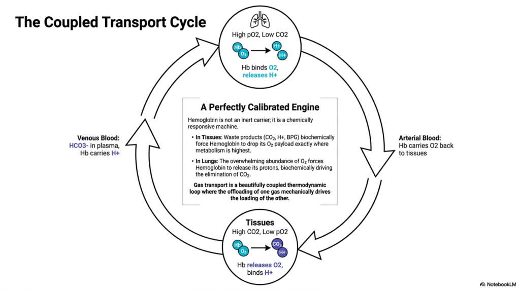
The entirety of Hemoglobin Oxygen Transport functions as a beautifully calibrated, closed-loop thermodynamic engine. This final slide synthesizes the entire process, demonstrating how gas transport is not just a series of isolated chemical reactions, but a fully coupled cycle. In peripheral tissues, the accumulation of waste products physically forces the protein to release its payload precisely where metabolism is highest. This illustrates that the carrier is not merely an inert vessel, but a highly responsive machine.
The continuous loop of Hemoglobin Oxygen Transport relies on the principle that the offloading of one gas mechanically drives the loading of another. In arterial blood, the protein carries vital fuel to tissues, while in venous blood, it actively helps transport protons back to the lungs. This deeply coupled nature of Hemoglobin Oxygen Transport guarantees that cellular respiration and pulmonary exhalation are perfectly synchronized. An imbalance in one half immediately alters the other.
Ultimately, students must view this system as a masterpiece of evolutionary biochemistry. The overwhelming abundance of gas in the lungs forces the elimination of waste, while the abundance of waste in the tissues forces the release of fuel. This elegantly balanced thermodynamic loop requires no external energy input beyond the concentration gradients generated by breathing and metabolism. Understanding this perfectly calibrated engine is the absolute key to mastering the complexities of human physiology and systemic respiratory mechanics.
Viewing the erythrocyte as a reciprocating biological piston makes the system’s mechanical genius incredibly clear. The continuous cycling between tense and relaxed molecular states drives the massive physiological shifts required for human life. Medical professionals apply these exact principles daily when managing patients on ventilators or treating severe metabolic disorders. The fundamental rules governing this microscopic cycle ultimately determine the health, resilience, and viability of the entire macroscopic human organism.
Please read our Content Disclaimer Statement.
Check out our social media channels: