95. Digestion and Resorption: Guide to Enzymatic Hydrolysis
Think of the human body as a highly advanced refinery. It takes in complex raw materials and breaks them down into usable fuel. This slide deck explores the fundamental biochemical mechanisms that power this process. Students will discover how enzymes dismantle large macromolecules and how cells transport these nutrients to sustain life. The following explanations will guide learners through the essential concepts of this vital biological system.
Slide 1: Introduction to Digestion and Resorption
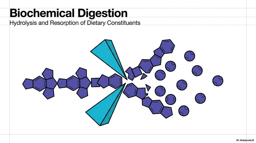
The visual represented here serves as a foundational model for understanding Digestion and Resorption. At its core, the human body must convert massive, complex dietary structures into simple, usable molecular units. This initial illustration highlights the central biochemical theme of breaking down large polymer chains into individual monomers. A stylized enzyme acts as a wedge, representing the vital biological scissors that make nutrient acquisition possible. Without this precise dismantling mechanism, the energy locked within our food would remain completely inaccessible to human cells, rendering the entire feeding process useless for sustaining life.
The physiological process of Digestion and Resorption is not just a mechanical event but a highly orchestrated biochemical cascade. Large macromolecules ingested during a standard meal are far too bulky to pass through the intricate intestinal lining. Therefore, the digestive system employs specific molecular tools to sever the chemical bonds holding these complex chains together. The visual captures the exact moment of enzymatic cleavage, where a larger dietary constituent is split into scattered, resorbable fragments. This fundamental transformation stands as the crucial first step in cellular nourishment and systemic energy distribution.
By studying this opening diagram, students can clearly visualize the ultimate goal of Digestion and Resorption. The image emphasizes that large structural chains must become single, isolated units before they can safely enter the bloodstream. The scattered spheres in the graphic represent the final biochemical cleavage products ready for cellular uptake. This elegant, simplified view prepares medical learners for the more complex enzymatic reactions and intricate transport mechanisms detailed in subsequent sections. Mastering this broad conceptual framework makes it easier for future healthcare professionals and scientists to grasp the specific, detailed biochemical pathways.
This introductory slide sets a clear academic tone for the entire presentation. It strips away the anatomical complexity of the gastrointestinal tract to focus purely on the molecular interactions at play. By highlighting the interaction between the enzyme and the substrate, the image distills a massive physiological event into a single, understandable biochemical reaction.
Slide 2: The Biochemical Imperative of Digestion and Resorption
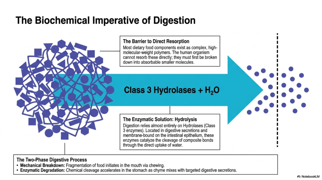
The biochemical imperative behind Digestion and Resorption centers on a major physiological barrier. Most dietary food components exist as complex, high-molecular-weight polymers. The human organism simply cannot absorb these massive structures directly across the intestinal barrier. To solve this problem, the body relies on a specific class of enzymes known as Class 3 Hydrolases. These specialized proteins catalyze the cleavage of composite bonds through the direct uptake of water, a process formally known as hydrolysis. This action transforms impenetrable polymers into smaller, absorbable molecules.
Understanding the two distinct phases of Digestion and Resorption is essential for medical students. The journey begins with mechanical breakdown in the mouth, where chewing fragments the food and increases its surface area. This physical disruption prepares the ingested material for the intense chemical processing that follows. Once the fragmented food reaches the stomach, it mixes with targeted digestive secretions to form chyme. Here, enzymatic degradation accelerates, utilizing acidic environments and specialized hydrolases to begin dismantling the complex macromolecules into their fundamental building blocks.
The integration of these mechanical and chemical steps highlights the sheer efficiency of Digestion and Resorption. Membrane-bound enzymes on the intestinal epithelium further refine this process, ensuring that every dietary polymer is broken down entirely. Water plays a crucial role here, as hydrolases require water molecules to break the chemical bonds linking the polymer subunits. Without this continuous enzymatic hydrolysis, nutrient extraction would halt entirely. The arrow in the diagram illustrates this decisive transformation, showing a chaotic tangle of polymers crossing a threshold to become ordered, resorbable particles.
Ultimately, this slide emphasizes that structural modification is a strict prerequisite for cellular uptake. The depicted barrier line represents the critical transition point where digestion ends, and resorption begins. By relying on hydrolases and water, the digestive tract ensures a steady supply of bioavailable nutrients. This systematic breakdown guarantees that the body can extract the maximum amount of energy and building materials from every ingested meal.
Slide 3: Protein Digestion and Resorption Mechanics
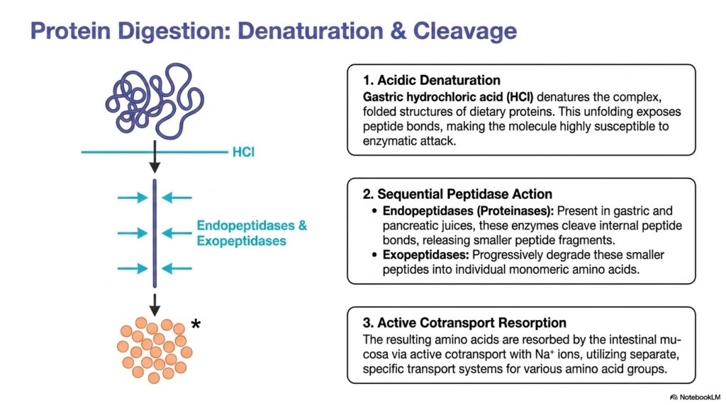
The specific pathway for protein Digestion and Resorption involves sequential stages of structural dismantling. Dietary proteins enter the stomach as complex, highly folded three-dimensional structures. Gastric hydrochloric acid initiates the first critical step by denaturing these proteins. This acidic environment forces the complex molecules to unfold, losing their protective shapes. Unfolding exposes the hidden peptide bonds holding the amino acid chain together. Once these bonds become accessible, the denatured protein chain becomes highly susceptible to subsequent enzymatic attack, setting the stage for precise chemical cleavage.
Following denaturation, the actual cleavage phase of Digestion and Resorption relies on a coordinated team of enzymes. Endopeptidases, also known as proteinases, are present in both gastric and pancreatic juices. These specialized enzymes attack the internal peptide bonds of the unfolded protein, chopping the long chain into smaller, manageable peptide fragments. Next, exopeptidases take over the process. These enzymes work at the ends of the fragments, progressively degrading the smaller peptides into individual, monomeric amino acids. This sequential teamwork ensures complete breakdown of the complex dietary protein.
The final stage of protein Digestion and Resorption involves active cellular transport. The resulting individual amino acids must cross the intestinal mucosa to be useful to the body. Enterocytes accomplish this via active cotransport mechanisms utilizing sodium ions. The cells maintain separate, highly specific transport systems to handle various distinct groups of amino acids. This sodium-dependent transport requires cellular energy, highlighting that the body actively pulls these vital nutrients inside rather than relying on passive diffusion, ensuring efficient nutrient acquisition.
This slide illustrates the beauty of sequential biochemical processing. The visual transition from a tangled coil to a straight line, and finally to individual orange spheres, perfectly models the protein degradation journey. Medical students must recognize that a failure at any stage, whether insufficient acid production or enzyme deficiency, disrupts the entire chain of nutrient acquisition. Proper protein breakdown is absolutely fundamental to maintaining muscle mass, enzyme production, and overall cellular repair.
Slide 4: Carbohydrate Digestion and Resorption Pathways
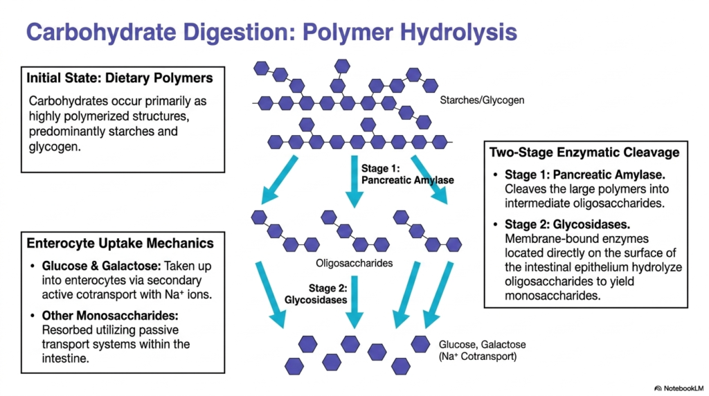
Carbohydrate Digestion and Resorption presents a unique biochemical challenge due to the massive size of dietary polymers. Most carbohydrates consumed in a standard diet exist as highly polymerized structures, predominantly starches and glycogen. These massive chains of sugar molecules must undergo a systematic, two-stage enzymatic cleavage before they can provide energy to human cells. The visual demonstrates the initial state of these polymers, highlighting the extensive web of glycosidic bonds that link the individual sugar units together. Breaking these bonds requires specific and highly coordinated enzymatic interventions.
The two-stage enzymatic cleavage is central to carbohydrate Digestion and Resorption. Stage one utilizes pancreatic amylase, a powerful enzyme that attacks the large dietary polymers. This initial hydrolysis cleaves the massive starches into intermediate, shorter chains called oligosaccharides. However, these oligosaccharides are still too large for cellular uptake. Stage two employs glycosidases, which are membrane-bound enzymes located directly on the brush border surface of the intestinal epithelium. These specialized enzymes perform the final hydrolysis, snipping the oligosaccharides into single, absorbable monosaccharides like glucose and galactose.
Understanding enterocyte uptake mechanics completes the picture of carbohydrate Digestion and Resorption. Once the enzymes produce free monosaccharides, the intestinal cells must transport them inward. Glucose and galactose enter the enterocytes through a specialized secondary active cotransport system that relies on sodium ions. This active pulling mechanism ensures that vital energy sources are efficiently gathered even against a concentration gradient. Meanwhile, other monosaccharides utilize passive transport systems to cross the intestinal barrier. This dual-transport approach guarantees maximum extraction of dietary sugars.
The schematic on this slide provides a clear visual map of progressive degradation. The transformation from complex interconnected webs to isolated hexagonal units illustrates the efficiency of pancreatic and intestinal enzymes working in tandem. For healthcare students, mastering this pathway is crucial for understanding metabolic disorders, diabetes management, and nutritional deficiencies. The complete conversion of starch to glucose is a cornerstone of human energy metabolism.
Slide 5: Nucleic Acid Digestion and Resorption
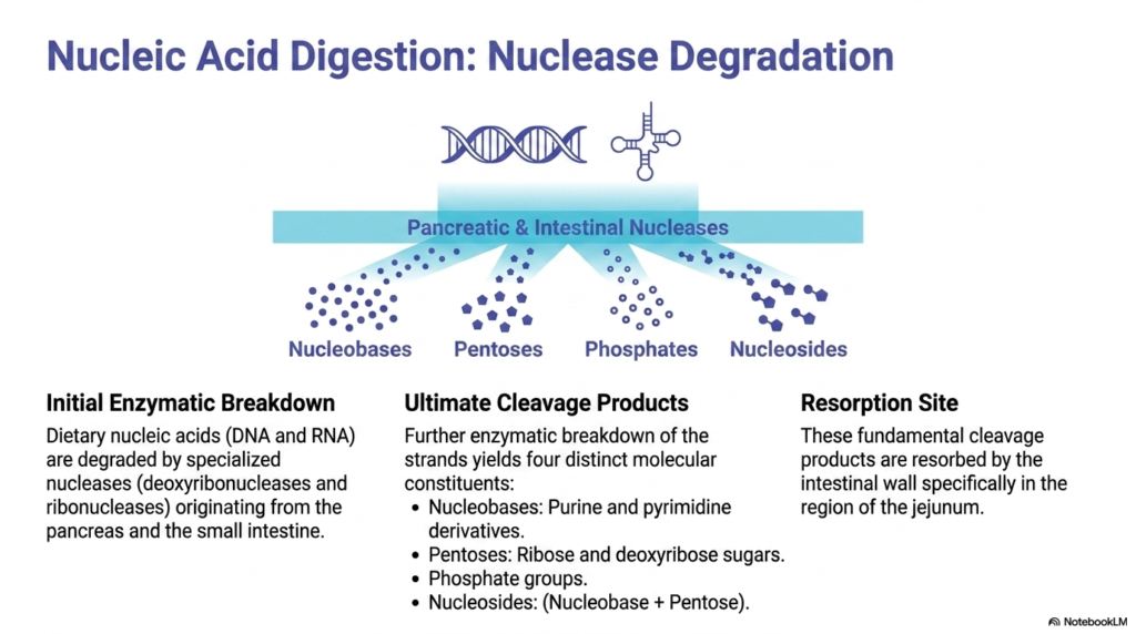
While often overshadowed by macronutrients, nucleic acid Digestion and Resorption is an essential biochemical process. The human diet contains cellular material from plants and animals, meaning DNA and RNA are regularly consumed. These complex genetic blueprints must be dismantled into smaller components to be recycled by the human body. The initial enzymatic breakdown relies on specialized nucleases, specifically deoxyribonucleases and ribonucleases. These powerful enzymes originate from both the pancreas and the small intestine, working together to shear the long nucleic acid strands into manageable fragments.
The ultimate cleavage products of nucleic acid Digestion and Resorption are diverse and biochemically valuable. As the enzymes continue their degradation work, the nucleic acid strands yield four distinct molecular constituents. The process generates nucleobases, which include purine and pyrimidine derivatives. It also releases pentoses, specifically ribose and deoxyribose sugars, alongside free phosphate groups. Furthermore, the breakdown produces nucleosides, which are combinations of a nucleobase and a pentose sugar. These fundamental building blocks form a pool of raw materials ready for cellular absorption.
The primary site for nucleic acid Digestion and Resorption occurs deeper in the intestinal tract. These fundamental cleavage products are absorbed by the intestinal wall specifically within the region of the jejunum. The slide’s visual elegantly demonstrates the transition from a recognizable double helix structure to a scattered array of diverse molecular shapes. This imagery reinforces the concept that complex genetic information is ultimately reduced to chemical components during digestion, ready to be repurposed by human cells.
For medical and biochemistry students, this pathway illustrates the incredible recycling capability of the human body. We do not incorporate foreign DNA into our own genome; instead, we strip it down for parts. The resulting nucleobases and sugars provide essential materials for synthesizing our own cellular DNA, RNA, and vital energy carriers like ATP. Understanding this degradation pathway completes the comprehensive picture of dietary macromolecule processing.
Slide 6: Lipid Digestion and Resorption: Phase I
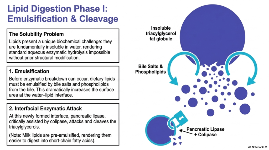
The initial phase of lipid Digestion and Resorption introduces a unique solubility problem. Dietary lipids are fundamentally insoluble in water, presenting a major obstacle in the aqueous environment of the gastrointestinal tract. Standard aqueous enzymatic hydrolysis is completely impossible without prior structural modification of these fat molecules. If left untreated, lipids would form massive, impenetrable globules, severely restricting enzyme access. To overcome this, the digestive system must first physically alter the state of the lipids before any chemical breakdown can occur.
To solve the solubility issue during lipid Digestion and Resorption, the body relies on emulsification. Before enzymatic breakdown begins, dietary lipids are attacked by bile salts and phospholipids secreted from the gallbladder. These biological detergents interact with the large, insoluble triacylglycerol fat globules, breaking them apart into millions of microscopic droplets. This crucial emulsification process dramatically increases the total surface area at the water-lipid interface. Milk lipids provide an exception, as they arrive pre-emulsified, rendering them significantly easier to digest into short-chain fatty acids.
Once emulsification is complete, the chemical phase of lipid Digestion and Resorption can proceed. At the newly expanded water-lipid interface, specialized enzymes launch a targeted interfacial attack. Pancreatic lipase is the primary enzyme responsible for this chemical degradation. However, lipase cannot function efficiently alone; it requires the critical assistance of colipase to anchor it to the lipid droplet surface. Together, this enzyme complex aggressively cleaves the triacylglycerols, breaking down the stubborn fat molecules into smaller, manageable chemical components.
The diagram provides a striking visual representation of this physical and chemical transformation. The large dark circle represents the inaccessible fat globule, while the curving arrows demonstrate the disruptive action of bile salts. Seeing the massive globule shatter into smaller spheres helps students visualize the massive increase in surface area. This preparatory stage is absolutely critical, as without successful emulsification, the subsequent action of pancreatic lipase would be severely crippled, leading to severe nutritional deficiencies.
Slide 7: Lipid Digestion and Resorption: Phase II
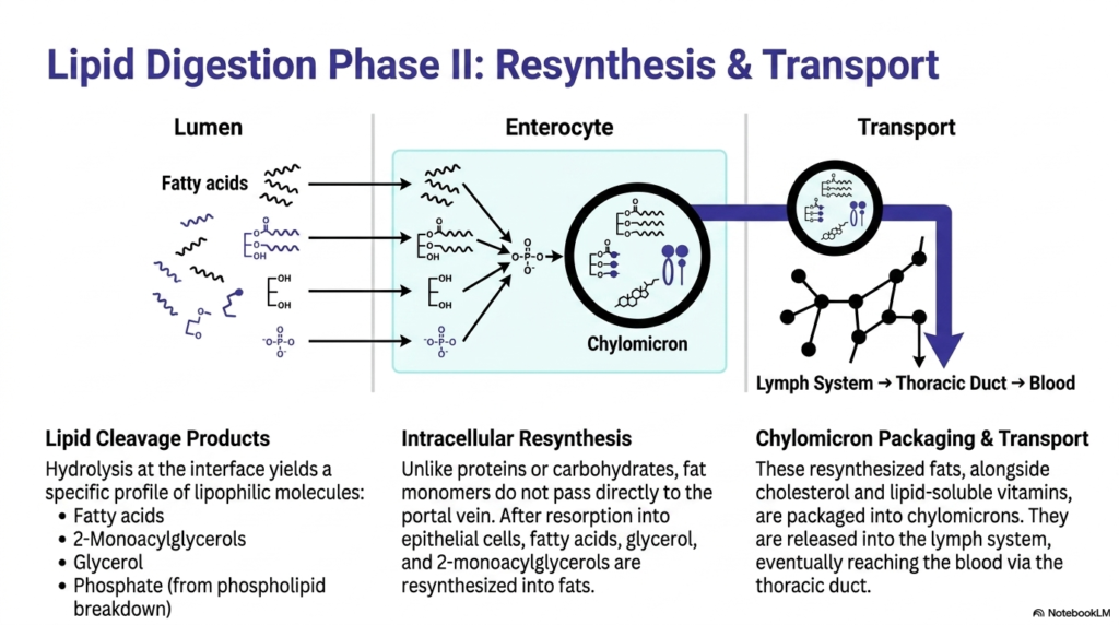
The second phase of lipid Digestion and Resorption focuses on intracellular processing and systemic transport. The hydrolysis at the water-lipid interface yields a very specific profile of lipophilic molecules. These lipid cleavage products include free fatty acids, 2-monoacylglycerols, glycerol, and free phosphate resulting from phospholipid breakdown. These components are small enough to pass from the intestinal lumen into the enterocyte. However, unlike hydrophilic nutrients, these fat monomers undergo a remarkable transformation once inside the epithelial cell, completely changing their transport destiny.
A defining characteristic of lipid Digestion and Resorption is the process of intracellular resynthesis. Unlike proteins or carbohydrates that pass straight through the cell, fat monomers do not pass directly to the portal vein. After entering the enterocyte, the free fatty acids, glycerol, and 2-monoacylglycerols are rapidly reassembled. The cellular machinery stitches these fragments back together, resynthesizing them into complex fats. This internal rebuilding step is entirely unique to lipid metabolism and is essential for the subsequent packaging and transport phases.
The final step in lipid Digestion and Resorption involves specialized chylomicron packaging and transport. The newly resynthesized fats, alongside dietary cholesterol and lipid-soluble vitamins, are bundled into large transport vesicles called chylomicrons. Because these bundles are too large and lipophilic for standard blood transport, they are extruded from the enterocyte into the lymphatic system. They travel through the lymph network, eventually reaching systemic blood circulation via the thoracic duct. This complex detour ensures that dense lipid packages enter the bloodstream safely.
This slide beautifully maps out the lumen-to-blood journey of a fat molecule. The visual separation into three distinct zones—Lumen, Enterocyte, and Transport—clarifies a notoriously complex topic for medical scholars. Tracing the breakdown of molecules on the left to their reconstruction and packaging on the right solidifies the concept of resynthesis. Understanding this lymphatic bypass is crucial for grasping how the body manages cholesterol distribution and avoids overwhelming the liver with sudden influxes of dietary fats.
Slide 8: Inorganic Components in Digestion and Resorption
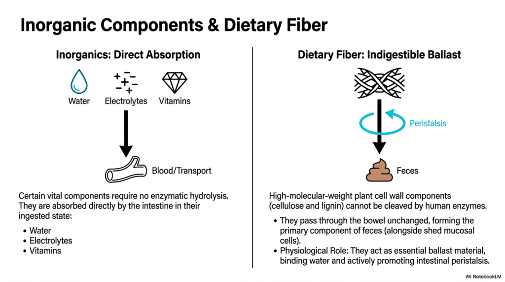
Not all dietary materials require complex breakdown during Digestion and Resorption. Certain vital components, primarily inorganic substances, entirely bypass the need for enzymatic hydrolysis. Water, various essential electrolytes, and vitamins are small and simple enough to be absorbed directly by the intestine in their original, ingested state. They move efficiently from the intestinal lumen straight into the bloodstream for rapid systemic transport. This direct absorption pathway is critical for maintaining immediate cellular hydration and preserving a delicate electrolyte balance across the organism.
In contrast to directly absorbed nutrients, dietary fiber plays a paradoxical role in Digestion and Resorption. Dietary fibers consist of high-molecular-weight plant cell wall components, predominantly cellulose and lignin. The human digestive tract completely lacks the specific enzymes required to cleave these tough structural carbohydrates. Consequently, these materials pass through the entire length of the bowel completely unchanged. They form the primary solid component of feces, mixing alongside shed mucosal cells that are naturally expelled from the intestinal lining during normal physiological turnover.
Despite providing no caloric value, fiber is crucial for optimal Digestion and Resorption. Because it remains indigestible, fiber acts as an essential dietary ballast material. As it travels unchanged through the intestines, this bulk material binds significant amounts of water. This water retention swells the fiber mass, which in turn physically stretches the intestinal walls. This stretching action actively promotes robust intestinal peristalsis, the muscular contractions that keep digesting food moving smoothly through the gastrointestinal tract.
The visual contrast on this slide highlights the dual nature of our diet. On one side, vital life-sustaining molecules like water and ions slip effortlessly into the blood. On the other side, indestructible plant fibers act as a mechanical brush and bulk agent. For future healthcare providers, understanding the physiological role of this indigestible ballast is essential for addressing gastrointestinal motility disorders and advising patients on basic digestive health and bowel regularity.
Slide 9: The Transport Divide in Digestion and Resorption
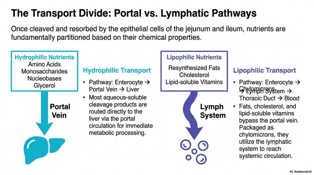
After structural breakdown, a critical sorting process defines the final stages of Digestion and Resorption. Once cleaved and absorbed by the epithelial cells of the jejunum and ileum, nutrients face a fundamental physiological fork in the road. The body partitions these absorbed molecules strictly based on their inherent chemical properties. The defining factor is water solubility. This strict chemical division ensures that nutrients are routed through the safest and most efficient vascular pathways to support ongoing systemic metabolism and organ function.
The handling of hydrophilic nutrients demonstrates the direct route of Digestion and Resorption. Aqueous-soluble cleavage products, including amino acids, monosaccharides, nucleobases, and glycerol, prefer water-based environments. Their transport pathway runs straight from the enterocyte directly into the portal vein. The portal vein acts as a nutrient superhighway, routing these dissolved molecules straight to the liver. This direct delivery system allows the liver to perform immediate metabolic processing, sorting, and storage of these vital water-soluble building blocks before they reach general circulation.
Conversely, lipophilic nutrients require an entirely different pathway during Digestion and Resorption. Resynthesized fats, structural cholesterol, and lipid-soluble vitamins simply cannot dissolve in the aqueous environment of portal blood. Therefore, they completely bypass the portal vein. Packaged safely within chylomicrons inside the enterocyte, these fat-soluble molecules are extruded into the lymphatic system. They travel slowly through the lymphatic vessels, ultimately entering systemic circulation at the thoracic duct. This lymphatic detour prevents massive lipid droplets from clogging the tiny capillaries of the liver.
The bold arrows on this slide create a highly effective visual summary of systemic distribution. The clear split between the blue portal pathway and the purple lymphatic route simplifies a complex anatomical concept. Medical students must master this transport divide, as it explains why certain oral medications are metabolized quickly by the liver while others take a slower, fat-dependent route. Recognizing this chemical sorting mechanism is fundamental to pharmacology and advanced clinical nutrition.
Slide 10: Diagnostic Matrix for Digestion and Resorption
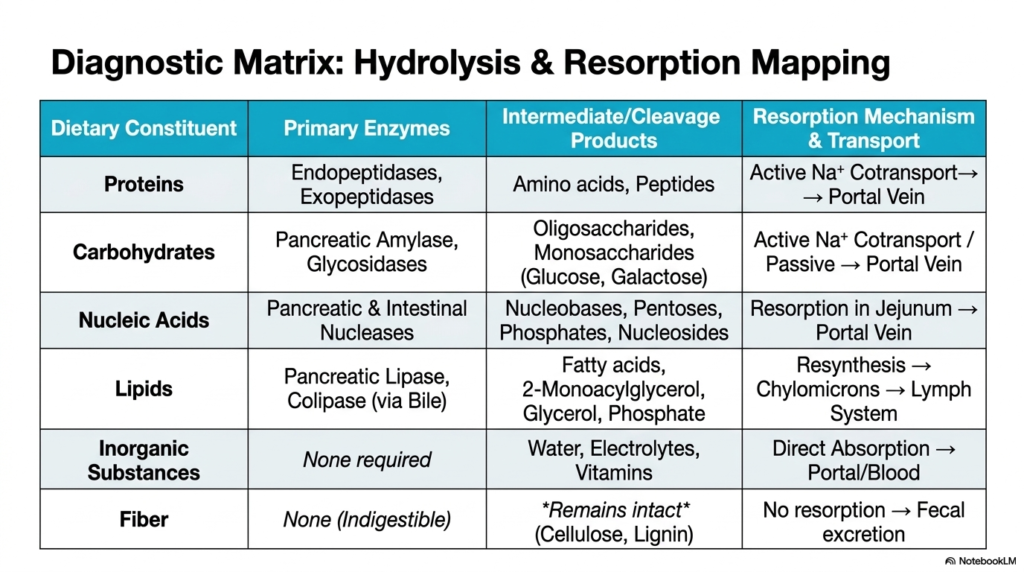
This final slide serves as a comprehensive diagnostic matrix for all processes of Digestion and Resorption. It elegantly consolidates the vast amount of biochemical data discussed previously into one highly accessible reference table. Students can instantly map any dietary constituent to its specific primary enzymes, intermediate cleavage products, and final transport mechanism. This structured approach allows researchers to trace the lifecycle of proteins, carbohydrates, nucleic acids, lipids, and inorganic substances across a single, unified visual framework, reinforcing the overarching biological concepts.
Reviewing the specific rows highlights the diverse strategies of Digestion and Resorption. For instance, proteins require endopeptidases to become amino acids, which then utilize active sodium cotransport to reach the portal vein. Carbohydrates rely on amylase and glycosidases to become monosaccharides, also heading to the portal vein. Nucleic acids are dismantled by nucleases into bases and pentoses for jejunal uptake. Meanwhile, lipids require a complex interaction with lipase and bile, resulting in chylomicron packaging and a distinct journey through the lymphatic system.
The matrix also emphasizes the exceptions within Digestion and Resorption. The bottom two rows clearly define the behavior of inorganic substances and fiber. Inorganics like water and vitamins demand zero enzymatic effort, passing directly into the blood. Conversely, dietary fiber remains entirely intact, ignoring enzymes completely to serve as vital fecal bulk. Having these exceptions listed alongside the complex enzymatic pathways provides a perfect contrast, ensuring medical scholars understand the entire spectrum of dietary processing from mouth to excretion.
For exam preparation and clinical application, this matrix is an invaluable study tool. It synthesizes multiple distinct physiological chapters into a single, cohesive snapshot of human metabolism. By memorizing these core relationships between substrate, enzyme, product, and pathway, future physicians can rapidly diagnose malabsorption syndromes, enzyme deficiencies, and specific nutrient transport failures. This table represents the ultimate culmination of biochemical study applied to human digestion.
Please read our Content Disclaimer Statement.
Check out our social media channels: