98. Intestinal Resorption: The Guide to Nutrient Transport
Every day, the human body transforms complex meals into fundamental biochemical building blocks, fueling cellular processes. Nutrient transport is the critical bridge between digestion and cellular energy. This slide deck explores the highly regulated cellular machinery that makes life possible. The core purpose of this presentation is to detail the precise molecular pathways governing nutrient uptake. By mastering these mechanisms, students will understand how microscopic protein channels and energy gradients sustain human physiology and prevent severe malabsorption diseases.
Slide 1: Introduction to Intestinal Resorption
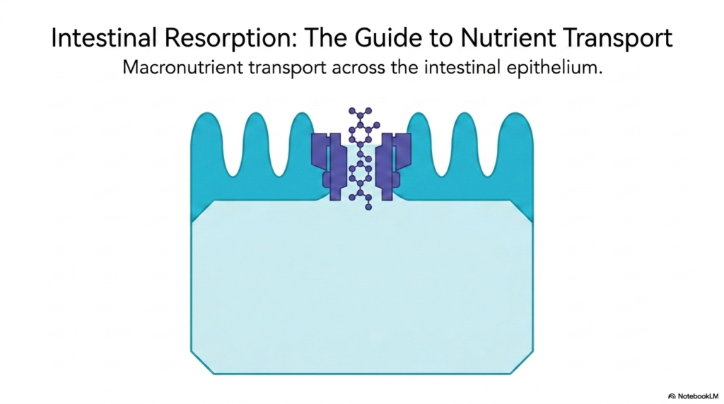
The study of intestinal resorption begins with understanding the grand scale of macronutrient transport across the intestinal epithelium. Intestinal resorption is not merely a passive biological phenomenon but a highly orchestrated cellular symphony. At the boundary between the external environment and the internal bloodstream lies a single layer of epithelial cells. These enterocytes act as the ultimate gatekeepers for all ingested nutrients. Without efficient intestinal resorption, the rigorous breakdown of food into molecules would be entirely wasted.
The visual representation highlights the microvilli, which are crucial for maximizing the surface area available for nutrient uptake. The biological necessity for such a massive surface area stems from the enormous volume of molecules that must cross the epithelial barrier daily. Countless transport proteins populate this membrane, each designed to capture specific dietary components. The efficiency of intestinal resorption relies entirely on this architectural marvel, ensuring that no valuable caloric or structural building block is lost to the environment.
Furthermore, a deep dive into these pathways reveals how evolutionary pressures have perfected energy extraction. Biochemistry students will recognize that every transporter depicted functions with incredible precision and specificity. By exploring these exact mechanisms, the foundation is laid for understanding pathological states like celiac disease or acute nutrient intolerances.
Ultimately, mastering the concept of intestinal resorption provides the biochemical framework necessary for advanced medical and physiological studies. It bridges the critical gap between basic luminal digestion and advanced cellular metabolism. The epithelial layer is a dynamic, living membrane that constantly adapts to dietary intake, representing the precise intersection of biochemistry and anatomy.
Slide 2: The Cellular Checkpoint of Intestinal Resorption
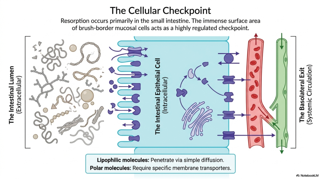
The small intestine serves as the primary anatomical site for intestinal resorption. Within this organ, the immense surface area provided by the brush-border mucosal cells functions as a highly regulated cellular checkpoint. This biological border separates the extracellular intestinal lumen from the systemic circulation. During intestinal resorption, every single molecule must be sorted, processed, and transported across this formidable cellular barrier. The enterocyte determines exactly what enters the internal environment.
A fundamental principle governing this checkpoint is the chemical nature of the molecules themselves. Lipophilic molecules, such as certain vitamins and fats, possess the physical ability to penetrate the cell membrane via simple diffusion. Their hydrophobic nature allows them to slip through the lipid bilayer unhindered. However, the vast majority of dietary nutrients are polar. For these water-soluble molecules, intestinal absorption requires specialized, complex membrane transporters.
These protein transporters act as exclusive cellular gateways, ensuring that essential polar nutrients can successfully breach the hydrophobic core of the cell membrane. Once inside the intestinal epithelial cell, the biochemical journey is far from over. Nutrients must transit through the intracellular space, often undergoing further processing or specialized routing. The final hurdle is the basolateral exit, where molecules cross the basal membrane to enter the systemic circulation.
Blood vessels quickly sweep these newly absorbed nutrients away, maintaining a steep concentration gradient that favors continuous uptake. This relentless flow is what sustains the energy requirements of peripheral tissues. Understanding this checkpoint is paramount for comprehending pharmacology, as oral medications must possess specific properties to bypass or utilize these exact mechanisms during intestinal resorption.
Slide 3: Principles of Membrane Transport in Intestinal Resorption
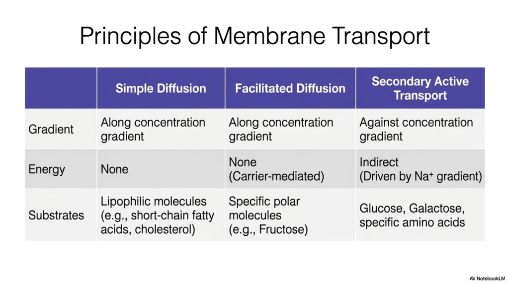
To comprehend the mechanics of intestinal resorption, one must first master the core principles of membrane transport. The biological membranes of the enterocyte present a significant barrier, and nature employs three primary strategies to overcome it. The most basic of these is simple diffusion. Molecules move passively along their concentration gradient without any cellular energy expenditure. Lipophilic substrates, such as short-chain fatty acids, utilize this straightforward pathway to enter the cell during intestinal resorption.
When molecules are polar, simple diffusion is no longer a viable option. This physical limitation necessitates facilitated diffusion. This process still relies on movement down a concentration gradient but requires a dedicated carrier protein. Fructose provides a classic biochemical example of a specific polar molecule relying on facilitated diffusion. While no direct energy is consumed, the carrier protein undergoes conformational changes to shuttle the molecule, highlighting the specialized architecture fundamental to intestinal resorption.
The most complex and biochemically fascinating mechanism is secondary active transport. Unlike diffusion, this process moves critical substrates like glucose, galactose, and specific amino acids against their concentration gradients. To achieve this thermodynamically unfavorable movement, the cell harnesses indirect energy driven by a pre-established sodium ion gradient. The kinetic energy released as sodium flows back into the cell drags the nutrient molecule along.
This reliance on secondary active transport demonstrates the incredible efficiency of human cellular machinery. By linking the uptake of vital nutrients to a robust ionic gradient, the enterocyte guarantees that the body extracts every last bit of fuel from ingested food. This elegant coupling of electrochemical forces and nutrient transport represents a cornerstone concept in understanding intestinal resorption.
Slide 4: The Engine Driving Intestinal Resorption
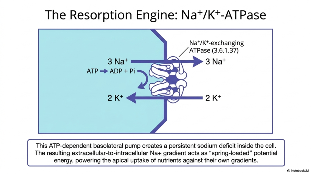
At the very heart of intestinal resorption lies a powerful cellular engine: the Na+/K+-ATPase pump. Located exclusively on the basolateral membrane of the enterocyte, this ATP-dependent enzyme is the true unsung hero of nutrient transport. The pump actively ejects three sodium ions out of the cell while pulling two potassium ions inside. This unequal exchange, driven by the hydrolysis of ATP into ADP and inorganic phosphate, is an absolute requirement for sustaining intestinal resorption.
The relentless activity of the Na+/K+-ATPase creates a persistent, profound sodium deficit within the intracellular fluid. Because the extracellular fluid contains high sodium levels, a steep extracellular-to-intracellular chemical and electrical gradient is established. This gradient acts as spring-loaded potential energy. It is this exact reservoir of potential energy that secondary active transporters on the apical membrane tap into. Without this basolateral pump, the apical transporters would completely stall.
Therefore, the entire process of intestinal resorption for carbohydrates and proteins is heavily dependent on this single enzyme. When the secondary active transporters allow sodium to rush back into the cell, the potential energy is converted into kinetic work, dragging glucose or amino acids against their own gradients. This ingenious biological design ensures that the cell burns ATP at one strategic location to power multiple different transport systems.
Clinically, the significance of the Na+/K+-ATPase cannot be overstated. Any metabolic poison, lack of oxygen, or pathophysiological state that depletes cellular ATP will rapidly shut down this essential pump. Once the pump stops, the sodium gradient dissipates, and the secondary active transport of vital nutrients during intestinal resorption grinds to an absolute halt.
Slide 5: Carbohydrate Cleavage Prior to Intestinal Resorption
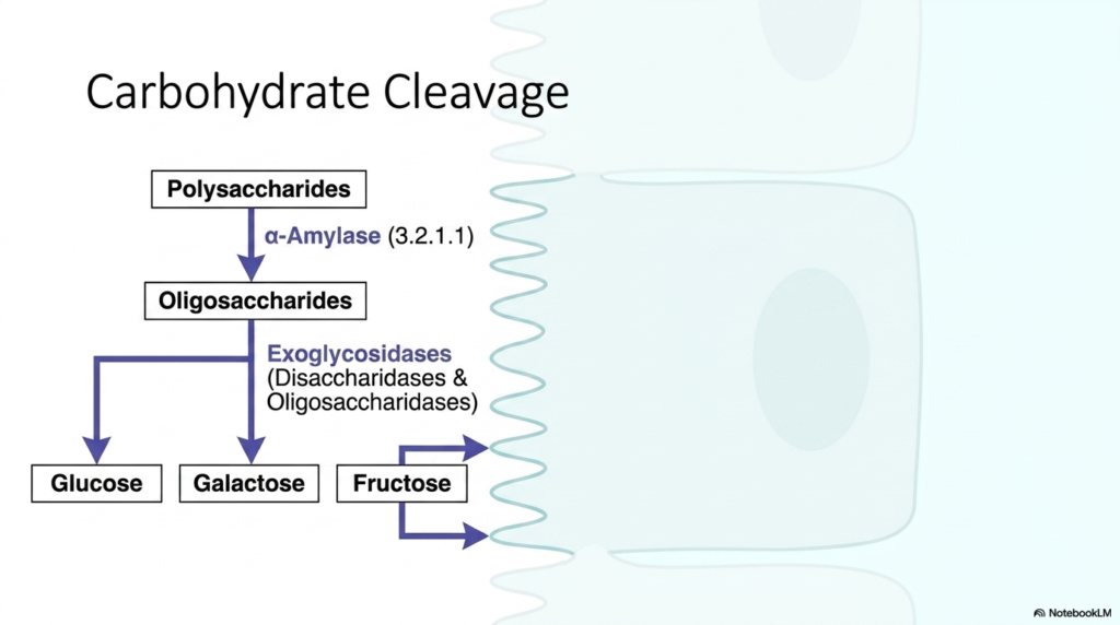
Before intestinal resorption can begin, complex dietary macromolecules must be meticulously dismantled into their simplest monomeric forms. Carbohydrate cleavage is a prime example of this required preliminary enzymatic degradation. Dietary carbohydrates primarily arrive in the form of massive polysaccharides, such as starch and glycogen. These molecular behemoths are far too large to interact with any membrane transporter, necessitating a highly coordinated sequence of hydrolytic events.
The breakdown initiates with the enzyme alpha-amylase, which specifically targets internal alpha-1,4-glycosidic bonds. This initial cleavage rapidly reduces the massive polysaccharides into much smaller oligosaccharides. However, these intermediate molecules are still too complex for cellular uptake. The final, critical stage of preparation for intestinal resorption occurs precisely at the cellular boundary, carried out by a specialized group of enzymes known as exoglycosidases.
These exoglycosidases, which include various disaccharidases and oligosaccharidases, are anchored directly to the brush border of the intestinal epithelium. By situating these enzymes on the microvilli, the digestive system ensures that the final monomeric products—glucose, galactose, and fructose—are generated exactly where transport proteins await them. This spatial coupling of enzymatic hydrolysis and membrane transport maximizes the efficiency of intestinal resorption.
The biochemical precision of this process is truly remarkable. Each brush border enzyme possesses a highly specific active site tailored for distinct glycosidic linkages. Deficiencies in any of these exoglycosidases, such as lactase, lead directly to profound gastrointestinal distress as unabsorbed sugars draw water into the lumen and ferment. Thus, mastering carbohydrate cleavage provides the essential biochemical context for understanding intestinal resorption.
Slide 6: Monosaccharide Entry During Intestinal Resorption
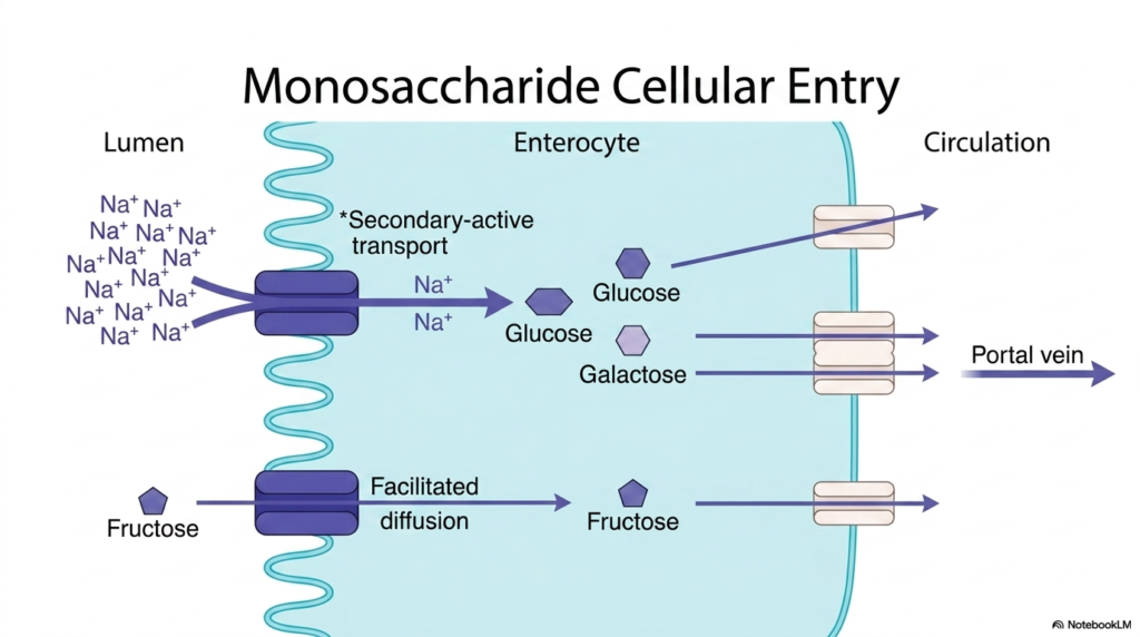
Once carbohydrate macromolecules are successfully reduced to monomers, the critical phase of monosaccharide cellular entry begins. This stage represents the definitive act of intestinal resorption for dietary sugars. The architecture of the enterocyte membrane features distinct transport pathways optimized for different monosaccharides. Glucose and galactose, the most crucial energy substrates, are transported via secondary active transport. This mechanism is directly driven by the high extracellular concentration of sodium ions.
As sodium ions flow down their electrochemical gradient into the enterocyte, they provide the necessary kinetic force to pull glucose and galactose against their respective concentration gradients. This co-transport mechanism guarantees that intestinal resorption remains highly efficient even when luminal sugar concentrations drop after a meal. Conversely, the monosaccharide fructose utilizes an entirely different entry strategy, crossing the apical membrane via facilitated diffusion.
After these three monosaccharides have entered the intracellular space of the enterocyte, they face one final biological barrier before systemic distribution. They must exit the basolateral membrane to enter the bloodstream. Regardless of how they initially entered the cell, glucose, galactose, and fructose all rely on facilitated diffusion transporters located on the basolateral side to exit during intestinal absorption.
The ultimate destination for these newly absorbed monosaccharides is the portal vein, which swiftly ferries them directly to the liver. The liver acts as the primary metabolic clearinghouse, regulating systemic blood sugar levels. A thorough understanding of this transport machinery reveals the elegant precision of human metabolism and highlights the pharmacological targets designed to modulate sugar uptake.
Slide 7: Proteolytic Degradation and Intestinal Resorption
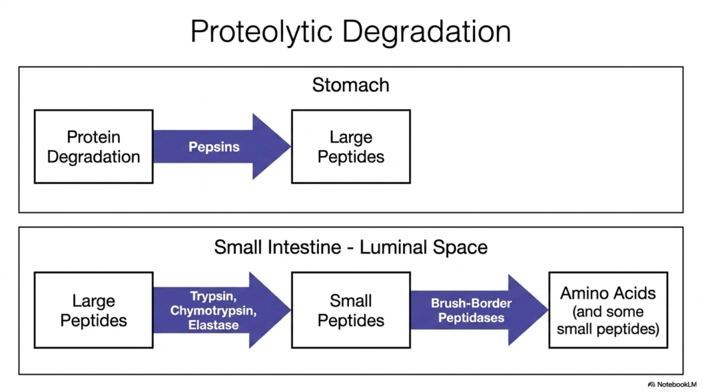
Just as complex carbohydrates require extensive hydrolysis, dietary proteins must undergo rigorous proteolytic degradation to become eligible for intestinal resorption. Proteins represent highly complex, three-dimensional structures held together by tough peptide bonds. The digestive system deploys a sequential arsenal of proteases to systematically dismantle these macromolecules. This necessary dismantling begins in the harsh, acidic environment of the stomach, where pepsins initiate the breakdown of intact proteins into large peptides.
Upon leaving the stomach, these large peptides enter the luminal space of the small intestine, the primary theater for intestinal resorption. Here, the pancreas secretes a powerful cocktail of enzymes, including trypsin, chymotrypsin, and elastase. These potent proteases exhibit distinct specificities for different amino acid residues, rapidly cleaving the large peptides into much smaller peptide chains. This massive coordinated enzymatic attack prepares the molecules for cellular transport.
The ultimate step before intestinal resorption occurs at the cellular surface itself. Anchored to the microvilli are specialized brush-border peptidases. These enzymes perform the final, precise cleavages required to reduce small peptides into individual, free amino acids. Notably, some very small dipeptides and tripeptides escape this final cleavage and remain intact. This dual availability provides the enterocyte with diverse substrates for transport.
The necessity of this staged proteolytic cascade lies in the absolute physical limitations of cellular membrane transporters. Only monomeric amino acids and the smallest of peptides can physically fit through the transport channel pores. Without this comprehensive degradation sequence, the immense nutritional value locked within dietary proteins would simply pass through the digestive tract unutilized.
Slide 8: Group-Specific Transport in Intestinal Resorption
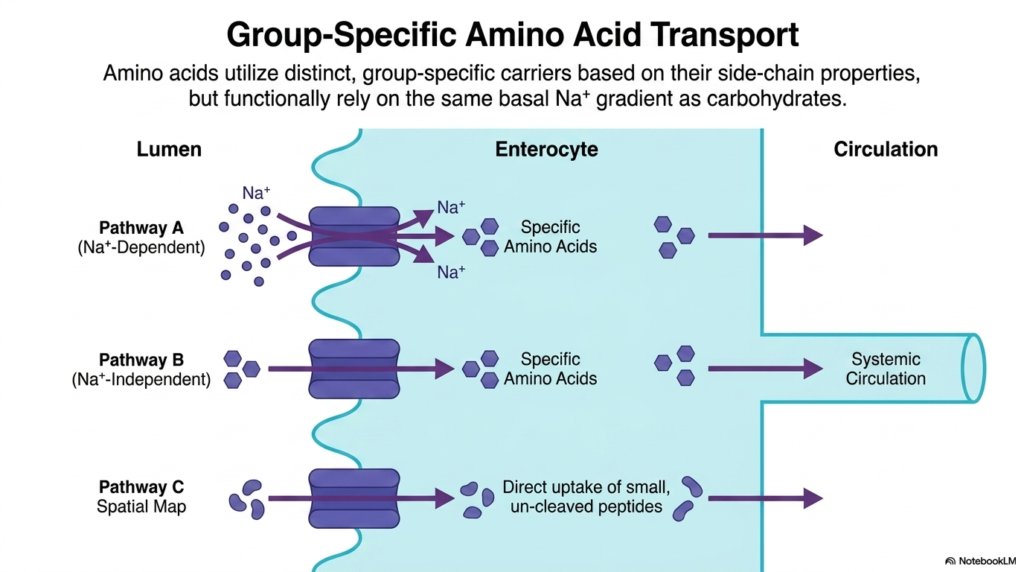
Following thorough proteolytic degradation, the resulting amino acids face the challenge of crossing the enterocyte membrane. Intestinal resorption of amino acids is vastly more complex than that of carbohydrates due to the sheer chemical diversity of the twenty distinct amino acid side chains. To manage this molecular variety, the enterocyte membrane deploys distinct, group-specific carriers that categorize amino acids based on their specific chemical properties.
A major route for amino acid entry is Pathway A, which represents sodium-dependent secondary active transport. Much like the uptake of glucose, this pathway relies fundamentally on the basal sodium gradient established by the Na+/K+-ATPase pump. The kinetic energy of sodium rushing into the cell pulls specific amino acids against their concentration gradients, ensuring highly efficient intestinal resorption of vital protein building blocks.
However, not all amino acids rely on sodium for intestinal resorption. Pathway B illustrates sodium-independent transport mechanisms. These specific carriers facilitate the entry of certain amino acids based on distinct structural properties without tapping into the sodium gradient. Furthermore, Pathway C reveals a fascinating alternate route: the direct uptake of small, un-cleaved peptides. Specific transporters can shuttle intact di- and tripeptides directly into the enterocyte.
Regardless of which entry pathway is utilized, the final step requires all free amino acids to cross the basolateral membrane. This exit into the systemic circulation is primarily achieved via specialized facilitated diffusion transporters. By utilizing multiple, parallel transport strategies, the human digestive system maximizes its capacity to capture diverse structural materials.
Slide 9: The Emulsification Challenge in Intestinal Resorption
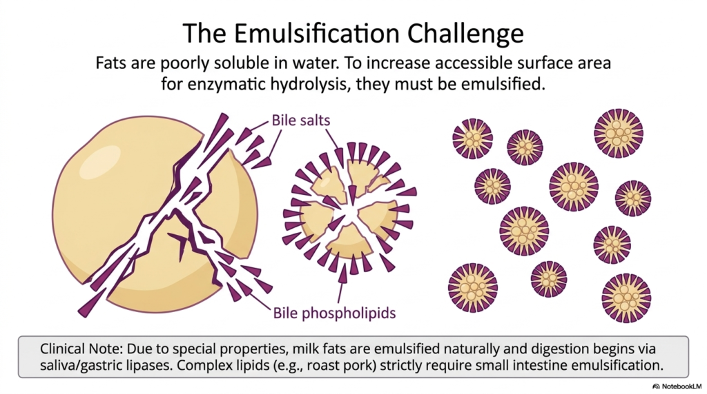
The digestion and subsequent intestinal resorption of dietary fats present a unique and profound biochemical challenge. Unlike carbohydrates and proteins, fats are highly lipophilic and poorly soluble in the aqueous environment of the gastrointestinal tract. Consequently, dietary lipids naturally clump together into large, intractable fat globules. This physical aggregation presents a massive problem for water-soluble digestive enzymes, which can only interact with the fat molecules at the outermost surface.
To overcome this physical barrier and enable eventual intestinal resorption, the body employs a physical processing step known as emulsification. The liver synthesizes bile salts and bile phospholipids, which are subsequently secreted into the duodenum. These molecules act as powerful biological detergents. When the large fat globules encounter these bile components, they are shattered into countless microscopic droplets, massively expanding the total accessible surface area.
This dramatic increase in surface area is an absolute prerequisite for efficient enzymatic hydrolysis. Without successful emulsification, the lipases would take an impossibly long time to digest the fat, and intestinal resorption of lipids would fail. A crucial clinical note highlights that human milk fats possess unique properties allowing them to be naturally emulsified and partially digested by saliva and gastric lipases.
In stark contrast, complex adult dietary lipids strictly require the heavy-duty emulsification power provided by the small intestine and biliary system. Understanding this physical transformation from giant globules to microdroplets is essential for biochemistry students, as it bridges the gap between gross anatomy and the molecular mechanisms of intestinal absorption.
Slide 10: Triacylglycerol Hydrolysis and Intestinal Resorption
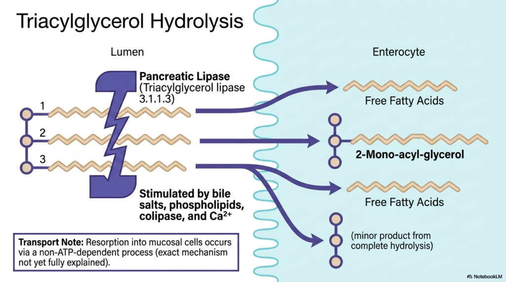
Once dietary fats have been successfully emulsified into tiny droplets, the crucial enzymatic stage of triacylglycerol hydrolysis commences. This chemical breakdown is mandatory for the intestinal resorption of complex lipids. The primary workhorse of this process is pancreatic lipase, officially classified as triacylglycerol lipase. This potent enzyme is secreted into the intestinal lumen, where it seeks out the vastly expanded surface area of the emulsified fat droplets to initiate catalysis.
Pancreatic lipase cannot function optimally on its own during intestinal resorption. Its full hydrolytic activity is robustly stimulated by the presence of bile salts, phospholipids, and essential cofactors like colipase and calcium ions. Once activated, the enzyme targets the specific ester bonds of the triacylglycerol molecule. The biochemical action of pancreatic lipase selectively cleaves the fatty acid chains located at the outer positions of the glycerol backbone.
This highly specific enzymatic cleavage results in a distinct set of breakdown products. The primary outputs are two free fatty acid chains and a single 2-mono-acyl-glycerol molecule. A very minor amount of complete hydrolysis may occur, producing a free glycerol molecule, but the dominant products are the free fatty acids and the mono-acylglycerol. These resulting fragments are finally ready to undergo intestinal resorption.
A critical transport note regarding lipid intestinal resorption is the method of cellular entry. Unlike the energy-demanding secondary active transport required for most polar nutrients, the resorption of these lipid breakdown products into the mucosal cells occurs via a non-ATP-dependent process. These lipophilic molecules readily diffuse across the apical membrane of the enterocyte into the intracellular space.
Slide 11: The Structural Divergence of Lipid Intestinal Resorption
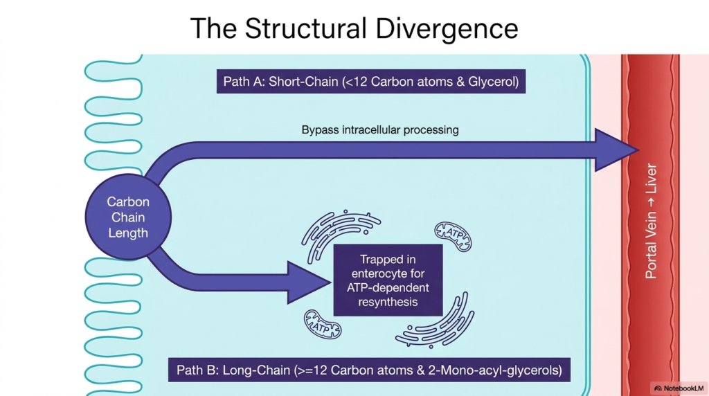
A fascinating biochemical divergence occurs immediately after lipid breakdown products cross the enterocyte membrane. The specific pathway taken during the intracellular phase of intestinal resorption is entirely dictated by the physical carbon chain length of the fatty acids involved. This structural divergence represents a highly sophisticated biological sorting mechanism that directs different types of cellular fuel to distinct anatomical destinations based solely on molecular geometry.
Path A illustrates the journey of short-chain fatty acids, defined as those possessing fewer than twelve carbon atoms, as well as free glycerol. Because these smaller molecules maintain a higher degree of water solubility, they can bypass complex intracellular processing entirely. During this specific route of intestinal resorption, these short-chain molecules diffuse swiftly straight through the enterocyte, exiting directly into the portal vein for rapid transport.
Conversely, Path B dictates a dramatically different fate for long-chain fatty acids containing twelve or more carbon atoms. These larger, highly hydrophobic molecules are effectively trapped within the enterocyte upon entry. Their extreme insolubility in aqueous environments prohibits them from simply diffusing into the bloodstream. Therefore, this specific pathway of intestinal resorption necessitates a complex, ATP-dependent resynthesis process within the cellular organelles.
This strict separation of pathways ensures that highly lipophilic molecules do not haphazardly enter and clog the delicate aqueous portal circulation. By forcing long-chain lipids to undergo specialized intracellular packaging, the enterocyte protects systemic hemodynamics while simultaneously processing the dense caloric energy acquired through intestinal resorption.
Slide 12: Intracellular Resynthesis in Lipid Intestinal Resorption
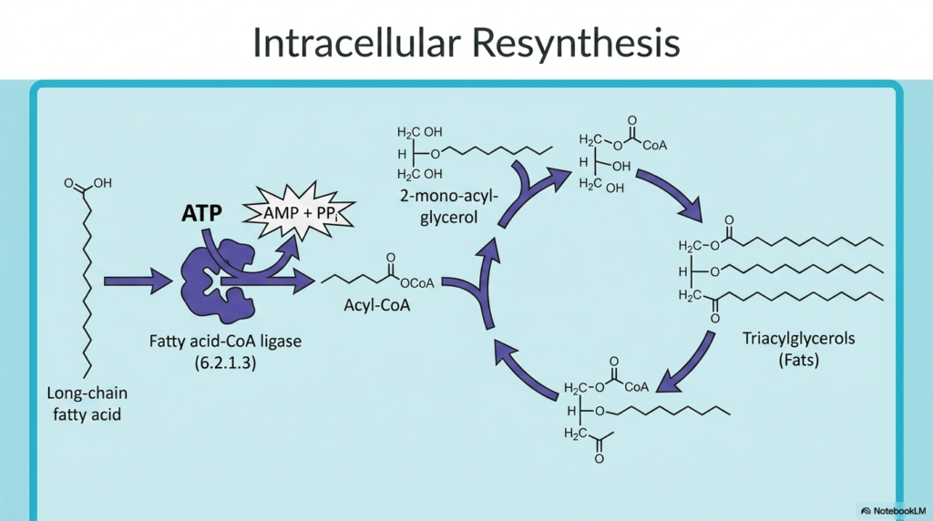
For long-chain fatty acids, simply crossing the apical membrane is only the first hurdle in the process of intestinal resorption. Because these highly hydrophobic molecules are trapped within the enterocyte, the cell must execute an energy-intensive intracellular resynthesis to prepare them for safe export. This complex biochemical choreography takes place primarily within the smooth endoplasmic reticulum of the mucosal cell, rebuilding the complex fats just dismantled in the lumen.
The resynthesis pathway begins with the mandatory activation of the free, long-chain fatty acids. An enzyme known as fatty acid-CoA ligase utilizes the energy from ATP hydrolysis to attach a Coenzyme A molecule to the fatty acid, generating a reactive Acyl-CoA intermediate. This ATP-dependent activation step is critical, as it provides the thermodynamic driving force required to rebuild ester bonds during intestinal absorption.
Once activated, the Acyl-CoA molecules are systematically combined with the previously absorbed 2-mono-acyl-glycerol molecules. A sequence of specific acyltransferase enzymes facilitates the stepwise addition of fatty acid chains back onto the glycerol backbone. This sequential esterification rapidly regenerates complete triacylglycerols within the intracellular space. The cell expends significant ATP to perform this rebuilding process during intestinal resorption.
This required resynthesis represents a fascinating paradox within the framework of intestinal resorption. The digestive system expends enormous effort to break down triacylglycerols in the lumen just to permit membrane transport, only to immediately burn cellular energy reconstructing those exact same molecules. This is an evolutionary necessity for safely absorbing water-insoluble caloric energy.
Slide 13: Packaging and Lymphatic Export in Intestinal Resorption
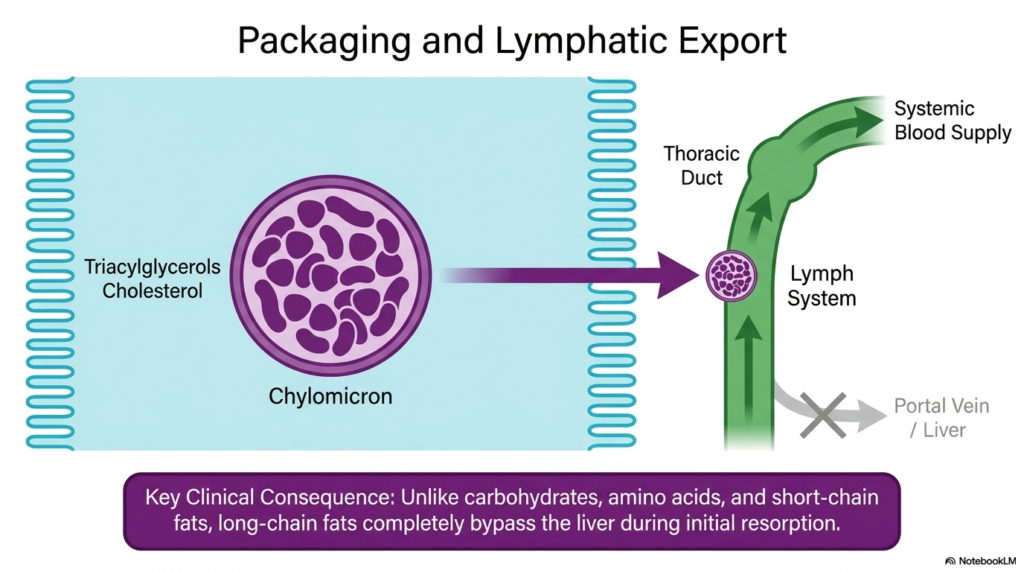
After the enterocyte has painstakingly resynthesized complex triacylglycerols, the final challenge of lipid intestinal resorption is exporting these highly insoluble molecules out of the cell. They cannot simply be dumped into the aqueous extracellular fluid. Instead, the newly formed triacylglycerols, along with dietary cholesterol, are meticulously assembled into massive, water-soluble lipoprotein particles known as chylomicrons. This specialized packaging process occurs within the Golgi apparatus.
The architecture of the chylomicron features a highly hydrophobic core packed tightly with triacylglycerols, surrounded by a hydrophilic outer shell. This brilliant structural design allows the enormous lipid payload to remain stably suspended within biological fluids. Once the chylomicron is fully assembled, the enterocyte secretes the particle across the basolateral membrane via exocytosis, completing the intracellular phase of lipid intestinal resorption.
Because chylomicrons are incredibly large structures, they are physically incapable of penetrating the tight junctions of the intestinal blood capillaries. Consequently, they must be exported into the highly permeable lymphatic vessels located within the intestinal villi. These lymphatic vessels coalesce and eventually drain into the thoracic duct. The thoracic duct then empties this lipid-rich lymph directly into the systemic blood supply.
A key clinical consequence of this specific pathway is that these long-chain fats completely bypass the liver during initial intestinal resorption. This anatomical detour allows peripheral tissues, such as skeletal muscle and adipose tissue, to have the first physiological opportunity to extract energy and structural lipids directly from a fatty meal before hepatic clearance occurs.
Slide 14: Master Synthesis of Intestinal Resorption
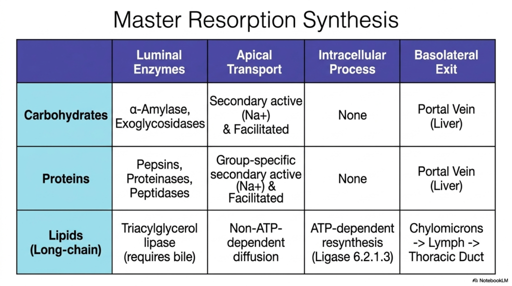
The final slide presents a comprehensive master synthesis of the biochemical mechanics governing intestinal resorption. This comparative table serves as a vital tool for biochemistry students to consolidate the distinct, parallel pathways utilized by the three primary macronutrients. By aligning these distinct processes side-by-side, the overarching elegance and specialization of the human digestive tract become unmistakably clear, highlighting biological efficiency.
Carbohydrate intestinal resorption relies entirely on luminal alpha-amylase and brush-border exoglycosidases for breakdown. Their apical transport is heavily dependent on sodium-driven secondary active mechanisms and facilitated diffusion. Because monosaccharides are highly water-soluble, they require absolutely no complex intracellular processing. They pass directly through the enterocyte, exit the basolateral membrane, and enter the portal vein to be transported straight to the liver for metabolic sorting.
Similarly, intestinal protein absorption uses robust luminal enzymes like pepsin and peptidases to generate free amino acids and small peptides. Their cellular entry leverages group-specific, sodium-dependent secondary active transport and facilitated pathways. Like carbohydrates, these polar molecular building blocks require zero intracellular resynthesis. They swiftly exit the enterocyte basolaterally and join the carbohydrates within the portal vein.
In stark contrast, intestinal absorption of long-chain lipids requires triacylglycerol lipase and the crucial assistance of bile for luminal breakdown. While their apical entry is a simple non-ATP-dependent diffusion, their intracellular journey is highly complex. They undergo strict ATP-dependent resynthesis into triacylglycerols and are meticulously packaged into large chylomicrons, exiting into the lymphatic system and completely bypassing the portal vein.
Please read our Content Disclaimer Statement.
Check out our social media channels: