102. Hemoglobin: Structural Biology and Biochemical Mechanisms
The ability of a single breath to sustain every cell in the human body relies on an elegant molecular machine. This mechanism perfectly balances oxygen capture and tissue release. This presentation explores the structural biology and dynamic systems of this critical respiratory protein. The following slide deck breaks down its intricate design, from initial folding to complex allosteric regulation. By studying these core concepts, biochemistry students will grasp exactly how life is sustained at the cellular level.
Slide 1: Introduction to Hemoglobin: Structure, Allostery, and Oxygen Transport
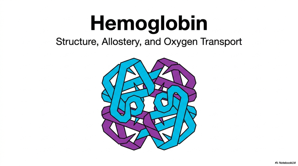
The journey into human metabolism begins with a deep dive into the primary oxygen-carrying protein of the blood. The opening slide introduces Hemoglobin, a remarkable metalloprotein essential for vertebrate life. This molecule is not merely a passive container for oxygen; it is a highly dynamic and responsive structure. Its primary mission is to pick up oxygen in the lungs and deliver it safely to distant tissues. To accomplish this, the protein relies on intricate structural biology and sophisticated regulatory mechanisms to ensure perfect timing.
Understanding Hemoglobin requires an appreciation for its three-dimensional architecture. The visual representation on this slide highlights a folded, multi-subunit complex. This beautiful symmetry is the foundation of its biological function. The protein is composed of four distinct polypeptide chains that weave together to form a highly ordered tetramer. This quaternary structure is what enables the molecule to perform its vital physiological duties. Without this specific arrangement, the protein could never meet the immense metabolic demands of a complex organism.
Beyond mere structure, the slide points to two other core concepts: allostery and oxygen transport. Allostery refers to the way this Hemoglobin molecule changes its shape in response to environmental cues. When one part of the molecule binds to a ligand, the rest of the structure feels the shift. This shape-shifting ability is what makes the protein a master of oxygen transport. It must bind oxygen tightly in the lungs but release it easily in the tissues.
These three pillars—structure, allostery, and transport—form the backbone of the entire presentation. Medical and biochemistry students must master these concepts to understand respiratory physiology. As the presentation progresses, each of these themes will be unpacked in detail. The interplay between the physical shape of the Hemoglobin molecule and its chemical environment dictates human survival. The upcoming slides will reveal exactly how this molecular machine operates under varying physiological conditions.
Slide 2: Hemoglobin and the Solution to Oxygen Solubility
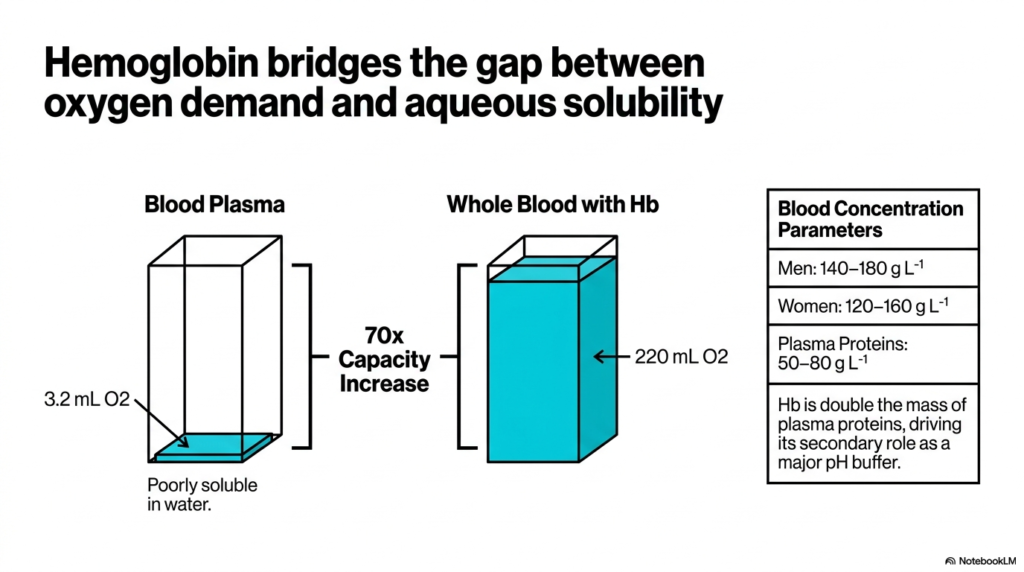
A fundamental physiological problem exists within the human body regarding oxygen delivery. Oxygen is a nonpolar gas, meaning it dissolves very poorly in the aqueous environment of blood plasma. If the circulatory system relied solely on dissolved oxygen, complex organisms could never survive. Slide two illustrates this challenge perfectly. Blood plasma alone can only carry a meager amount of oxygen, roughly 3.2 milliliters per unit volume shown. This extremely low solubility necessitates a specialized biochemical carrier system.
The biological solution to this solubility crisis is Hemoglobin. By packing red blood cells full of this specific protein, the oxygen-carrying capacity of whole blood increases dramatically. The graphic demonstrates a staggering seventy-fold increase in capacity compared to plasma alone. Whole blood equipped with this molecule can carry up to 220 milliliters of oxygen. This massive boost bridges the vital gap between the high oxygen demand of cellular metabolism and the physical limitations of water.
The concentration of this protein in the blood is remarkably high, which underscores its overall importance. In men, normal physiological parameters range from 140 to 180 grams per liter, while in women, the range is 120 to 160 grams per liter. When compared to typical plasma proteins, the mass of Hemoglobin is roughly double. This massive physical presence in the bloodstream is not just for carrying oxygen; it serves another critical biochemical purpose.
Because it exists in such high concentrations, Hemoglobin serves a secondary but equally vital physiological role. It acts as a major pH buffer within the bloodstream. The numerous amino acid residues on the surface of the protein can absorb or release protons as needed. This buffering capacity helps maintain the delicate acid-base balance required for cellular function. Thus, this single protein solves both the oxygen transport problem and helps stabilize blood chemistry, proving its immense value.
Slide 3: The Architectural Blueprint of the Hemoglobin Tetramer
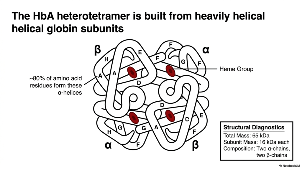
Moving deeper into the structural biology, slide three details the specific physical composition of the protein. Adult Hemoglobin, often referred to as HbA, is a heterotetramer. This means it is built from four separate polypeptide subunits that are not all identical to one another. The diagnostic box notes that the entire complex has a total mass of 65 kilodaltons. This mass is divided among the four individual subunits, each weighing approximately 16 kilodaltons.
The composition of this adult tetramer includes two alpha chains and two beta chains. The visual diagram showcases how these four subunits fit together into a compact, globular shape. A striking feature of these globin chains is their underlying secondary structure. Approximately eighty percent of the amino acid residues in each chain fold into rigid alpha-helices. These helical segments are named sequentially from A to H, creating a stable pocket for the active site.
Nestled carefully within the folds of each globin chain is a critical non-protein component called the heme group. Because the Hemoglobin tetramer has four separate globin subunits, it contains a total of four individual heme groups. Each of these groups serves as an isolated binding site for an oxygen molecule. Therefore, a single fully saturated tetramer can carry four molecules of oxygen at a time. The protective helical structure ensures these sites remain totally viable.
This highly ordered architecture is essential for the proper function of Hemoglobin. The specific interactions between the alpha and beta chains create the necessary tension and flexibility for the protein to operate. If the genetic code introduces a mutation that alters even one amino acid, this delicate helical structure can collapse. The resulting structural instability often leads to severe blood disorders. Understanding this precise heterotetrameric build is crucial for diagnosing these molecular diseases in clinical settings.
Slide 4: Hemoglobin and the Mechanics of Iron Coordination
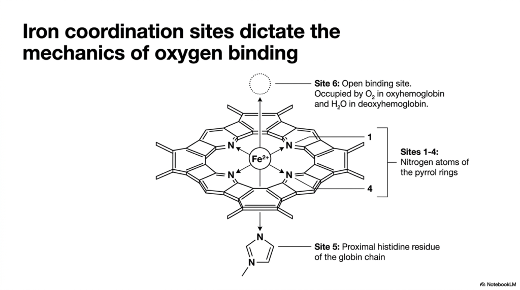
At the very heart of the oxygen transport process lies the heme group, beautifully showcased in slide four. The biological function of Hemoglobin depends entirely on the precise chemical coordination of a single central iron ion. This central ferrous iron is suspended within a large, flat, organic ring structure known as a porphyrin ring. The detailed diagram illustrates how the iron atom is held in place by multiple coordinate bonds, which dictate its unique chemical reactivity.
The chemical coordination mechanics involve six specific binding sites around the central iron atom. The first four sites are permanently occupied by the nitrogen atoms of the surrounding pyrrole rings that make up the porphyrin structure. These four strong bonds hold the iron firmly within the flat plane of the molecule. This arrangement prevents the iron from floating away and readies it for its primary task. The remaining two sites are situated exactly perpendicular to this flat plane.
The fifth coordination site connects the iron more directly to the protein structure. A specific amino acid residue, known as the proximal histidine, reaches out from the globin chain to bind the iron from below. This connection tethers the heme group securely to the Hemoglobin subunit. It also provides a vital mechanical link, meaning any movement of the iron atom will physically pull on the surrounding protein chain, triggering wider conformational shifts.
The final, sixth coordination site is the functional core of Hemoglobin. This is the open binding site where reversible oxygen attachment actually occurs. In the deoxygenated state, this site is either empty or weakly occupied by a simple water molecule. When blood reaches the lungs, an oxygen molecule swoops in and binds to this sixth site. The precise geometry of these six coordination sites is what makes life-sustaining oxygen transport chemically possible within the human body.
Slide 5: Protecting Hemoglobin from Irreversible Iron Oxidation
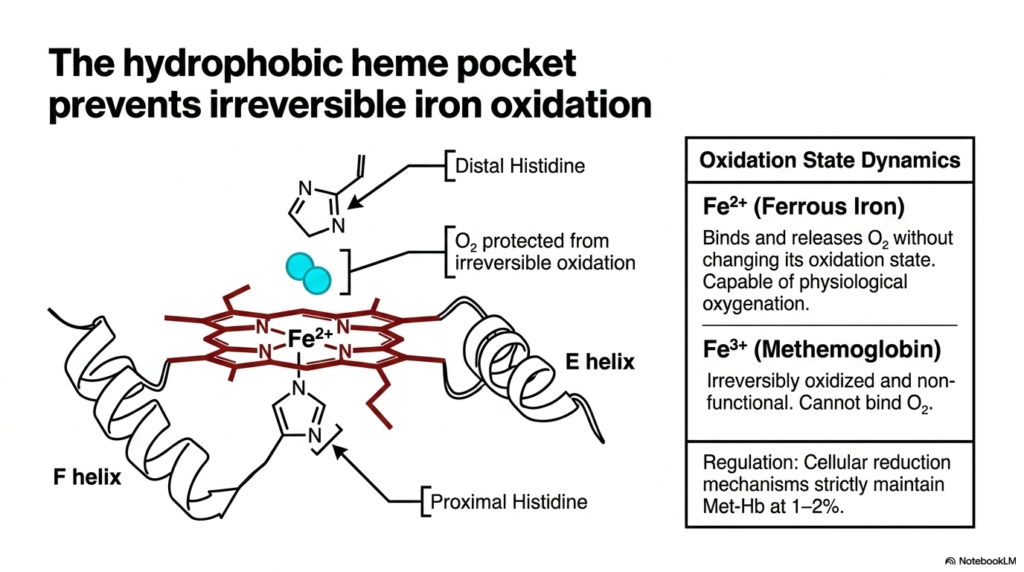
The binding of oxygen to iron is a notoriously risky chemical process. Slide five highlights a crucial protective mechanism engineered directly into the molecule’s structure. For Hemoglobin to function correctly, the central iron atom must remain exclusively in the ferrous state, carrying a plus-two charge. In this specific oxidation state, the iron can bind and release oxygen reversibly without undergoing a permanent chemical change. This specific state allows for continuous cycles of physiological oxygenation.
If the iron atom loses an electron and oxidizes to the ferric state, bearing a plus-three charge, it forms methemoglobin. This oxidized version is entirely non-functional because it strictly cannot bind oxygen. The Hemoglobin architecture prevents this disaster by creating a deep, highly hydrophobic heme pocket. By burying the iron deep within the water-repelling folds of the globin chains, the protein shields the reactive center from the surrounding aqueous environment, drastically slowing unwanted chemical oxidation.
A key player in this protective biochemical strategy is the distal histidine. While the proximal histidine binds directly to the iron, the distal histidine hovers just above the open binding site. When oxygen enters the pocket and binds to the iron, the distal histidine acts exactly like a protective lid. It forms a stabilizing hydrogen bond with the trapped oxygen, securing it in place and preventing it from stealing an electron from the vulnerable iron atom.
Despite these advanced structural safeguards, a small amount of Hemoglobin constantly falls victim to ambient oxidation. Under normal physiological conditions, cellular reduction mechanisms work tirelessly to convert useless methemoglobin back to the functional ferrous state. These dedicated enzymatic pathways strictly maintain the circulating levels of oxidized protein at a mere one to two percent. This delicate chemical balance ensures that the blood retains its maximum oxygen-carrying capacity at all times, preventing tissue suffocation and cellular death.
Slide 6: The Conformational States of Hemoglobin
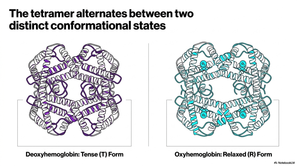
One of the most fascinating aspects of complex biochemistry is how protein shape strictly dictates function. Slide six introduces the core concept that Hemoglobin is not a static rock, but rather a shifting, moving molecular machine. The entire tetramer actively alternates between two distinct conformational states. These physical shapes are known broadly as the Tense form, or T-state, and the Relaxed form, or R-state. The transition between these two specific structures is the basis of efficient oxygen delivery.
The Tense form strictly represents the deoxygenated state of the molecule. In this configuration, the four individual subunits are tightly bound together by strong, non-covalent interactions. This rigid molecular structure naturally creates a vastly low affinity for oxygen. The protein automatically adopts this shape when it travels through the oxygen-deprived tissues of the body. The tight associations squeeze the central cavity, making it extremely difficult for incoming oxygen molecules to access the isolated iron-binding sites.
Conversely, the Relaxed form represents the fully oxygenated version of the molecule. When oxygen begins to bind, the strong internal bonds of the Hemoglobin T-state begin to rapidly break down. The subunits literally rotate and slide past one another, opening up the overall structure. This physical relaxation significantly increases the affinity of the remaining empty heme sites for oxygen. The protein naturally adopts this open configuration when it passes through the oxygen-rich environment of the human lungs.
The visual comparison on the slide clearly showcases the subtle but vital differences between the two functional tetramers. The physical shift from tense to relaxed is a cascading event, originally triggered by the simple attachment of a tiny gas molecule. By shifting cleanly between these two distinct conformational states, the protein acts as both an eager buyer and a willing seller of oxygen. This shape-shifting ability is the defining characteristic of this highly specialized respiratory pigment.
Slide 7: Analyzing Hemoglobin Through the Diagnostic Matrix
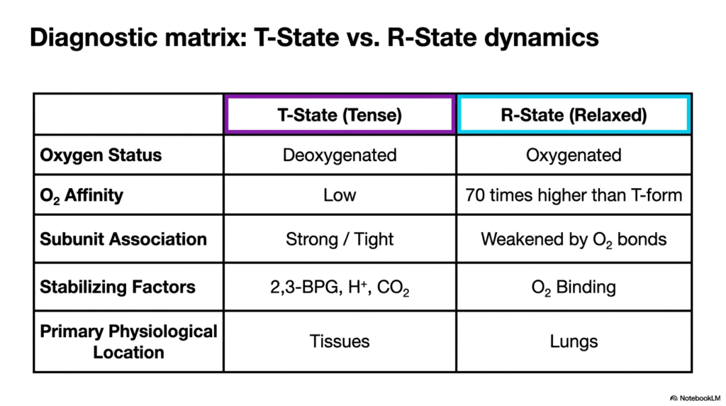
To truly master the complex function of Hemoglobin, students must compare its two primary states side by side. Slide seven provides a highly useful diagnostic matrix that contrasts the T-State and the R-State across several critical biological parameters. The first major difference is their basic oxygen status. The T-State is thoroughly deoxygenated and dominates in the tissues, while the R-State is fully oxygenated and is primarily located in the capillary beds of the lungs.
The most crucial parameter detailed in this matrix is overall oxygen affinity. The T-State exhibits a very low affinity for oxygen, which is absolutely perfect for unloading the vital cargo exactly where it is needed most. In stark contrast, the R-State has an oxygen affinity that is seventy times higher than the tense form. This massive functional difference ensures that Hemoglobin rapidly and completely saturates itself with oxygen the moment it enters the pulmonary circulation.
Subunit association clearly explains this dramatic shift in binding affinity. In the T-State, the physical bonds between the four polypeptide chains are strong and tight, severely restricting access to the internal binding sites. When the Hemoglobin molecule transitions to the R-State, these tight subunit associations are noticeably weakened by the formation of multiple oxygen bonds. The physical expansion of the molecule makes it exponentially easier for subsequent oxygen molecules to find and successfully attach to the iron.
Finally, the detailed matrix highlights the specific environmental factors that actively stabilize each structural form. The relaxed state is naturally stabilized simply by the presence and binding of oxygen itself. The tense state, however, requires specific external effectors to lock it into its rigid shape. These stabilizing factors specifically include protons, carbon dioxide, and 2,3-BPG. Understanding this comprehensive diagnostic matrix for Hemoglobin is essential for predicting exactly how the protein will behave under fluctuating metabolic conditions.
Slide 8: Hemoglobin and the Power of Cooperative Binding
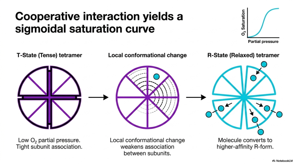
Slide eight perfectly illustrates a cornerstone concept in structural biochemistry known as cooperative binding. Because Hemoglobin has four completely separate binding sites, the attachment of oxygen to one site profoundly influences the remaining empty sites. This molecular teamwork yields a highly distinctive sigmoidal, or S-shaped, saturation curve. When the blood partial pressure of oxygen is very low, the tetramer rests comfortably in the tense state. Binding the very first molecule of oxygen is incredibly difficult.
The biochemical breakthrough happens once that first oxygen molecule finally forces its way into a restricted binding site. This initial attachment causes a small local conformational change in that specific subunit. Because the Hemoglobin chains are physically linked together, this small local movement actively tugs on the adjacent subunits. This internal molecular pull slightly weakens the tight association between the chains that defines the tense state. The protein begins to physically open up, paving the way.
As the strict structural associations rapidly weaken, the second and third oxygen molecules can bind much more easily. The complex molecule is actively converting to the higher-affinity relaxed form. By the time the fourth oxygen molecule approaches, the Hemoglobin tetramer is fully relaxed. The final binding site is completely exposed, and the final oxygen snaps directly into place with minimal energetic resistance. This cascading increase in structural affinity is the very definition of a cooperative interaction.
The distinctive sigmoidal saturation curve is the direct physiological result of this impressive structural teamwork. It ensures that the protein always remains highly sensitive to small changes in local oxygen concentration. In the lungs, the cooperative nature strictly guarantees that the molecule becomes fully saturated in a mere fraction of a second. In the tissues, the reverse process occurs just as cooperatively, allowing the vital protein to dump its cargo rapidly when the tissue pressure drops during exercise.
Slide 9: Allosteric Effectors Regulating Hemoglobin
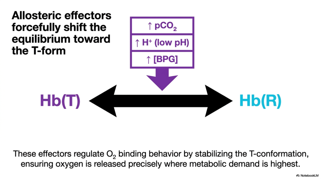
The beautiful molecular machinery of Hemoglobin requires strict metabolic regulation to function properly without error. Slide nine completely introduces allosteric effectors, which are specialized molecules that bind to the protein at specific sites other than the primary oxygen-binding pocket. These effectors essentially act as biological switches, forcefully shifting the delicate structural equilibrium back toward the tense form. The major effectors highlighted here are increased carbon dioxide, increased protons, and the metabolite BPG.
When muscular tissues are actively metabolizing, they predictably produce massive amounts of carbon dioxide and highly acidic protons as waste products. These allosteric effectors quickly enter the bloodstream and interact directly with the circulating red blood cells. By binding to very specific amino acid residues on the outside of the protein, these waste products force the tetramer to close up heavily. They strongly stabilize the T-conformation, fundamentally altering the oxygen binding behavior of the entire complex.
This forced structural shift serves an absolutely brilliant physiological purpose for the organism. As Hemoglobin travels directly into working muscle tissue, it immediately encounters high concentrations of these acidic effectors. The sudden stabilization of the tense state dramatically lowers the protein’s overall affinity for oxygen. Consequently, the molecule has absolutely no choice but to actively release its bound oxygen directly into the surrounding tissue. The metabolic waste products actually trigger their own relief by forcing oxygen delivery.
Without these critical regulatory effectors, the Hemoglobin protein would bind oxygen far too tightly and strictly refuse to release it to the starving tissues. The intricate balance between the relaxed and tense states is constantly modulated by these vital chemical signals. This ensures that molecular oxygen is released precisely where local metabolic demand is highest. Understanding advanced allosteric regulation is critical for grasping how the body dynamically responds to environmental stressors by using chemical signals to maximize efficiency.
Slide 10: Hemoglobin and the Structural Wedge of 2,3-BPG
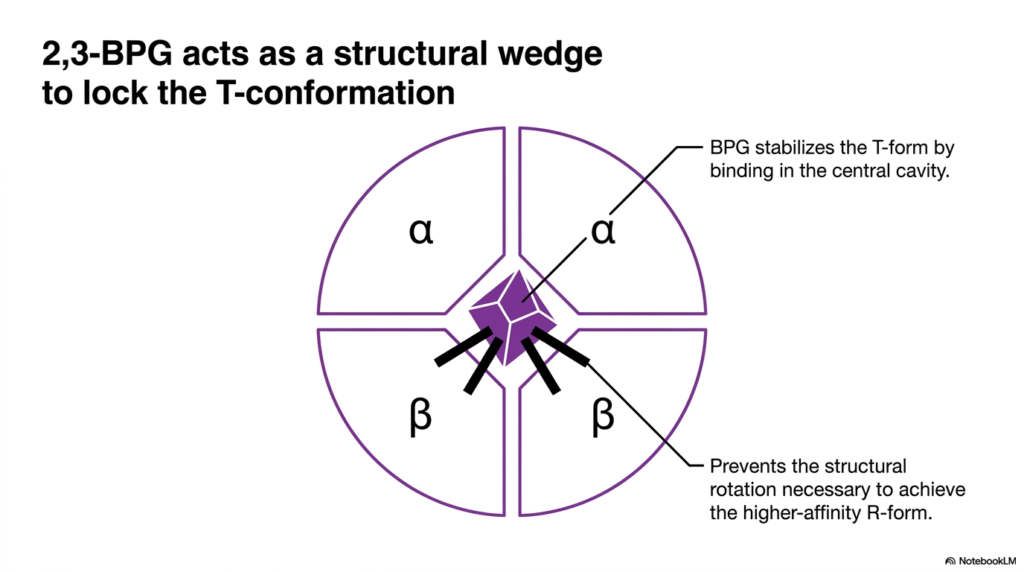
Of all the characterized allosteric effectors, 2,3-Bisphosphoglycerate, or simply BPG, plays a uniquely mechanical role in respiration. Slide ten specifically zeroes in on exactly how this small, highly negatively charged metabolite strictly dictates the shape of Hemoglobin. BPG remarkably does not bind to the outside of the protein like free protons or carbon dioxide do. Instead, it directly targets the very center of the tetramer. A central cavity strictly exists when the protein rests in its tense state.
The central cavity of the typical T-state is completely lined with positively charged amino acid residues. BPG, naturally carrying a very strong negative charge, is perfectly attracted to this internal pocket. Once inside, BPG literally acts as a physical structural wedge. It forms multiple strong salt bridges strictly with the surrounding beta chains, effectively tying them firmly together. This internal chemical bracing strongly stabilizes the tense form, locking the complex into its low-affinity configuration and preventing premature binding.
For Hemoglobin to successfully transition to the much higher-affinity relaxed state, the central cavity must physically shrink and collapse. However, the strong BPG wedge prevents the necessary structural rotation of the subunits. As long as BPG remains securely lodged in the center, the molecule immensely struggles to achieve the R-form. This strictly ensures that the protein does not accidentally re-bind the vital oxygen it just actively released into the tissues.
When the Hemoglobin-packed red blood cell finally returns to the incredibly oxygen-rich environment of the lungs, the massive influx of oxygen eventually overpowers the internal wedge. The overwhelming pressure of oxygen forces the resistant subunits to rotate, forcefully collapsing the central pocket and actively expelling the BPG molecule entirely. This mechanical loading and unloading of BPG is a primary mechanism for finely tuning oxygen delivery. It perfectly demonstrates the elegant relationship between structure and function in the intricate field of human biology.
Slide 11: The Bohr Effect on Hemoglobin Oxygen Release
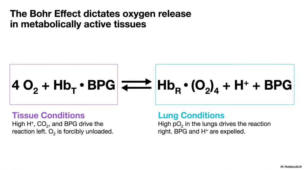
The vital intersection of foundational acid-base chemistry and systemic respiratory physiology is beautifully defined by the Bohr Effect. Slide eleven clearly illustrates the reversible chemical equation that completely dictates targeted oxygen release in metabolically active tissues. The complex equation demonstrates the direct molecular competition between oxygen molecules and acidic allosteric effectors for complete control over Hemoglobin. The delicate chemical balance of this exact reaction shifts dramatically depending entirely on the local chemical environment found within the circulatory system.
Under typical active tissue conditions, the local environment naturally becomes highly acidic and heavily rich in carbon dioxide due to continuous cellular respiration. The high concentration of free protons and the strong presence of BPG drive the chemical reaction forcefully to the left. The Hemoglobin protein is compelled to rapidly bind these numerous protons and BPG molecules. As a direct physical consequence, the entire tetramer is forced violently back into the tense state, and oxygen is forcefully unloaded.
The exact opposite biochemical phenomenon occurs immediately when the deoxygenated blood finally returns to the vast pulmonary circulation. Under normal lung conditions, the alveolar partial pressure of oxygen is overwhelmingly high. This massive chemical concentration gradient quickly drives the central reaction firmly to the right. Pure oxygen rapidly forces its way onto the empty iron-binding sites, actively causing the protein to transition fully into the relaxed state. As the structure shifts, the allosteric effectors lose their hold entirely.
The rapid physical expulsion of protons and BPG strictly in the lungs successfully completes the loop. The displaced protons typically combine with floating bicarbonate to eventually form carbon dioxide, which is then swiftly exhaled. The classic Bohr Effect perfectly ensures that Hemoglobin is remarkably sensitive to localized and changing metabolic needs. By directly linking overall oxygen affinity to local pH levels, the body guarantees prioritized oxygen delivery on a microscopic scale.
Slide 12: Hemoglobin Expression Shifts Across Human Ontogeny
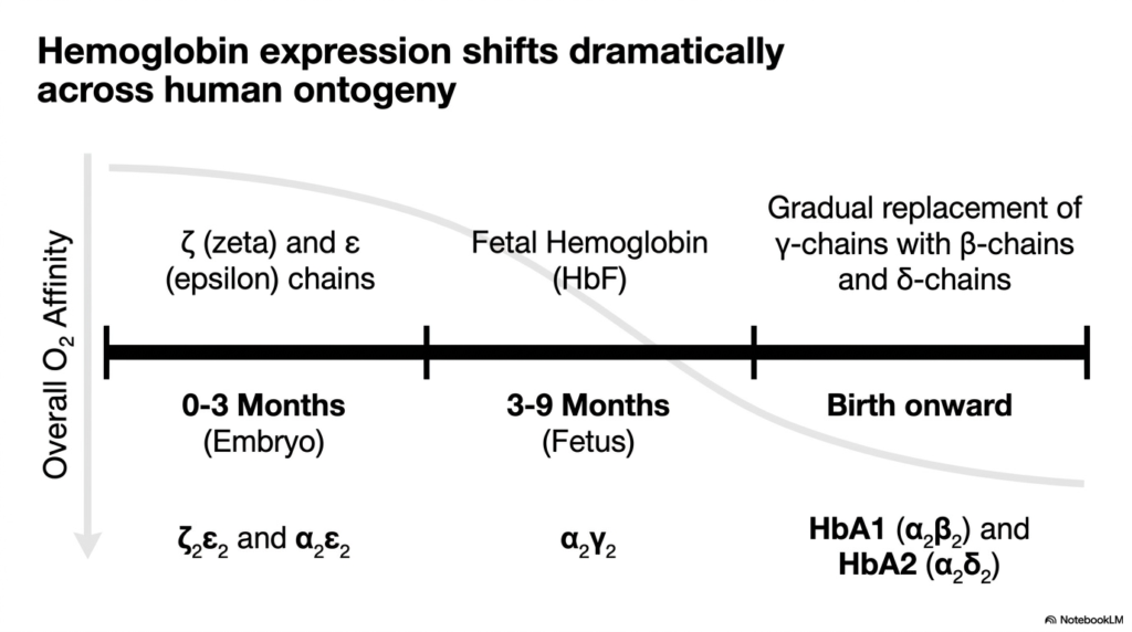
The physiological need for oxygen changes quite dramatically as a human dynamically develops from a tiny embryo into a fully grown adult. Slide 12 effectively charts how Hemoglobin expression shifts across human ontogeny to meet these continuously evolving demands. The human genome specifically contains multiple different genes for various globin subunits, and these genes are strictly turned on and off at highly specific stages of life. The detailed graph demonstrates a clear downward trend in overall oxygen affinity.
During the fragile first three months of life, the growing embryo relies strictly on unique chains known individually as zeta and epsilon. These specific embryonic variants naturally create a tetramer with the absolute highest possible affinity for oxygen. As the early embryo matures slowly into a fetus, gene expression shifts dramatically. The epsilon and zeta chains fade entirely away, actively replaced by newly synthesized alpha and gamma chains. This specific process creates Fetal Hemoglobin, which maintains a significantly higher affinity.
The relatively high oxygen affinity of these specific early developmental proteins is an absolute biological necessity for the growing child. Because the fetus absolutely cannot breathe air directly, it must efficiently steal oxygen completely from the mother’s blood supply. The fetal red blood cells must successfully pull oxygen aggressively across the placenta. If fetal Hemoglobin possessed the exact same low affinity as the standard adult version, the oxygen transfer would instantly fail, causing immediate and severe cellular hypoxia.
Around the exact time of natural birth, another massive genetic shift permanently occurs. The early gamma chains are gradually suppressed entirely and swiftly replaced by adult beta and delta chains. From birth onward, the predominant form permanently becomes adult HbA1, with very small, supportive amounts of HbA2. This final adult configuration completely provides the standard baseline oxygen affinity required for normal terrestrial respiration. The timeline strongly highlights the incredible adaptability of human molecular tools for developmental success.
Slide 13: Engineered Hemoglobin Isoforms for Fetal Survival
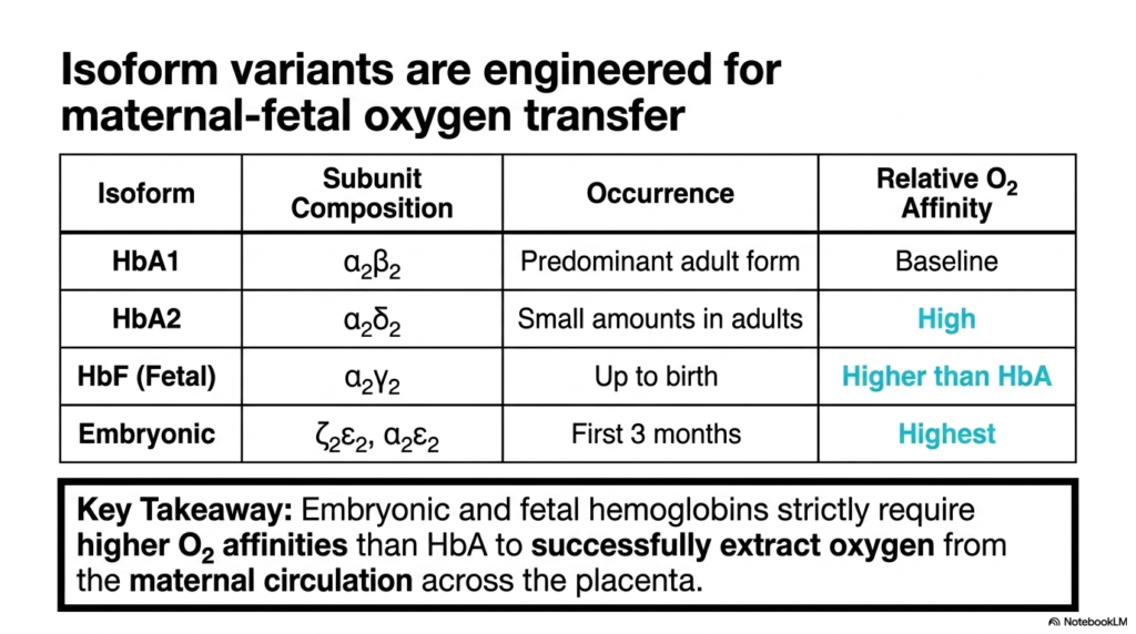
Building directly on the dynamic timeline of human development, slide thirteen carefully provides a highly detailed breakdown of the specific isoform variants. Hemoglobin is not merely a single, unchanging entity; it is effectively a large family of uniquely related tetramers precisely engineered for specific biological tasks. The comprehensive table clearly lists the exact subunit composition and the corresponding relative oxygen affinity for each major structural type. The primary adult form, known as HbA1, serves as the standard comparative baseline.
A secondary adult structural form, also known as HbA2, specifically contains two alpha and two delta chains. While it generally exists only in small amounts, it naturally plays a minor supportive role in overall adult oxygen transport. The most critical functional comparison, however, lies strictly between the mature adult isoforms and Fetal Hemoglobin. The specialized fetal version, composed entirely of two alpha and two gamma chains, is specifically designed to universally possess an oxygen affinity higher than the adult baseline.
As explicitly mentioned in the prominent key takeaway, the basic survival of the fetus strictly depends entirely on this distinct difference in binding strength. Maternal blood heavily pools in the placenta, strictly carrying adult red blood cells tightly packed with oxygen. When specialized fetal red blood cells securely enter this same arena, a molecular tug-of-war instantly begins. Because the fetal variant binds oxygen much more tightly, it successfully strips the oxygen directly away, transferring it cleanly across the placental barrier.
The distinct embryonic form, entirely utilizing zeta and epsilon chains, ultimately boasts the absolute highest relative affinity of all forms. This completely ensures survival during the earliest and entirely most vulnerable stages of embryonic vascular development. By cleverly utilizing vastly different genetic blueprints for Hemoglobin, the human body completely and elegantly solves the complex physical problem of oxygen sharing. It vividly represents evolutionary design tailored strictly for reproductive success and continuous fetal oxygenation.
Slide 14: Synthesis of the Hemoglobin Closed-Loop Mechanics
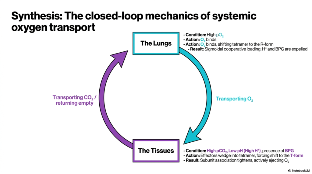
The final summary slide efficiently brings all the structural and complex chemical concepts strictly together to fully explain the closed-loop mechanics of systemic oxygen transport. Hemoglobin acts as a highly specialized, endlessly cycling cellular courier, constantly moving between two vastly different extreme physiological environments. The large diagram clearly illustrates this continuous, necessary journey between the active lungs and the peripheral tissues. At absolutely every step of the respiratory cycle, specific chemical conditions instantly trigger highly precise molecular actions.
In the lungs, the strictly prevailing environmental condition is an incredibly high partial pressure of fresh oxygen. This acts exactly as the primary initiating trigger for the entire respiratory cycle. The structural action is immediate: oxygen successfully binds tightly to the available heme pockets. This successful binding rapidly forces the entire Hemoglobin tetramer to completely undergo a physical shape shift into the relaxed R-form. The ultimate result is a rapid sigmoidal, cooperative loading process, while protons and BPG are completely expelled.
The fully loaded, oxygen-rich protein then travels entirely through the active arterial system until it successfully reaches the tiny capillaries of metabolically active tissues. Here, the local environmental conditions completely and instantly flip. The traveling protein abruptly encounters naturally high levels of waste carbon dioxide, distinctly low pH, and the prominent presence of BPG. These extreme conditions trigger a different set of molecular actions. The allosteric effectors violently wedge themselves securely into the sensitive tetramer.
As subunit association tightens immensely due to the effectors, the protein actively and forcefully ejects its vital oxygen cargo exactly where it is desperately needed most. Now completely empty, Hemoglobin assists gracefully in effectively transporting waste carbon dioxide slowly back to the waiting lungs, perfectly ready to strictly begin the vital cycle anew. This brilliant closed-loop system prominently highlights the remarkable foundational efficiency of complex human respiration, supplying the necessary fuel required for life and continuous cellular energy production.
Please read our Content Disclaimer Statement.
Check out our social media channels: