137. Neurotransmitter Biochemistry: How Chemical Messengers Direct Brain and Body Function
Every thought, sensation, and movement relies on a sophisticated chemical communication network. Billions of neurons continuously send microscopic signals across cellular gaps to coordinate complex human behavior. This blog post explores a foundational slide deck on neurochemistry to clarify how these signaling molecules work. College and medical students will discover the major structural classes of signaling compounds, explore their key physiological roles, and map the step-by-step biosynthetic pathways that create essential catecholamines in the human body.
Slide 1: Introduction to Neurotransmitters and Chemical Signaling
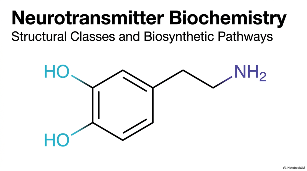
Neurochemistry begins with understanding how chemical messengers transmit signals across nerve cells. Central to nervous system communication, neurotransmitters serve as the molecular bridges that allow individual neurons to exchange vital information. The opening slide establishes the field’s foundation by focusing on the chemical structures and biosynthetic pathways that govern neuronal signaling. A prominent chemical structure displayed on the slide highlights dopamine, a vital biogenic amine that illustrates the precise atomic arrangement necessary for biological activity.
By categorizing these active molecules, researchers and medical students can systematically analyze how specific chemical properties dictate physiological responses. Endogenous neurotransmitters vary significantly in their molecular weight, ionic charge, and chemical stability, which directly influences their behavior within the synaptic cleft. For instance, small amino acid transmitters diffuse rapidly to produce fast synaptic responses, whereas larger neuropeptides provide sustained modulatory actions.
Biosynthetic pathways determine how precursor molecules, such as dietary amino acids, are enzymatically transformed into functional neurotransmitters within nerve cells. Enzymatic reactions modify side chains, add hydroxyl groups, or remove carboxyl groups to create active signaling ligands that bind specific receptors. Studying these molecular transitions provides crucial insight into clinical neurology, neuropharmacology, and cellular physiology across human tissue systems.
Slide 2: The Synaptic Mechanism of Neurotransmitters
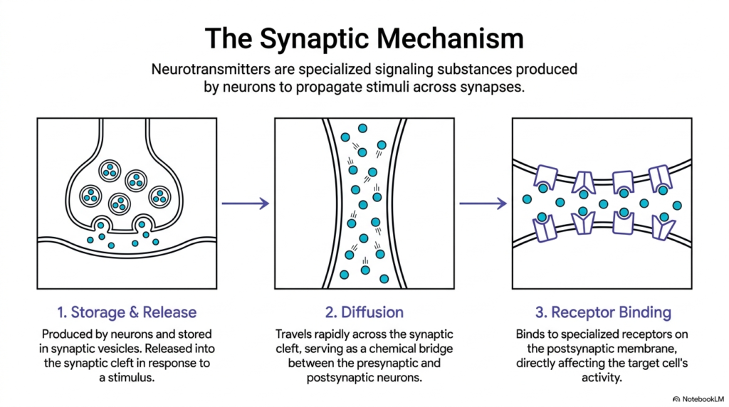
Signal transmission across neuronal junctions relies on a coordinated three-step sequence known as the synaptic mechanism. First, presynaptic neurons synthesize specialized chemical messengers called neurotransmitters and pack them into membrane-bound vesicles for safe storage. When an electrical action potential reaches the axon terminal, voltage-gated calcium channels open, triggering exocytosis. These synaptic vesicles fuse with the presynaptic membrane, discharging their chemical contents into the extracellular space. This controlled exocytosis converts electrical depolarizations into targeted chemical signals that propagate information between adjacent cells.
Second, the released neurotransmitters diffuse rapidly across the fluid-filled synaptic cleft, bridging the physical gap between neighboring neurons. Because the synaptic cleft measures only twenty to forty nanometers in width, diffusion occurs within microseconds. This rapid diffusion ensures nerve impulses travel seamlessly across synaptic junctions without significant delay. The cleft’s physical distance and structural environment determine how efficiently signaling molecules move from presynaptic release sites to postsynaptic targets.
Third, these diffuse neurotransmitters bind to specialized receptors on the postsynaptic membrane, triggering specific electrical or biochemical changes in the target cell. Depending on receptor type, binding opens ligand-gated ion channels or activates intracellular G-protein cascades. After signaling ends, enzymatic degradation or reuptake transporters clear neurotransmitters from the cleft, resetting the synapse for subsequent signaling events.
Slide 3: Structural Classification of Neurotransmitters
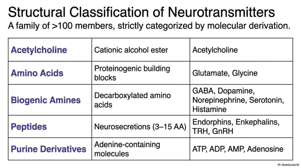
The human nervous system utilizes over one hundred distinct chemical messengers to regulate complex physiological processes. To organize this diverse group, biochemists strictly categorize neurotransmitters into five primary classes based on their molecular derivation and chemical structures. These categories include acetylcholine, amino acids, biogenic amines, neuropeptides, and purine derivatives. Each structural class possesses unique chemical properties that dictate how the molecules are synthesized, stored, and recognized by target membrane receptors.
Class I consists solely of acetylcholine, a cationic alcohol ester that plays a dominant role at neuromuscular junctions. Class II encompasses standard proteinogenic amino acids such as glutamate and glycine, which function as primary excitatory and inhibitory signals in the brain. Class III comprises biogenic amines formed through amino acid decarboxylation, including dopamine, norepinephrine, serotonin, histamine, and gamma-aminobutyrate. Class IV includes short-chain neuropeptides, while Class V covers adenine-containing purine derivatives such as ATP, ADP, AMP, and adenosine.
Classifying these diverse neurotransmitters by chemical identity allows medical students to predict their biological stability, receptor interaction mechanisms, and degradation pathways. For example, small-molecule biogenic amine neurotransmitters produce rapid synaptic responses, whereas peptide neurosecretions mediate slower, long-lasting neuromodulatory effects. Understanding these structural categories provides a logical framework for analyzing neurological function, drug mechanisms, and metabolic pathways in clinical medicine.
Slide 4: Class I Acetylcholine as Exemplary Neurotransmitters
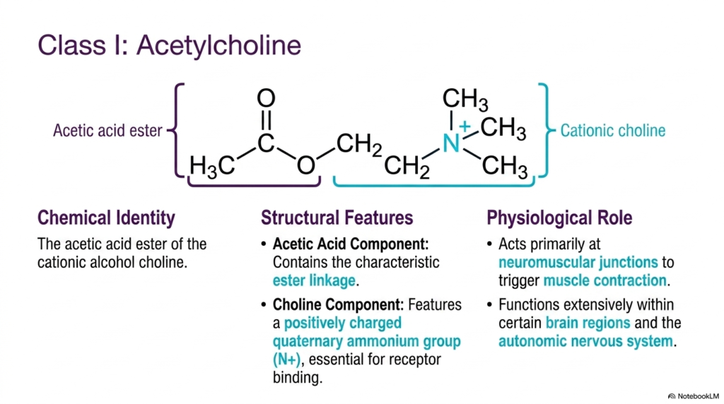
Acetylcholine occupies a unique position in neurochemistry as the only Class I chemical messenger. Chemically identified as the acetic acid ester of the cationic alcohol choline, its molecular structure features two distinct structural components. An acetic acid component provides a characteristic ester linkage, while a choline component contains a positively charged quaternary ammonium group. This positively charged quaternary nitrogen atom is essential for binding specifically to ionic and G-protein coupled cholinergic receptor sites on target cell membranes.
In the peripheral nervous system, acetylcholine functions as an essential neurotransmitter at neuromuscular junctions, triggering voluntary muscle contraction. When motor neurons release acetylcholine into the neuromuscular junction, the molecules bind to nicotinic receptors on muscle endplates, driving sodium influx and muscle depolarization. This cationic ester also acts extensively within the autonomic nervous system, serving as the preganglionic signaling agent for both sympathetic and parasympathetic divisions.
In the central nervous system, acetylcholine regulates cognitive processes, attention, and memory formation. Because acetylcholinesterase rapidly hydrolyzes the ester bond in the synaptic cleft, signaling by these neurotransmitters remains brief and tightly controlled. Students studying neuropharmacology learn that targeted inhibition of acetylcholinesterase enhances cholinergic signaling, providing therapeutic benefit in treating conditions such as myasthenia gravis and Alzheimer’s disease.
Slide 5: Class II Amino Acid Neurotransmitters in the CNS
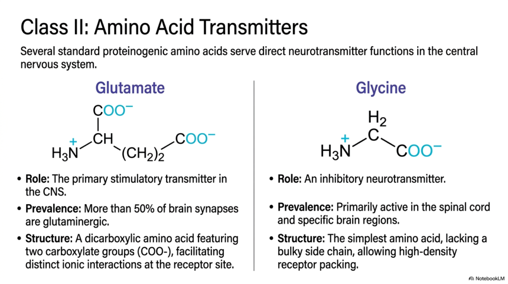
Class II chemical signaling agents consist of standard proteinogenic amino acids that function directly in central synaptic transmission. Glutamate and glycine represent the primary excitatory and inhibitory amino acid neurotransmitters in the human central nervous system. Glutamate is a dicarboxylic amino acid with two negative carboxylate groups that facilitate electrostatic interactions at postsynaptic receptor sites. Remarkably, over fifty percent of all central synapses rely on glutamatergic signaling to mediate synaptic plasticity, learning, and memory.
In contrast, glycine is an essential inhibitory signaling agent active primarily in the spinal cord and lower brainstem regions. Glycine is the simplest amino acid, lacking a bulky side chain, which allows tight, high-density packing within receptor binding pockets. When it binds to ionotropic glycine receptors, it triggers intracellular chloride influx, hyperpolarizing the postsynaptic membrane to prevent unwanted muscle spasms and uncoordinated nerve firing.
Together, these amino acid neurotransmitters maintain the precise physiological balance between excitation and inhibition throughout the brain and spinal cord. Medical students must master these basic structural features to understand how imbalances in amino acid neurotransmitters contribute to clinical neurological disorders. For instance, excessive glutamate release causes excitotoxic neuronal cell death, whereas deficient glycine activity leads to severe muscle hyperreflexia and motor disturbances.
Slide 6: Class III Biogenic Amine Neurotransmitters
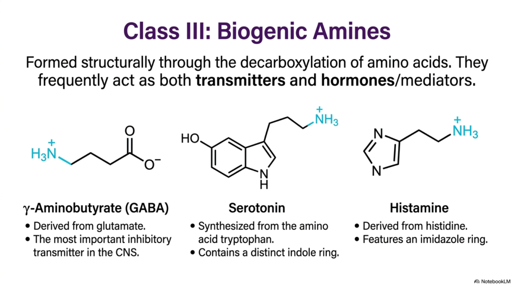
Class III signaling compounds, known as biogenic amines, form structurally through the enzymatic decarboxylation of specific amino acid precursors. These versatile biogenic amine neurotransmitters frequently function as both central synaptic messengers and systemic hormones or inflammatory mediators throughout the body. Three prominent examples in this class include gamma-aminobutyrate, serotonin, and histamine, each possessing a distinct chemical ring or aliphatic chain derived from its precursor amino acid.
Gamma-aminobutyrate, commonly called GABA, forms via the decarboxylation of glutamate and serves as the brain’s principal inhibitory neurotransmitter. Serotonin is synthesized from the essential amino acid tryptophan and contains a characteristic double-ring indole structure that interacts with diverse 5-HT receptor subtypes. Histamine is synthesized from histidine and features a distinct five-membered imidazole ring that regulates wakefulness, immune responses, and gastric acid secretion throughout peripheral tissues.
Understanding how biogenic amine neurotransmitters are synthesized helps students understand major mood circuits and neurological disease mechanisms. Because biogenic amine neurotransmitters modulate mood, sleep architecture, and autonomic tone, clinical drugs targeting their reuptake transporters represent cornerstone treatments in psychiatry. Selective serotonin reuptake inhibitors and antihistamines directly modify biogenic amine neurotransmitters to relieve depression, anxiety, and allergic symptoms in clinical patients, illustrating their immense therapeutic value.
Slide 7: Class IV Peptide Neurotransmitters and Neurosecretions
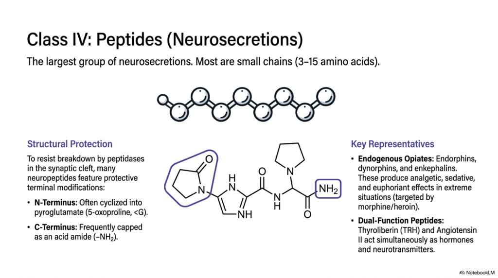
Class IV chemical messengers comprise the largest and most structurally diverse group of neurosecretory agents in the human nervous system. These peptide neurotransmitters consist of short amino acid chains, typically ranging from three to fifteen amino acid residues. Because peptidases in the synaptic cleft rapidly hydrolyze unprotected peptide bonds, many neuropeptides feature protective terminal chemical modifications. Their N-terminus is frequently cyclized into pyroglutamate, while their C-terminus is commonly capped with an acid amide group to resist enzymatic breakdown.
Key representatives of this class include endogenous opiate peptides such as endorphins, dynorphins, and enkephalins. These opiate neurotransmitters bind to specific G-protein-coupled opioid receptors in the brain and spinal cord to produce potent analgesic, sedative, and euphoriant effects. Their natural pain-relieving mechanisms serve as the primary pharmacological targets for powerful clinical analgesics like morphine and fentanyl during severe traumatic stress.
Other peptide neurotransmitters function as dual-action neurosecretions, including thyroliberin and angiotensin II, which act simultaneously as endocrine hormones and central messengers. Recognizing how structural modifications protect these peptide neurotransmitters helps medical students appreciate their prolonged signaling duration and modulatory influence over complex neuroendocrine behavior. Unlike small-molecule transmitters, neuropeptides are synthesized in the soma and transported along axons, reflecting a distinct presynaptic management mechanism.
Slide 8: Class V Purine Derivative Neurotransmitters
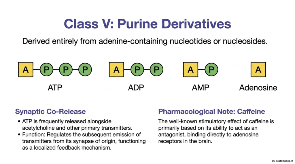
Class V chemical signaling agents consist of purine derivatives derived entirely from adenine-containing nucleotides or nucleosides. This specialized group includes adenosine triphosphate, adenosine diphosphate, adenosine monophosphate, and unphosphorylated adenosine. These purine neurotransmitters play crucial non-canonical roles in cell-to-cell signaling alongside their well-known metabolic functions in cellular energy transfer across peripheral and central synapses throughout the human body.
Adenosine triphosphate is stored in presynaptic vesicles and co-released alongside classical signaling agents such as acetylcholine and catecholamines. Once released into the synaptic cleft, purinergic neurotransmitters bind to ionotropic P2X or metabotropic P2Y receptors, regulating subsequent transmitter release as a localized feedback mechanism. Extracellular ecto-nucleotidases rapidly hydrolyze extracellular triphosphates down to adenosine, terminating purinergic stimulation and activating distinct P1 adenosine receptors on adjacent cell membranes.
Purinergic signaling provides a classic pharmacological example regarding the daily consumption of caffeine. Caffeine achieves its well-known alerting effect by acting as a competitive antagonist that binds directly to adenosine receptors in the central nervous system. By preventing endogenous adenosine from binding, caffeine blocks inhibitory purinergic tone, demonstrating how purine neurotransmitters regulate central arousal, fatigue, and cerebrovascular tone. Students appreciate that purine neurotransmitters coordinate both cellular energy status and neural excitation throughout human neural tissue networks in health and disease.
Slide 9: Catecholamine Biosynthesis in Neurotransmitters
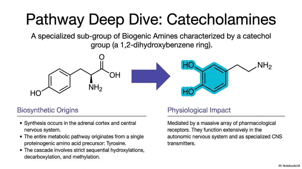
Catecholamines are a clinically vital subgroup of biogenic amines characterized by a catechol nucleus, a 1,2-dihydroxybenzene ring. These essential catecholamine neurotransmitters include dopamine, norepinephrine, and epinephrine, which govern motor control, sympathetic activation, and cognitive focus. Biosynthesis of catecholamines occurs in specialized central dopaminergic and adrenergic neurons, as well as in the chromaffin cells of the adrenal medulla, in response to physiological demands.
The entire biosynthetic pathway originates from a single proteinogenic amino acid precursor, L-tyrosine, derived from dietary sources or phenylalanine hydroxylation. Through a strict sequence of two enzymatic hydroxylations, a decarboxylation, and a terminal N-methylation, tyrosine transforms into functional catecholamine neurotransmitters. This tightly regulated metabolic cascade ensures that specific endocrine and neural tissues synthesize the exact chemical messenger required for physiological homeostasis.
Physiologically, these catecholamine neurotransmitters exert their effects through a widespread network of G-protein coupled adrenergic and dopaminergic receptors. They function extensively in the autonomic nervous system to drive fight-or-flight responses, regulate arterial blood pressure, and modulate central reward pathways. Mastering this metabolic pathway provides medical students with a necessary foundation for understanding neuropharmacology, shock management, and cardiovascular therapeutics. Each enzymatic step offers a specific target for clinical drug intervention in hypertensive emergencies and movement disorders.
Slide 10: Early Enzymatic Steps for Catecholamine Neurotransmitters
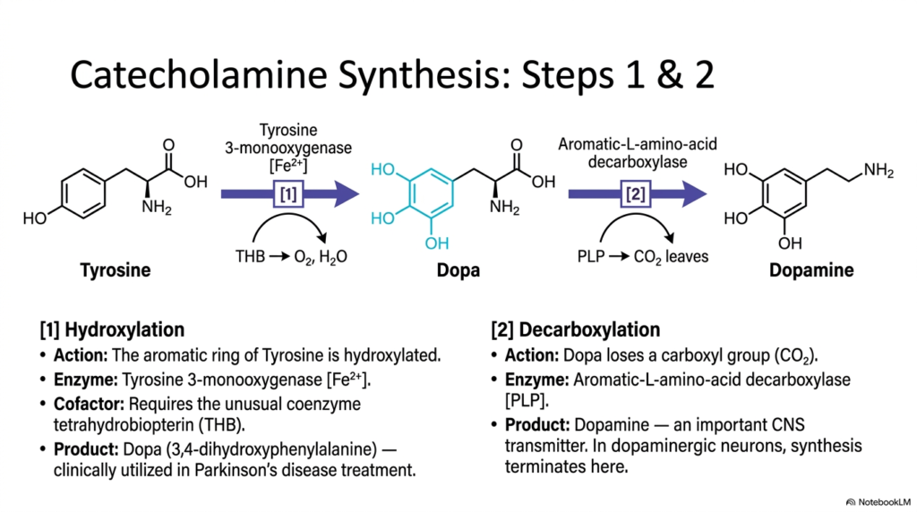
The catecholamine biosynthetic pathway begins with two precise enzymatic reactions that transform the precursor amino acid L-tyrosine into dopamine. In Step 1, the aromatic ring of tyrosine undergoes ring hydroxylation catalyzed by the enzyme tyrosine 3-monooxygenase, commonly called tyrosine hydroxylase. This rate-limiting enzymatic reaction requires ferrous iron and the coenzyme tetrahydrobiopterin as essential cofactors, adding a second hydroxyl group to form L-DOPA. L-dopa serves as a vital clinical intermediate administered to Parkinson’s disease patients because it crosses the blood-brain barrier to restore depleted dopamine levels.
In Step 2, L-DOPA undergoes rapid decarboxylation catalyzed by the enzyme aromatic L-amino acid decarboxylase. This reaction utilizes pyridoxal phosphate, an active vitamin B6 derivative, as a cofactor to cleave the carboxyl group, releasing carbon dioxide. The resulting product is dopamine, an essential central signaling agent involved in fine motor coordination, emotion, and executive reward processing. Dopamine acts as a major catecholamine neurotransmitter across specific receptor families in striatal and limbic neural circuits.
In dopaminergic neurons, synthesis terminates after Step 2 because these cells lack downstream converting enzymes, so they release dopamine as a primary neurotransmitter. Understanding these initial steps helps students appreciate how enzymatic cofactors and rate-limiting steps dictate the synthesis of essential catecholamine neurotransmitters in medical biochemistry. Disruptions in cofactor availability or enzyme activity significantly alter central dopamine levels, predisposing individuals to psychiatric and movement disorders.
Slide 11: Final Synthesis Steps for Adrenergic Neurotransmitters
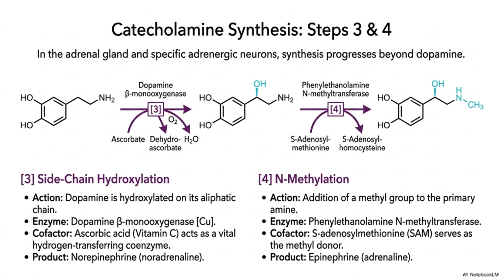
In the adrenal medulla and specialized noradrenergic neurons, catecholamine synthesis progresses beyond dopamine through two additional enzymatic steps. In Step 3, dopamine undergoes side-chain hydroxylation catalyzed by the membrane-bound enzyme dopamine beta-monooxygenase inside storage vesicles. This enzyme contains essential copper ions and requires ascorbic acid, or vitamin C, as a hydrogen-transferring coenzyme to yield norepinephrine. Norepinephrine functions as a major adrenergic neurotransmitter in sympathetic postganglionic fibers and central alertness centers like the locus coeruleus.
In Step 4, norepinephrine undergoes N-methylation catalyzed by phenylethanolamine N-methyltransferase within adrenal chromaffin cell cytoplasm. This reaction transfers a methyl group from S-adenosylmethionine to the primary amine, converting norepinephrine into epinephrine, commonly known as adrenaline. S-adenosylmethionine serves as the methyl donor, yielding S-adenosylhomocysteine as a byproduct after methylation. Glucocorticoids from the adrenal cortex stimulate this methyltransferase enzyme, coupling stress responses directly to hormone production in chromaffin tissue.
These final enzymatic conversions transform basic catecholamines into potent systemic stress hormones and specialized central adrenergic neurotransmitters. Medical students studying emergency physiology learn how released epinephrine triggers airway relaxation, elevated cardiac output, and glycogenolysis during acute stress. Together, norepinephrine and epinephrine prepare the human body to handle severe physical threats through coordinated autonomic activation driven by these adrenergic neurotransmitters.
Slide 12: How Structural Diversity Shapes Neurotransmitters
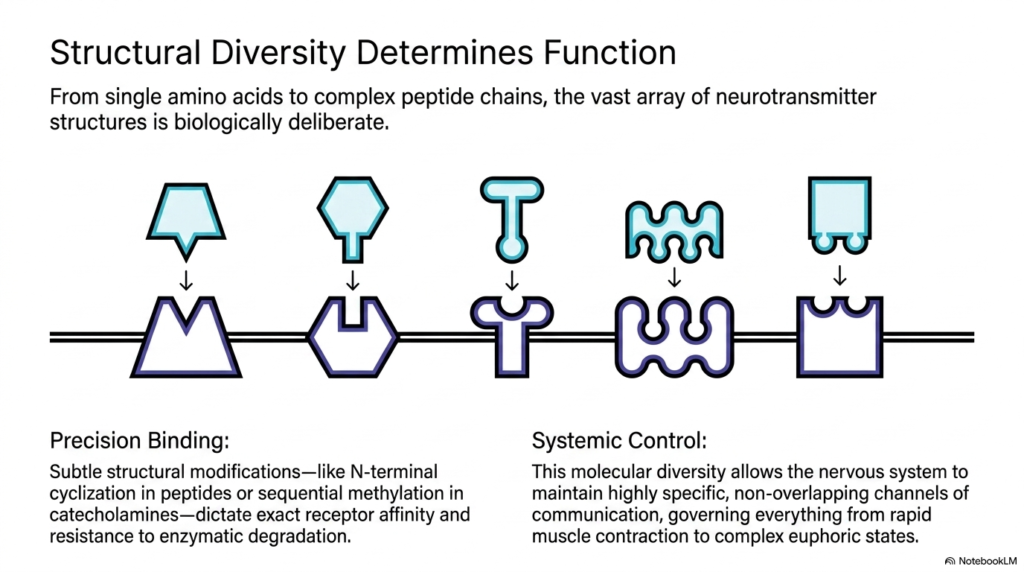
The remarkable structural diversity observed among signaling molecules reflects a precise evolutionary strategy for maintaining cellular control. From simple single amino acids to complex peptide chains, every structural motif in neurotransmitters serves a deliberate biological purpose. Subtle chemical modifications, such as N-terminal cyclization in neuropeptides or sequential methylation in catecholamines, dictate precise receptor-binding affinity. These structural variations also determine how rapidly extracellular enzymes break down signaling molecules within the synaptic cleft.
This wide molecular variety enables the human nervous system to operate highly specific, non-overlapping channels of chemical communication. Small amino acid neurotransmitters drive rapid point-to-point synaptic transmission, whereas modified peptide neurosecretions provide sustained modulatory influence over broad neuronal networks. Consequently, distinct structural classes govern everything from lightning-fast muscle contractions at neuromuscular junctions to complex emotional states in higher cortical regions.
Understanding how chemical structure dictates biological function helps college and medical students master core neurochemical principles. By linking chemical structure to receptor activation and enzymatic pathways, future healthcare professionals can better understand psychiatric drug actions and neurotransmitter-linked neurological diseases. This comprehensive structural perspective underpins modern neuropharmacology, rational drug design, and clinical neuroscience studying endogenous neurotransmitters in human medical and clinical practice.
Please read our Content Disclaimer Statement.
Check out our social media channels: