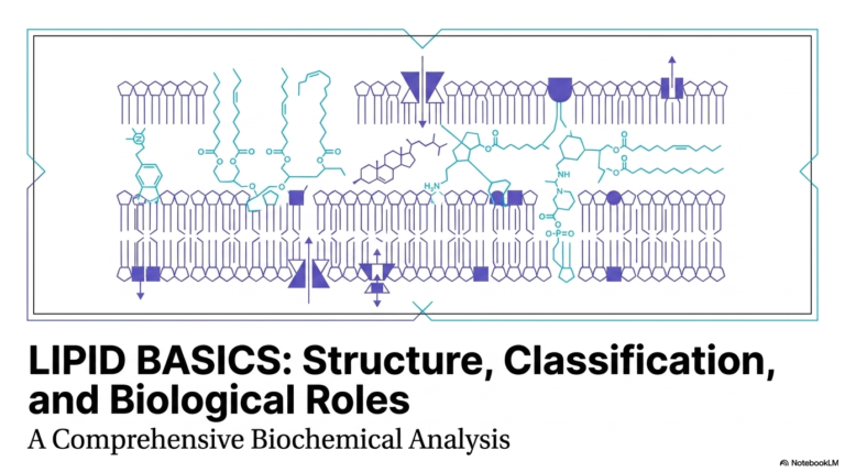
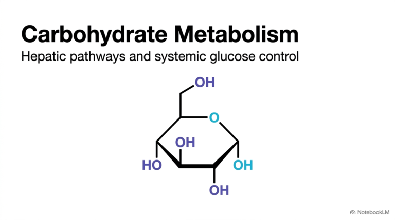
136. Biochemical Electrical Signaling: How Resting and Action Potentials Power Neural Communication
Have you ever wondered how your brain sends instructions to your muscles in a fraction of a millisecond? The secret lies in electrical bioenergetics across cell membranes. Living cells maintain delicate charge gradients to convert chemical energy into fast electrical signals. This slide deck breaks down the fundamental biochemistry behind these cellular phenomena. By examining ion channels, enzyme pumps, and thermodynamic forces, students can master how nerve cells generate and transmit rapid signals.
Slide 01: Biochemical Foundations of Resting and Action Potentials
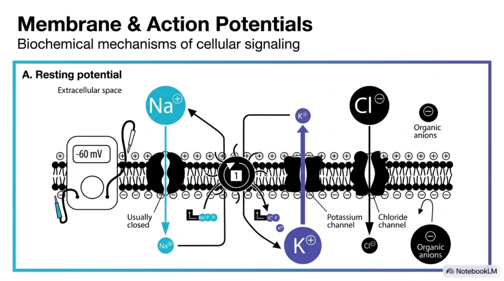
Cell membranes serve as dynamic electrical barriers that control signal generation. At the fundamental level, Resting and Action Potentials depend on the precise distribution of key ions across the lipid bilayer. Intracellular fluid contains high concentrations of potassium ions and negatively charged organic proteins. Conversely, extracellular fluid contains abundant sodium and chloride ions. This uneven chemical balance creates a baseline electrical gradient across the membrane, typically measuring around negative sixty millivolts inside relative to the outside environment.
Specialized integral membrane proteins regulate this delicate balance. Transmembrane ion channels permit specific charged particles to pass through the hydrophobic membrane core. While sodium channels remain mostly closed at rest, potassium leak channels allow potassium ions to move outward down their concentration gradient. Meanwhile, energy-dependent pumps actively maintain concentration differences. Understanding resting and action potentials requires analyzing how these selective protein channels coordinate ion movements to transform stored chemical energy into electrical signals.
Furthermore, large organic anions trapped inside the cytoplasm contribute significantly to the internal negative charge. Because these bulky proteins cannot cross the lipid membrane, they establish a permanent negative interior voltage. Chloride ions also help stabilize the resting membrane voltage. Together, these molecular components establish the foundation for cellular excitability. Mastering the biochemistry of Resting and Action Potentials provides essential insight into how nerve cells initiate rapid communication throughout the entire nervous system.
Slide 02: Uneven Ion Distribution in Resting and Action Potentials
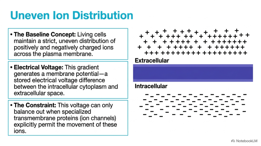
Living cells maintain a strict separation of charged particles across their plasma membrane. In the context of Resting and Action Potentials, this uneven ion distribution creates a stored bioelectrical voltage. The extracellular space contains a high density of positive charges, primarily from sodium ions. Inside the cell cytoplasm, negative charges dominate because immobile intracellular proteins carry them. This physical separation of opposite charges builds an electrical gradient across the thin lipid bilayer, forming a potential reservoir ready to do cellular work.
This voltage difference across the membrane represents potential energy. For Resting and Action Potentials, the stored membrane voltage remains stable only because the hydrophobic lipid bilayer prevents ions from crossing freely. Charged ions cannot dissolve through the nonpolar lipid interior without structural help. Consequently, the cell stores electrical energy much like a biological battery, holding positive and negative charges apart until specific membrane pathways open.
Ion movement occurs only when specialized transmembrane proteins permit transport across the membrane. These selective ion channels act as the cell’s gatekeepers. When channels open, stored electrical and chemical gradients drive rapid ion fluxes across the membrane. Without these regulated protein doors, cells could not discharge or control their stored voltage. Studying how uneven ion distributions create voltage helps explain the fundamental mechanism governing Resting and Action Potentials in excitable tissues.
Slide 03: Maintaining the Resting State in Resting and Action Potentials
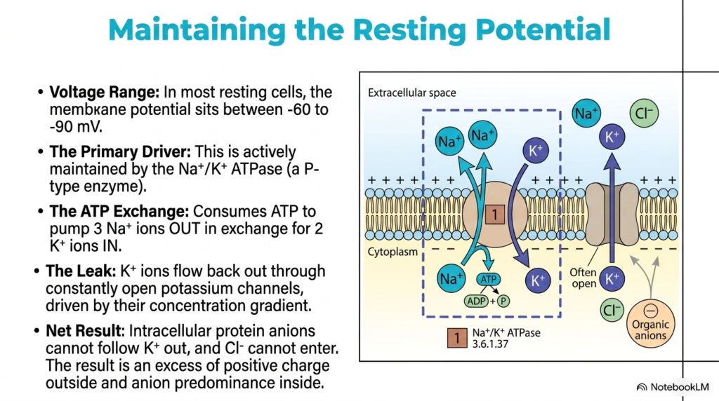
Maintaining a stable baseline voltage requires active enzyme transport working alongside passive leakage. In the study of Resting and Action Potentials, most resting cells maintain a membrane voltage between negative sixty and negative ninety millivolts. The sodium-potassium ATPase pump, a P-type transport enzyme embedded in the plasma membrane, primarily drives this baseline state. This active transporter consumes adenosine triphosphate to pump three sodium ions out of the cell while bringing two potassium ions inside against their concentration gradients.
Because the pump moves three positive charges out for every two positive charges brought in, it creates a net outward movement of positive charge. Additionally, constantly open potassium leak channels allow potassium ions to diffuse back out of the cell down their chemical gradient. As positive potassium exits, large intracellular protein anions remain trapped inside because they cannot cross the lipid bilayer. Chloride ions also remain predominantly outside, creating a net excess of positive charge on the external membrane face.
This continuous balance between active pumping and passive leakage preserves the negative resting state necessary for cellular signaling. Without the sodium-potassium ATPase constantly consuming ATP energy, concentration gradients would gradually dissipate, destroying cellular excitability. Consequently, active transport establishes the vital electrochemical baseline required for Resting and Action Potentials. Mastering this pump-and-leak balance explains how excitable cells store energy to trigger future bioelectrical events. Exploring Resting and Action Potentials demonstrates how ATP-driven transport maintains life-sustaining cellular gradients.
Slide 04: Thermodynamic Driving Forces of Resting and Action Potentials
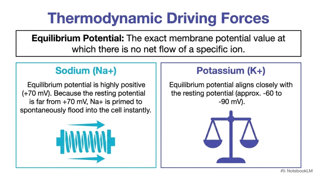
Thermodynamic forces dictate how individual ions move across the cell membrane. When analyzing Resting and Action Potentials, the equilibrium potential represents the exact membrane voltage where chemical diffusion balances electrical driving forces, resulting in no net ion movement. Each ion species possesses its own equilibrium potential determined by its concentration gradient across the membrane. Comparing an ion’s equilibrium potential to the actual membrane voltage reveals the thermodynamic driving force pushing that ion across the lipid bilayer.
Sodium ions experience an extraordinarily strong thermodynamic force at baseline during Resting and Action Potentials. The equilibrium potential for sodium sits at about +70 mV. Because the resting membrane voltage sits near negative sixty millivolts, sodium ions remain far from their thermodynamic equilibrium. This large voltage difference creates a strong gradient, driving sodium ions to flood into the cytoplasm as soon as sodium-selective channels open.
In contrast, potassium ions operate near thermodynamic balance during baseline conditions. The equilibrium potential for potassium ranges between negative sixty and negative ninety millivolts, aligning closely with the resting membrane potential. Consequently, potassium ions experience minimal net driving force at rest compared to sodium. Understanding these distinct thermodynamic driving forces provides crucial mathematical insight into how selective ion shifts drive Resting and Action Potentials during cellular activation.
Slide 05: Channel Gating Mechanisms in Resting and Action Potentials
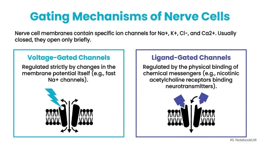
Nerve cells rely on specialized channel gating mechanisms to control ion movement. In the study of Resting and Action Potentials, cell membranes contain distinct channel proteins for sodium, potassium, chloride, and calcium ions. Under resting conditions, these gated channels remain predominantly closed, opening only briefly in response to specific environmental triggers. The two primary categories of gated ion channels are voltage-gated channels and ligand-gated channels, each serving a unique role in signal transmission across the membrane.
Voltage-gated channels respond directly to alterations in the electrical field across the plasma membrane. For example, fast voltage-gated sodium channels contain charged amino acid residues that shift when the internal membrane potential becomes more positive. This structural movement snaps the channel gate open, permitting rapid ion passage. These voltage-sensitive channels play a central role in generating fast electrical spikes across excitable membranes during cellular responses involved in Resting and Action Potentials.
Conversely, ligand-gated channels open when specific chemical messengers bind. Nicotinic acetylcholine receptors are classic ligand-gated channels that open when neurotransmitters bind to extracellular receptor sites. Once opened, these channels allow ions to cross the membrane, converting chemical signals into electrical voltage shifts. Both channel gating mechanisms cooperate seamlessly to initiate and regulate Resting and Action Potentials throughout the central and peripheral nervous systems.
Slide 06: The Chemical Stimulus in Resting and Action Potentials
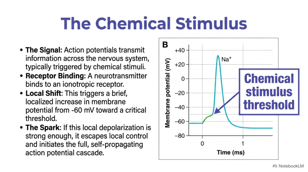
Action potential cascades begin with localized chemical stimuli at specialized synapses. Within the framework of Resting and Action Potentials, chemical signals transmit vital information across neural networks. The signaling sequence starts when a pre-synaptic nerve cell releases neurotransmitters into the synaptic cleft. These chemical messengers bind to ionotropic receptors on the postsynaptic cell membrane, initiating a local influx of positive ions that shifts the local membrane potential away from the resting baseline.
This initial ligand binding produces a brief, localized shift in membrane voltage. Positively charged ions entering through ligand-gated channels cause local depolarization, moving the internal voltage from negative sixty millivolts toward a critical threshold value. If the chemical stimulus remains weak, the voltage shift stays localized and decays without triggering a full electrical response. However, strong stimuli push the membrane voltage past the critical threshold required for Resting and Action Potentials.
Crossing the threshold sparks a dramatic, self-propagating electrical event. Once local voltage reaches threshold, neighboring voltage-gated channels snap open, escape local chemical control, and launch a rapid bioelectrical spike. Understanding how chemical stimuli cross the threshold barrier is essential for mastering Resting and Action Potentials. This critical threshold mechanism ensures that cells filter out minor background noise while reliably propagating meaningful biological signals.
Slide 07: Depolarization Mechanics in Resting and Action Potentials
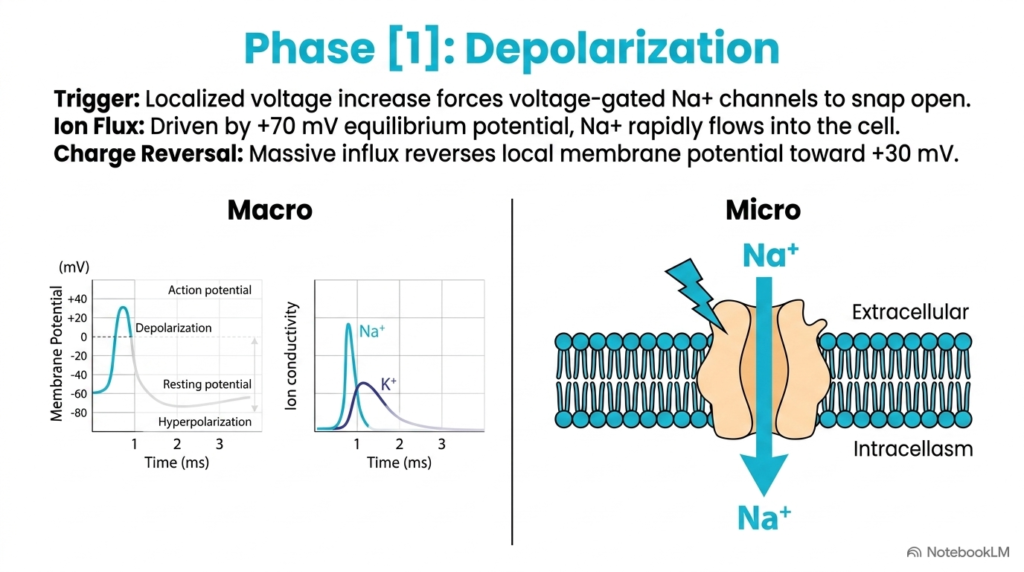
The first major functional phase of an electrical spike is depolarization. In the dynamic cycle of Resting and Action Potentials, depolarization represents a massive, rapid influx of positive charge into the cell. Once a chemical or electrical stimulus forces the membrane potential to threshold, voltage-gated sodium channels rapidly snap open. Driven by a positive seventy millivolt equilibrium potential, sodium ions flood into the cytoplasm down both chemical and electrical gradients.
This explosive inward sodium current reverses the charge across the lipid bilayer. Within one millisecond, the internal membrane potential surges from negative sixty millivolts past zero to about positive thirty millivolts. On a microscopic scale, thousands of individual sodium channels open simultaneously, drastically increasing membrane conductance to sodium. The rapid influx overwhelms passive leak currents, completely depolarizing the local membrane region during Resting and Action Potentials.
Graphing this phase demonstrates a steep upward voltage spike accompanied by a sharp peak in sodium conductivity. Because sodium ions carry positive charge inward, the cell interior temporarily becomes positively charged relative to the exterior. This explosive phase illustrates how voltage-gated channel kinetics drive rapid state changes. Depolarization forms the initial rising phase essential to understanding Resting and Action Potentials in excitable tissues.
Slide 08: Peak Potential Kinetics in Resting and Action Potentials
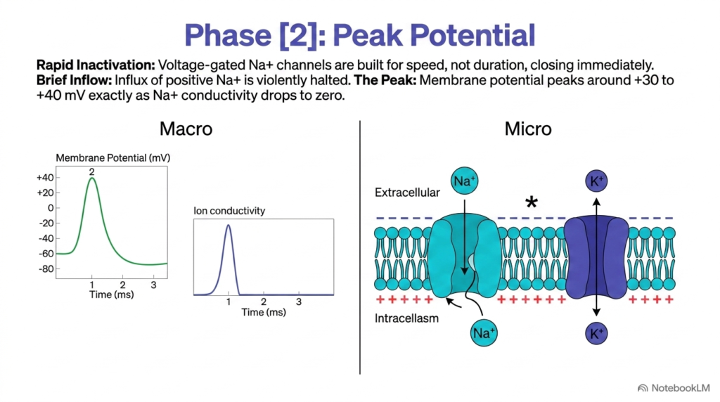
Following rapid depolarization, the electrical spike reaches its maximum voltage at peak potential. In the progression of Resting and Action Potentials, phase two marks the crucial transition where sodium influx abruptly ceases. Voltage-gated sodium channels possess an intrinsic ball-and-chain inactivation gate that closes automatically shortly after opening. This rapid inactivation halts positive sodium entry, capping the membrane potential at about +30 to +40 mV during electrical activation.
At peak potential, membrane conductivity to sodium drops precipitously to zero. Although the electrical driving force pushing sodium inward remains present, closed inactivation gates physically block sodium ions from entering the cytoplasm. At the same time, the highly positive internal voltage begins to alter the conformation of voltage-gated potassium channels, preparing them to open. This precise timing prevents the cell voltage from reaching sodium’s theoretical equilibrium potential of positive seventy millivolts during Resting and Action Potentials.
Understanding peak potential reveals how structural channel kinetics limit signal duration. Automatic sodium channel inactivation ensures the electrical pulse remains a brief, millisecond-long event rather than a sustained voltage shift. This rapid termination mechanism is vital for maintaining high-frequency signaling. Analyzing channel inactivation at peak potential provides deeper insight into the cyclical nature of Resting and Action Potentials.
Slide 09: Repolarization Dynamics in Resting and Action Potentials
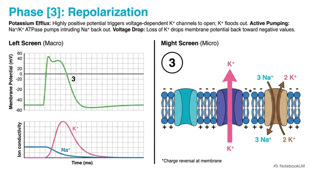
Once the membrane reaches peak potential, it must restore its negative internal charge through repolarization. In the cycle of Resting and Action Potentials, phase three restores the negative internal voltage through rapid ion movement. The highly positive internal charge triggers voltage-dependent potassium channels to open fully. Driven by both concentration gradients and electrical repulsion, potassium ions flood outward into the extracellular space, carrying positive charge away from the cell interior.
As potassium leaves the cytoplasm, the internal membrane potential drops rapidly back toward negative values. At the same time, active transport enzymes work continuously in the background. The sodium-potassium ATPase pump actively transports intruding sodium ions out of the cytoplasm while bringing potassium ions in, restoring proper ion concentrations across the lipid bilayer. This combination of passive potassium efflux and active enzymatic pumping rapidly clears positive charge from the cell interior during Resting and Action Potentials.
On conductance graphs, repolarization features a dramatic rise in potassium conductivity alongside a steep decline in membrane voltage. The rapid departure of positive potassium ions successfully reverses the positive overshoot, bringing the membrane back toward its baseline electrical state. Mastering the biochemical mechanisms of repolarization is fundamental to comprehending how cells reset after firing, illustrating the tight coordination underlying Resting and Action Potentials.
Slide 10: Hyperpolarization Overshoot in Resting and Action Potentials
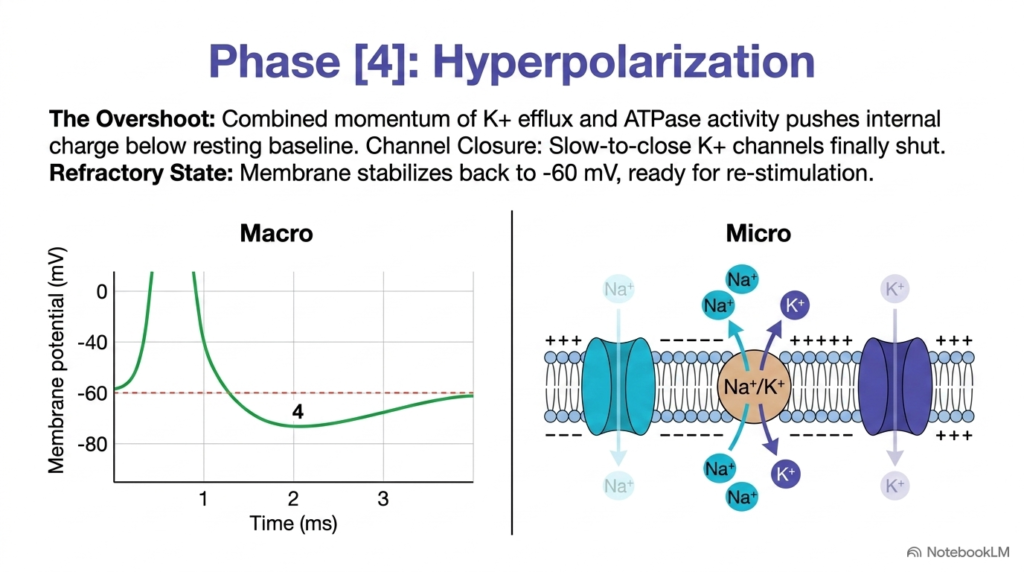
The final phase of the electrical signaling cycle is hyperpolarization. In the complete sequence of Resting and Action Potentials, hyperpolarization occurs when the internal membrane voltage temporarily drops below its normal resting baseline. Because voltage-gated potassium channels close slowly, potassium ions continue leaking out of the cell even after the membrane potential reaches negative sixty millivolts. This lingering potassium efflux creates a temporary negative overshoot down toward negative eighty millivolts across the lipid membrane.
During this hyperpolarized state, the cell enters a refractory period where re-stimulation is extremely difficult. The excessive internal negativity, combined with inactivated sodium channels, prevents immediate firing of a second electrical spike. Active sodium-potassium ATPase pumps, along with the eventual closure of slow potassium channels, gradually restore the membrane potential back to its stable resting baseline of negative sixty millivolts during Resting and Action Potentials.
Hyperpolarization serves a critical physiological function by preventing backward signal propagation and limiting overall firing frequency. Once potassium channels fully close and ion distributions normalize, the cell exits the refractory state and stands ready for new bioelectrical stimulation. Understanding how slow channel kinetics produce this negative overshoot completes the full cycle of Resting and Action Potentials, demonstrating how cells maintain directional signal transmission across neural circuits.
Slide 11: Depolarization Wave Propagation in Resting and Action Potentials
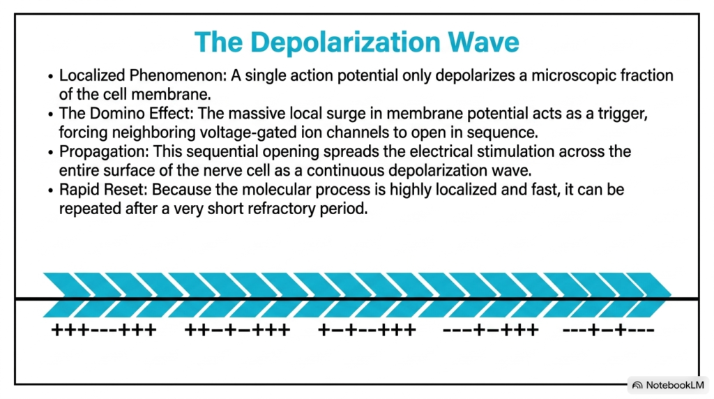
Action potentials do not remain stationary; they travel along cell membranes as continuous depolarization waves. When examining Resting and Action Potentials, a single electrical spike initially depolarizes only a microscopic patch of the plasma membrane. However, the sudden surge of positive sodium ions in that local region creates internal electrical currents that spread passively to adjacent membrane segments, raising neighboring areas to threshold as the signal propagates.
This passive current flow creates a biological domino effect along the nerve axon. As neighboring membrane regions reach threshold voltage, adjacent voltage-gated sodium channels open in sequence. This orderly progression propagates the electrical signal smoothly across the entire surface of the nerve cell as a self-sustaining depolarization wave. The signal travels long distances without losing amplitude because each membrane segment actively generates its own spike during Resting and Action Potentials.
Furthermore, the refractory period behind the advancing wave prevents the signal from traveling backward. Because recently depolarized membrane segments remain temporarily hyperpolarized and inactivated, the electrical wave moves strictly in one direction. Rapid membrane reset kinetics allow the cell to conduct subsequent signals after a brief delay. Analyzing wave propagation illustrates how localized Resting and Action Potentials convert isolated molecular shifts into long-distance biological signals.
Slide 12: Quantitative Dynamics Synthesis of Resting and Action Potentials
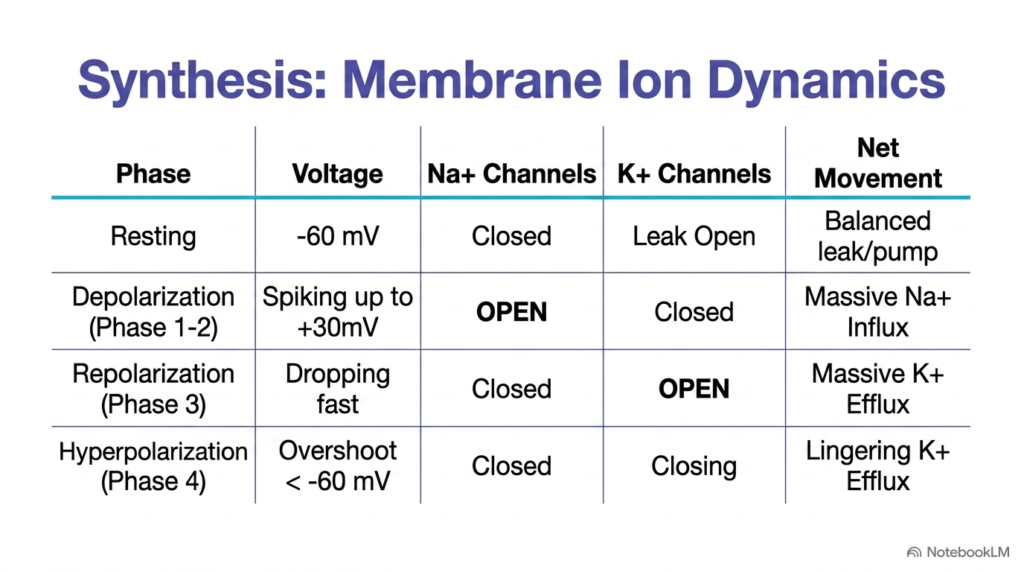
Synthesizing membrane ion dynamics provides a clear quantitative overview of cellular signaling. Comparing the stages of Resting and Action Potentials reveals how distinct voltage levels correlate with channel conformations and net ion movements. At rest, the membrane maintains a negative sixty millivolts with closed sodium channels and open potassium leak pathways, creating a balanced baseline maintained by active transport and passive leakage across the lipid bilayer.
During depolarization, the membrane voltage spikes rapidly to about +30 mV as voltage-gated sodium channels open while potassium channels remain closed, driving a massive inward sodium influx. In repolarization, the voltage drops sharply as sodium channels close and voltage-gated potassium channels open, causing a massive outward potassium efflux. Finally, hyperpolarization produces a temporary overshoot below negative sixty millivolts as potassium channels slowly shut, leaving lingering potassium efflux until baseline conditions return during Resting and Action Potentials.
This structured synthesis highlights the elegant molecular coordination driving bioelectrical signals across biological membranes. Each distinct phase depends on precise timing, conformational channel changes, and thermodynamic driving forces. By mastering how channel gating, voltage shifts, and ion fluxes align across every phase, students gain a comprehensive mastery of Resting and Action Potentials in excitable biological systems throughout human physiology.
Please read our Content Disclaimer Statement.
Check out our social media channels: