128. Biochemistry of Muscle Contraction: How Molecular Engines Generate Mechanical Force
Every movement, from a heartbeat to a sprint, relies on remarkable microscopic engines working inside our cells. How do single protein molecules convert chemical energy into powerful mechanical force? This slide deck offers a comprehensive walk-through of the structural framework, biochemical actors, and kinetic cycles behind skeletal movement. Designed for college and medical students, this visual guide breaks down complex protein interactions into clear steps, demonstrating how molecular events produce macroscopic movement.
Slide 1: Introduction to Muscle Contraction and the Sliding Filament Model
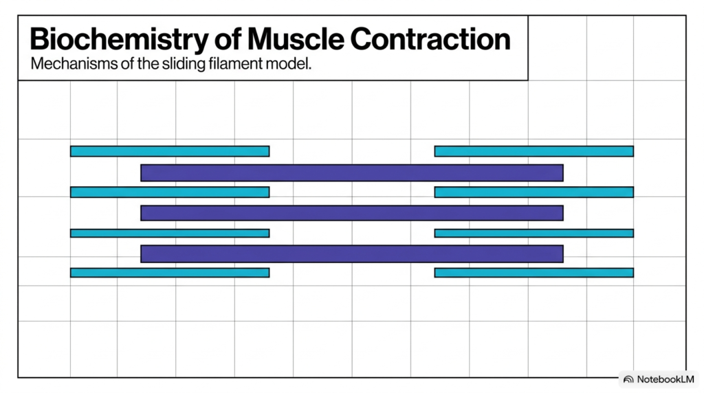
The opening slide provides a foundational visual introducing the core biochemical mechanisms that drive Muscle Contraction across human tissue. At the cellular level, biological movement relies on the sliding filament model, a physiological framework describing how interdigitated protein strands slide past one another to generate physical tension. When studying Muscle Contraction, college and medical school students must observe how thick filaments and thin filaments align in parallel arrays within the contractile apparatus.
In this visual overview, thick filaments composed of myosin sit positioned alongside thin filaments made of actin. During Muscle Contraction, these overlapping protein bands slide inward toward each other without physically shortening or compressing the protein strands themselves. This fundamental sliding mechanism serves as the master blueprint for voluntary skeletal movements and involuntary cardiac and smooth muscle actions throughout the human body.
Grasping this precise spatial arrangement is essential because physiological force originates directly from this organized architecture. By establishing this structural framework, the slide sets the stage for exploring how nanoscale protein interactions scale up into powerful cellular contractions. Ultimately, mastering Muscle Contraction requires appreciating both the structural geometry and the energetic steps that pull these filaments together smoothly during active tension generation.
Slide 2: Tissue Taxonomy and Universal Principles of Muscle Contraction
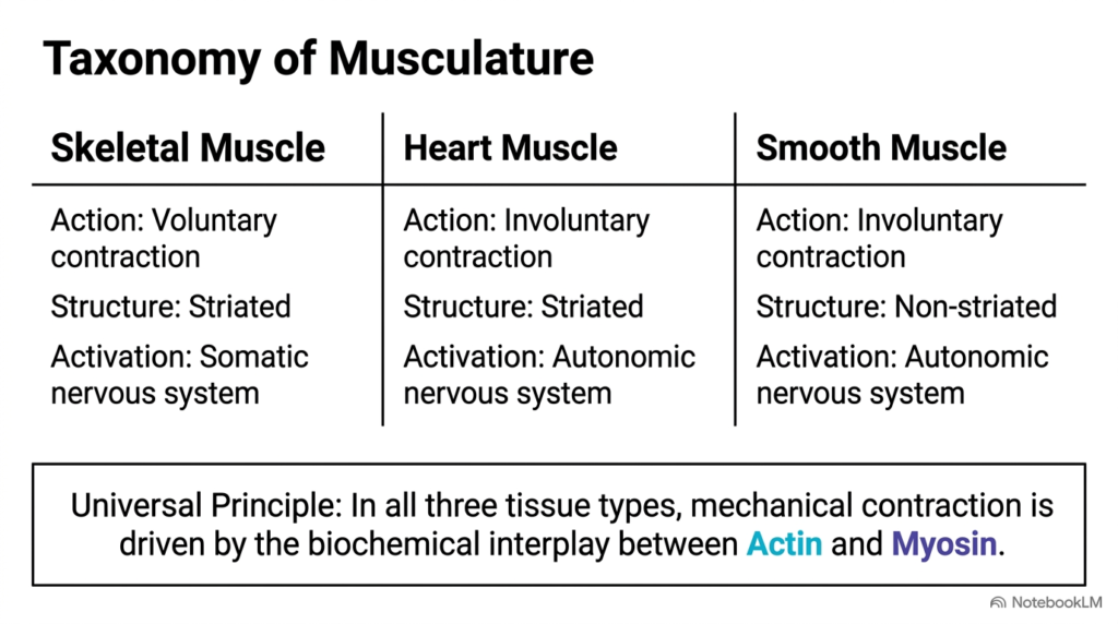
Human anatomy classifies muscle tissue into three distinct structural categories, yet each category relies on similar biochemical rules during Muscle Contraction. Skeletal muscle is under voluntary somatic control and shows clear microscopic striations from aligned contractile proteins. Heart muscle is also striated but contracts involuntarily under autonomic nervous system control. Smooth muscle lacks striations and contracts involuntarily in hollow organs like blood vessels and the digestive tract.
Despite these structural and neurological differences, a single universal principle governs every form of Muscle Contraction across human biology. Mechanical force generation always depends on the precise molecular interplay between actin and myosin filaments. Whether pumping blood through cardiac chambers or lifting heavy weights with skeletal limbs, cells harness these two primary proteins to drive structural movement and generate physical tension across varied anatomical contexts.
Recognizing this shared molecular engine helps students connect basic cellular biology with broad physiological systems. While upstream neural excitation and calcium regulation mechanisms vary across tissue types, the underlying chemistry of Muscle Contraction remains remarkably conserved throughout the human body. Understanding this unified pathway is essential for medical students analyzing cardiovascular, gastrointestinal, and musculoskeletal disorders across clinical practice.
Slide 3: Hierarchical Structural Organization of Muscle Contraction
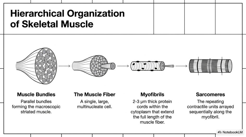
To understand how microscopic molecular events produce macroscopic body movements, students must examine the hierarchical organization that supports muscle contraction. Intact skeletal muscle consists of organized parallel bundles termed fascicles, which contain thousands of individual muscle fibers surrounded by supportive connective tissue sheaths. Each individual muscle fiber functions as a single, large multinucleate cell specialized for generating significant physical force during strenuous muscular exertion.
Inside the cytoplasm of these muscle fibers lie dense, thread-like protein cords called myofibrils, measuring roughly two to three micrometers in diameter. These myofibrils extend continuously along the full length of the muscle fiber and house repeating functional units known as sarcomeres. The sarcomere is the fundamental contractile unit where muscle contraction occurs at the subcellular level during active excitation.
Arrayed end to end, millions of individual sarcomeres work in series to shorten the entire muscle fiber during activation. This nested structural hierarchy ensures that tiny nanoscale shifts within individual sarcomeres accumulate efficiently across the entire cell length. Consequently, this multi-layered architecture drives the large-scale Muscle Contraction observed during athletic performance, posture maintenance, and routine daily physical activities throughout life.
Understanding this multi-level arrangement helps students see how cellular anatomy connects directly to macroscopic physiological function. When an action potential spreads through the cell, synchronized shortening across every nested level produces the overall tension required for coordinated body movement.
Slide 4: Sarcomere Topology and Banding Patterns in Muscle Contraction
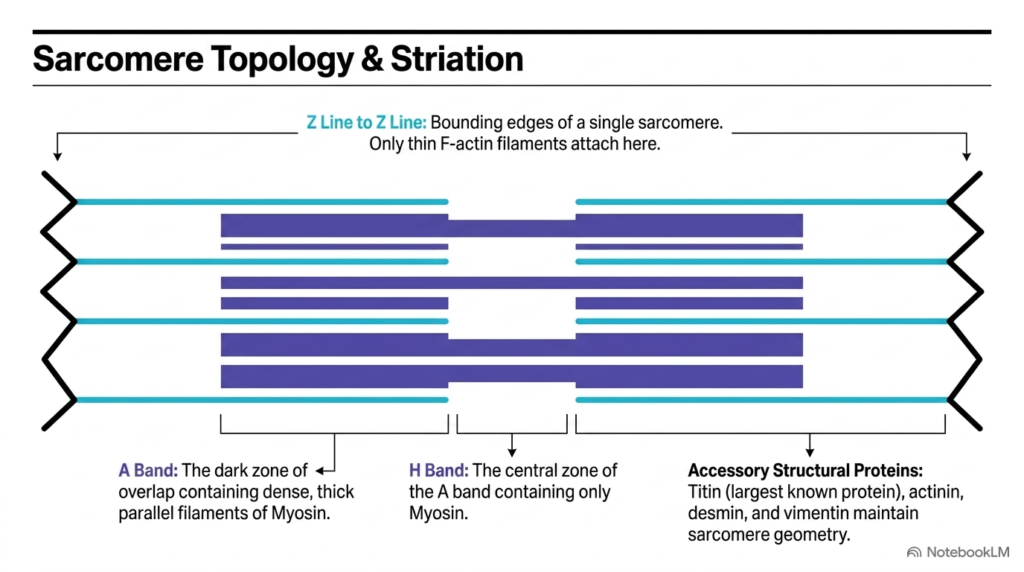
Understanding sarcomere geometry and banding patterns is essential to studying the structural basis of Muscle Contraction. The Z line defines the boundary of a single sarcomere and serves as the anchor site where thin actin filaments attach securely. Between two adjacent Z lines, overlapping protein filaments form distinct microscopic bands visible under light and electron microscopy in striated muscle tissue under polarized illumination.
The dark A band marks the region containing dense, parallel thick filaments composed of myosin, including regions where thick and thin filaments overlap. Within the central region of the A band sits the H band, a lighter zone consisting exclusively of myosin without actin overlap. During active Muscle Contraction, these bands change their relative dimensions as thin filaments slide deeper toward the central M line, progressively shortening the H band.
Accessory structural proteins preserve this precise geometrical architecture throughout repeated cycles of mechanical stress. Giant titin molecules, the largest known proteins in human biology, work alongside actinin, desmin, and vimentin to maintain proper filament alignment and elastic tension. These stabilizing elements protect the sarcomere from structural disruption during intense Muscle Contraction and passive tissue stretching in physiological conditions.
These structural landmarks provide essential clinical references when analyzing muscle biopsies and diagnostic imaging. Disruption of these distinct banding patterns often indicates underlying muscular dystrophies or metabolic myopathies in human patients.
Slide 5: Thick Filament Architecture and Myosin in Muscle Contraction
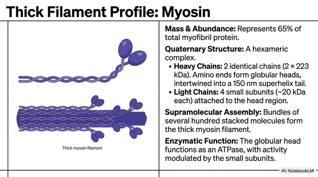
Thick filaments make up about 65% of total myofibril protein and play a central role in Muscle Contraction. These macromolecular structures consist of myosin, a massive hexameric protein complex containing two identical heavy chains and four smaller light chains. The elongated amino-terminal regions of the heavy chains form two globular heads connected to a long, flexible alpha-helical coiled-coil tail that measures 150 nanometers in length.
Hundreds of individual myosin molecules assemble in a staggered, bipolar arrangement to construct the thick filament body. The protruding globular heads extend outward in a helical pattern toward adjacent thin filaments, perfectly positioned to form cross-bridges during Muscle Contraction. Small light chain subunits attach near these globular heads to modulate enzymatic activity and structural stability during active force production across human muscle tissue.
Biochemically, the globular myosin head functions as an active adenosine triphosphatase enzyme capable of energy transduction. By binding and hydrolyzing adenosine triphosphate, myosin converts stored chemical energy directly into mechanical work. This enzymatic cleavage drives the force-generating structural steps required for effective Muscle Contraction in human skeletal muscle fibers during athletic exertion.
This structural symmetry ensures that force generation occurs in opposite directions on either side of the bare central zone. Consequently, thick filaments pull opposing thin filaments toward the center of the sarcomere with equal mechanical force.
Slide 6: Thin Filament Structure and Actin Dynamics in Muscle Contraction
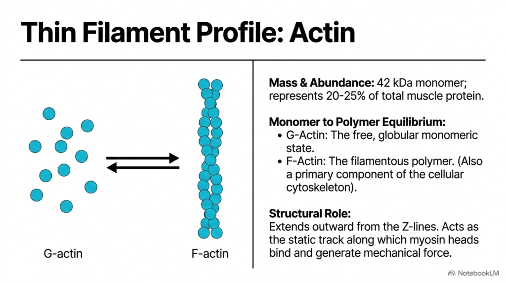
Thin filaments provide the essential structural track required for filament sliding during Muscle Contraction. Making up twenty to twenty-five percent of total muscle protein, thin filaments consist primarily of actin, a forty-two kilodalton protein. Actin exists in a dynamic monomer-to-polymer equilibrium between individual globular subunits and long fibrous helical strands within the cell cytoplasm under normal physiological conditions.
Free globular actin monomers, known as G-actin, polymerize into long double-helical filaments termed F-actin. These F-actin polymers attach directly to the Z lines and extend outward into the central region of the sarcomere. During Muscle Contraction, F-actin serves as the static structural track along which protruding myosin heads bind and generate mechanical pull toward the central M line.
While actin filaments also help maintain general cell shape and division in non-muscle cells, their role in contractile muscle tissue is highly specialized. By presenting specific binding sites for myosin heads, F-actin enables the conversion of chemical energy into linear mechanical force during Muscle Contraction across skeletal muscle fibers throughout human physiological movement.
Polarity within the F-actin strand directs myosin heads to move exclusively toward the Z line during active cycles. This intrinsic structural polarity ensures that sliding always occurs in the correct direction during force development.
Slide 7: Regulatory Tropomyosin and Troponin Complexes in Muscle Contraction
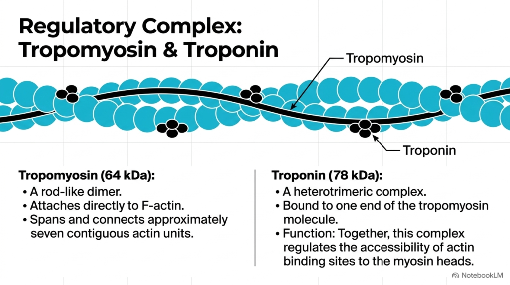
Uncontrolled interactions between actin and myosin would continuously consume cellular energy, making precise biochemical regulation vital for controlled Muscle Contraction. Two key regulatory proteins, tropomyosin and troponin, form a cooperative complex along the thin filament surface. Tropomyosin exists as a rod-like dimer that wraps around F-actin, physically blocking myosin-binding sites across seven contiguous actin monomers in resting muscle tissue.
Bound to one end of each tropomyosin dimer sits troponin, a heterotrimeric protein complex composed of three distinct subunits. Together, troponin and tropomyosin act as a molecular switch that prevents myosin heads from contacting actin when the muscle is relaxed, thereby inhibiting unscheduled Muscle Contraction and conserving vital cellular adenosine triphosphate supplies for future physical demands.
When calcium ions flood the sarcoplasm following electrical membrane depolarization, calcium binds directly to troponin, causing a conformational shift that pulls tropomyosin away from active actin sites. This structural movement exposes the binding sites, permitting cross-bridge formation and initiating the cascade of Muscle Contraction in active muscle fibers.
This tightly regulated calcium switch allows muscles to respond instantly to neural signals while remaining completely quiet during resting periods. Medical students must recognize that alterations in troponin subunits serve as vital diagnostic markers for cardiac injury.
Slide 8: The Sliding Filament Mechanism Driving Muscle Contraction
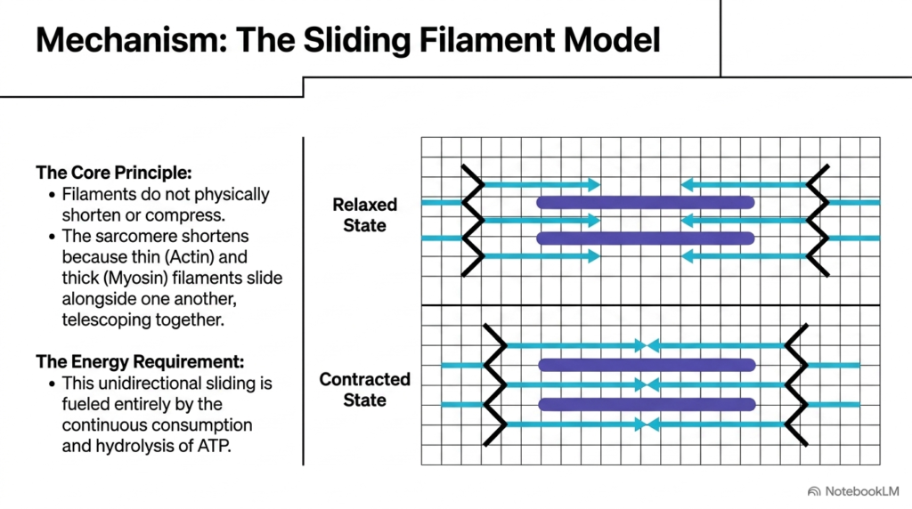
The sliding filament model provides the foundational mechanical principle for explaining how Muscle Contraction shortens muscle tissue. A crucial insight of this biochemical model is that individual actin and myosin filaments never physically shorten or compress during movement. Instead, the entire sarcomere shortens because thin filaments and thick filaments slide alongside one another in a telescoping fashion.
In a relaxed muscle, thick and thin filaments partially overlap, leaving a wide central H band. As active Muscle Contraction proceeds, cycling myosin heads pull thin filaments inward toward the central M line, narrowing the H band and drawing opposing Z lines closer together, thereby reducing total sarcomere length across the entire myofibril length.
This unidirectional sliding motion requires continuous chemical energy input to maintain cycle turnover and force output. Every step of filament movement relies on the steady consumption and enzymatic hydrolysis of adenosine triphosphate, ensuring that Muscle Contraction remains a tightly regulated, energy-dependent physiological process throughout active physical exercise.
As thin filaments slide deeper toward the center, the overall width of the A band remains entirely constant. This key experimental observation confirmed that filament sliding, rather than protein folding, drives muscle shortening.
Slide 9: ATP Binding, Cleavage, and Plasticity in Muscle Contraction
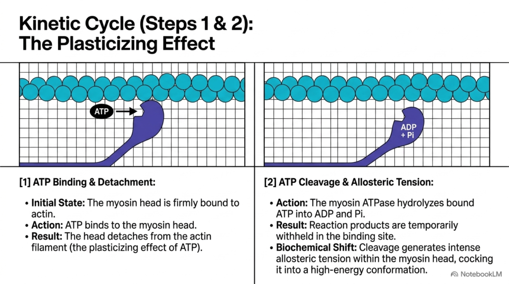
The initial steps of the cross-bridge kinetic cycle demonstrate how adenosine triphosphate prepares the myosin head for Muscle Contraction. In the starting state, the myosin head sits firmly attached to an actin monomer in a low-energy rigor state. When a fresh molecule of adenosine triphosphate binds to the myosin head, it induces a conformational change that weakens myosin affinity for actin, causing immediate detachment. This event is known as the plasticizing effect of ATP during Muscle Contraction.
Following detachment, the active site within the myosin head hydrolyzes bound adenosine triphosphate into adenosine diphosphate and inorganic phosphate. Crucially, the myosin head retains both cleavage products within its binding pocket rather than releasing them into the surrounding cytoplasm.
This enzymatic cleavage step stores free energy by generating intense allosteric tension within the protein structure, cocking the myosin head into a high-energy conformation. Fully cocked and energized, the myosin head stands prepared to form a new cross-bridge and drive the next phase of Muscle Contraction during active physical movement.
If cellular adenosine triphosphate becomes depleted, myosin heads remain permanently locked to actin in the rigor state. This biochemical phenomenon explains the muscle stiffness observed in rigor mortis following systemic metabolic failure.
Slide 10: The Myosin Power Stroke and Force Generation in Muscle Contraction
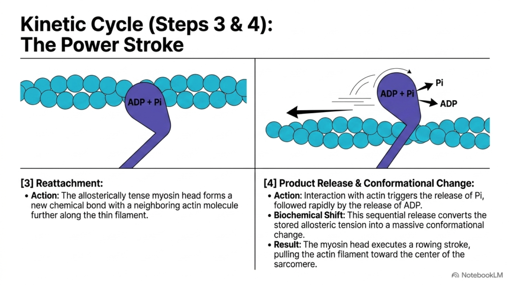
After activation and cocking, the myosin head forms a new chemical cross-bridge with an actin monomer farther along the thin filament. This binding event triggers the central mechanical phase of Muscle Contraction, widely known as the power stroke. Interaction with actin prompts the immediate release of inorganic phosphate, followed rapidly by the release of adenosine diphosphate.
The sequential release of these reaction products releases the stored allosteric tension within the myosin neck domain. This dramatic conformational shift causes the myosin head to pivot forcefully like an oar, pulling the attached actin filament roughly ten nanometers toward the center of the sarcomere during Muscle Contraction.
By converting stored chemical energy into mechanical displacement, the power stroke generates actual physical tension within the muscle fiber. Once the stroke completes, the myosin head remains tightly bound in a low-energy state until a new ATP molecule arrives to reset the cross-bridge cycle during ongoing Muscle Contraction.
This mechanical stroke generates approximately two to five piconewtons of force per individual myosin head. When thousands of heads operate simultaneously, these minute force vectors integrate into substantial mechanical power across the entire fiber.
Understanding this kinetic cycle allows medical students to grasp how pharmacological agents and metabolic inhibitors affect contractile force. By targeting specific steps in product release, researchers can modulate muscular performance.
Slide 11: Summation of Molecular Events into Macroscopic Muscle Contraction
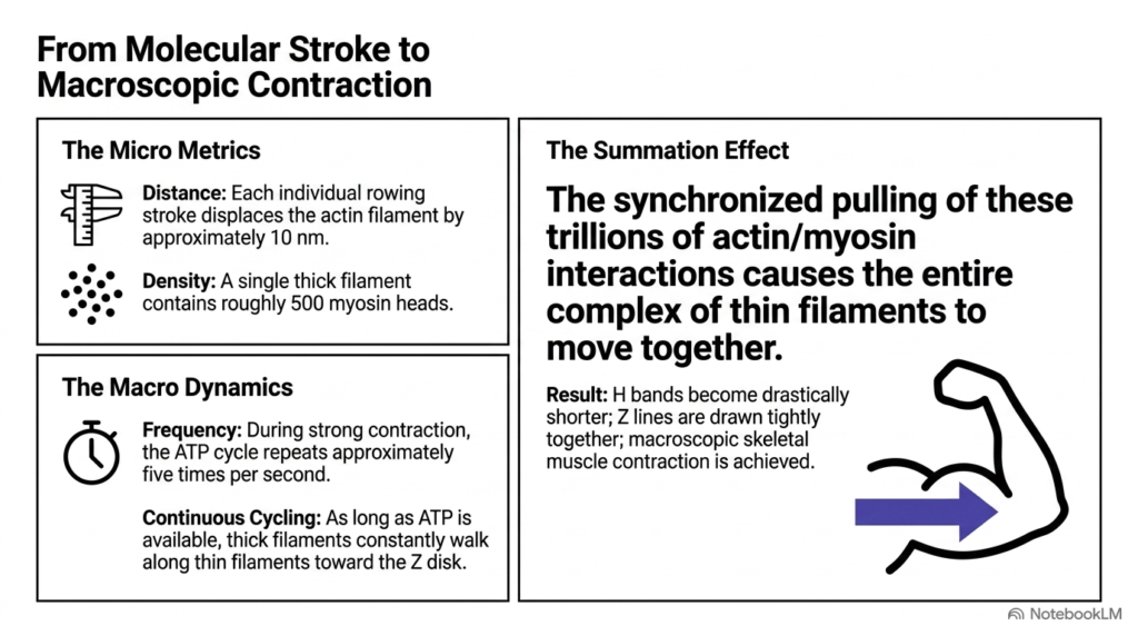
Translating individual molecular strokes into whole-body movement requires massive structural coordination during Muscle Contraction. On a microscopic scale, each single myosin power stroke displaces an actin filament by approximately ten nanometers. Because a single thick filament contains roughly five hundred myosin heads cycling several times per second, these tiny displacements compound rapidly during active Muscle Contraction across human muscle fibers in real time.
When millions of adjacent sarcomeres shorten simultaneously along a myofibril, their individual forces summate linearly along the entire length of the cell. Trillions of synchronized cross-bridge rowing strokes pull thin filaments toward sarcomere centers, drastically shortening H bands and drawing Z lines tightly together across all muscle fibers during active physiological contraction.
This collective molecular action transforms nanoscale protein conformational changes into powerful macroscopic force. Through this elegant summation effect, individual chemical reactions yield the smooth, coordinated Muscle Contraction that powers all human physical movement, locomotion, and athletic performance across daily biological functions.
Ultimately, this integrative cascade links sub-nanometer enzymatic events directly to whole-body biomechanics. Understanding these structural and chemical relationships equips future clinicians to evaluate neuromuscular performance, diagnose motor disorders, and treat complex muscle pathologies effectively in clinical medical practice.
Please read our Content Disclaimer Statement.
Check out our social media channels: