127. Understanding Renal Hormones: Erythropoietin, Calcitriol, and Renin
When most people think of the kidneys, they picture simple biological filters that clean blood and produce urine. However, these vital organs also function as a dynamic endocrine command center. Beyond removing metabolic waste, the kidneys synthesize essential signals that regulate red blood cell production, bone density, and arterial pressure. This slide deck breaks down the biochemical structures, activation triggers, and physiological pathways of kidney-derived chemical messengers. College and medical school students will explore how renal endocrinology maintains whole-body homeostasis.
Slide 1: Introduction to Renal Hormones and Endocrine Pathways
When exploring human physiology, students often focus on the kidneys as excretory organs that filter metabolic waste from the blood. However, these organs perform essential signaling duties that govern systemic balance across multiple tissues. Renal Hormones represent a critical bridge between filtration mechanics and whole-body chemical signaling.
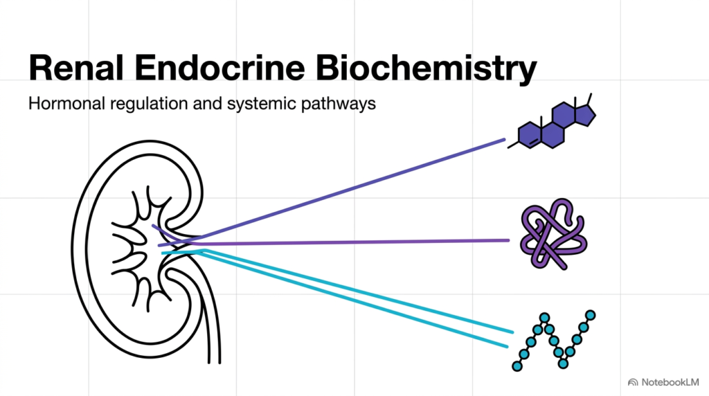
The human body relies on Renal Hormones to coordinate diverse physiological networks. Rather than secreting a single compound, the kidney synthesizes multiple distinct chemical classes. These include steroid derivatives like calcitriol, specialized peptide signals such as erythropoietin, and key enzymatic triggers like renin. Each chemical messenger leaves the kidney to act on specific target tissues across the body.
Understanding Renal Hormones requires analyzing how structural chemistry dictates physiological function. For instance, lipid-soluble steroid structures interact with intracellular receptors, whereas peptide chains bind cell-membrane targets. By releasing these diverse signals into circulation, the kidney maintains steady blood pressure, regulates oxygen distribution, and protects bone density.
Biochemists categorize these pathways to appreciate how local kidney mechanisms maintain global equilibrium. As students move through this slide deck, they will discover how each signaling molecule operates at the molecular level. Recognizing the key contributions of Renal Hormones is essential for mastering medical biochemistry and understanding complex human endocrine networks.
Slide 2: The Kidney as a Central Hub for Renal Hormones
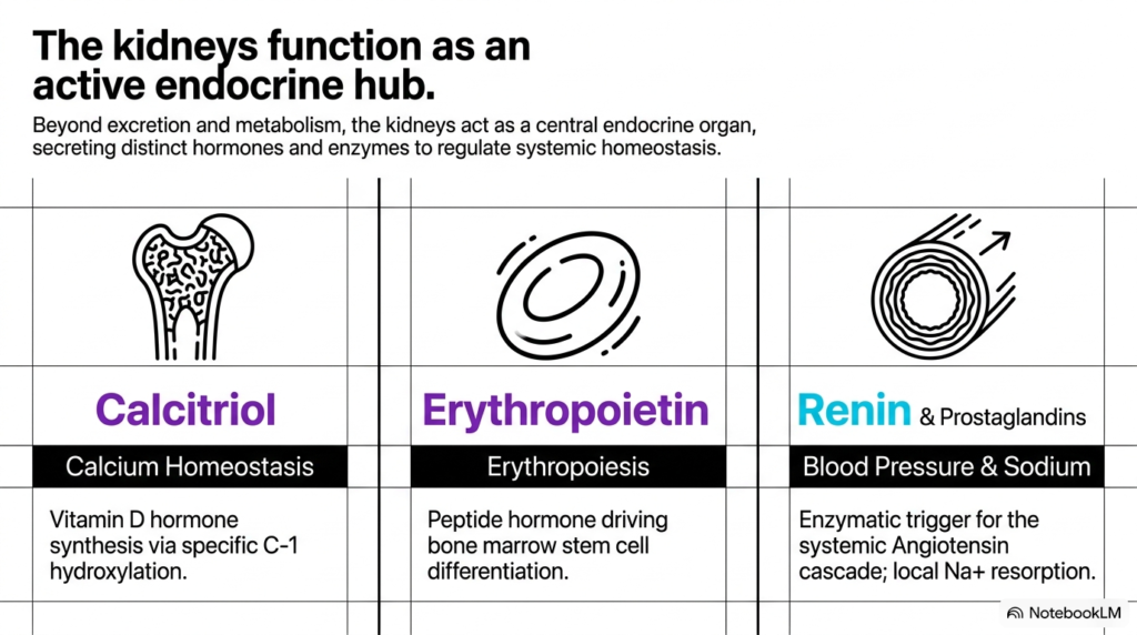
The kidney functions as a command center that continuously monitors fluid dynamics and nutrient balance throughout the circulation. Within this organ, specialized cellular populations produce Renal Hormones that address distinct physiological challenges. This active endocrine hub keeps systemic conditions within narrow functional boundaries.
Three major endocrine pathways showcase the versatility of Renal Hormones in human health. First, calcitriol governs calcium homeostasis through specific enzymatic modifications of vitamin D precursors. Second, erythropoietin acts directly on bone marrow stem cells to drive the differentiation of new erythrocytes. Third, renin functions alongside local prostaglandins to regulate sodium retention and systemic vascular tone.
Together, these signaling networks allow the kidney to respond dynamically to environmental and internal stresses. When tissue oxygen drops or blood pressure declines, the organ adjusts its output of Renal Hormones accordingly. Each pathway shows a precise mechanism in which local renal sensing triggers widespread physiological adjustments throughout the body.
For college and medical students, studying these three axes reveals how the kidney integrates metabolic demands across organ systems. The production of calcitriol, erythropoietin, and renin highlights the organ’s ability to communicate with the skeleton, circulatory system, and bone marrow. Mastering how Renal Hormones function within this central hub provides a strong foundation for understanding human endocrine regulation and systemic homeostasis.
Slide 3: Structural Activation of Calcitriol Among Renal Hormones
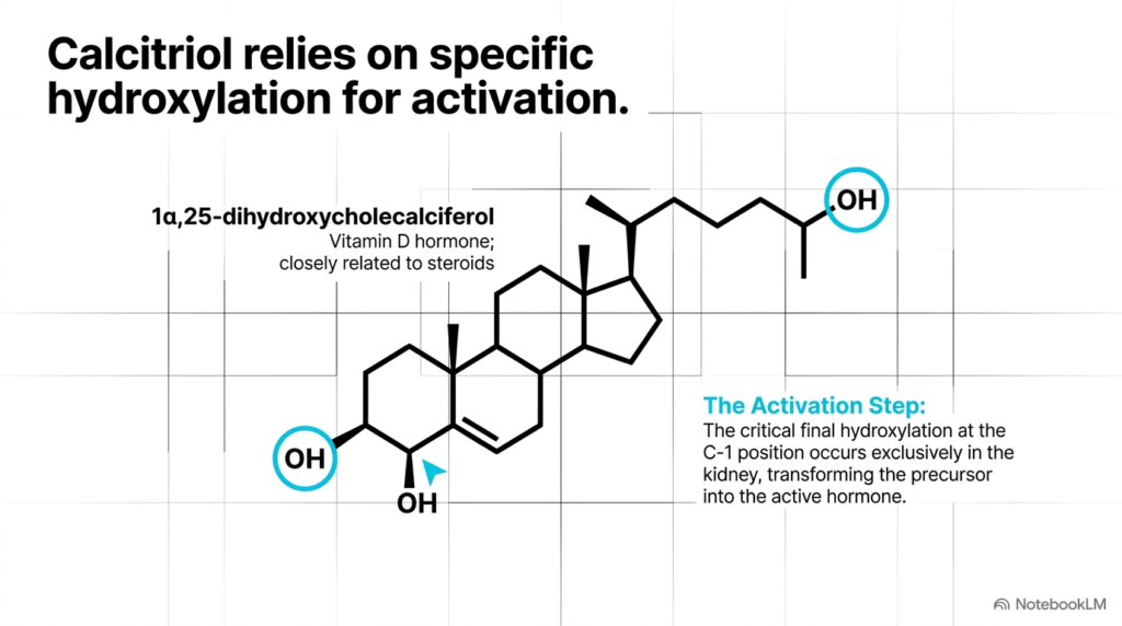
Calcitriol represents one of the most chemically distinct Renal Hormones produced by the human body. As the active form of vitamin D, known biochemically as 1alpha,25-dihydroxycholecalciferol, this molecule shares structural features with classical steroid signaling compounds. Its activation pathway requires precise sequential modifications across multiple organ systems to generate a functional signaling molecule.
The transformation of inactive precursors into active Renal Hormones hinges on a final, rate-limiting hydroxylation step. While early modifications occur in the liver, the crucial addition of a hydroxyl group at the carbon-1 position occurs exclusively in renal tissue. This enzymatic reaction alters the molecule’s spatial conformation, enabling it to bind nuclear receptors with exceptionally high affinity.
Without this specific renal hydroxylation, vitamin D precursors remain inactive and cannot maintain systemic mineral balance. The hydroxyl group at position one transforms a hydrophobic precursor into a potent signaling factor. This chemical specificity underscores how subtle structural changes govern hormone activity throughout human tissue.
Students analyzing steroid biochemistry will appreciate how the kidney enforces chemical activation. Calcitriol demonstrates how Renal Hormones undergo strict structural gating before entering systemic circulation. By mastering this carbon-1 hydroxylation step, future clinicians gain valuable insight into metabolic bone diseases, calcium regulation, and clinical Renal Hormones disorders.
Slide 4: Parathyrin Regulation of Calcitriol Synthesis and Renal Hormones
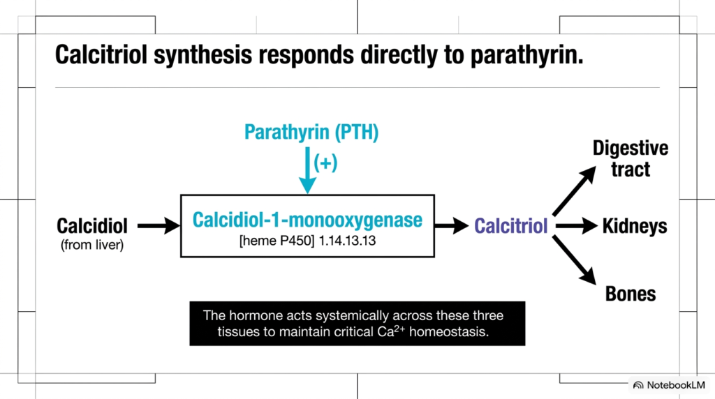
Calcitriol synthesis does not occur in isolation; it responds directly to systemic feedback loops involving parathyrin, also known as parathyroid hormone. When blood calcium levels fall, parathyrin stimulates the production of specific Renal Hormones to restore mineral equilibrium. This neuroendocrine axis demonstrates how endocrine glands communicate with renal tissue to preserve systemic stability.
Mechanistically, parathyrin acts directly on renal cells to increase calcidiol-1-monooxygenase activity. This enzyme, a specialized heme P450 protein, catalyzes the final step in activating Renal Hormones. Once synthesized, active calcitriol exits the kidney to exert profound effects on three primary target tissues: the digestive tract, the skeletal system, and the kidneys themselves.
In the digestive tract, calcitriol enhances intestinal absorption of dietary calcium. Within the bones, it mobilizes stored calcium reserves, while in the kidneys, it promotes renal calcium reabsorption. These coordinated actions ensure that plasma calcium concentration remains tightly controlled under varying dietary conditions and metabolic demands.
Understanding this regulatory loop highlights how Renal Hormones integrate multi-organ physiology. Parathyroid hormone provides the initial signal, but the kidney serves as the executing organ that generates active signaling molecules. College and medical students should recognize how Renal Hormones function within this classic example of negative feedback control across target organs.
Slide 5: Hypoxia Sensing and Erythropoietin Secretion via Renal Hormones
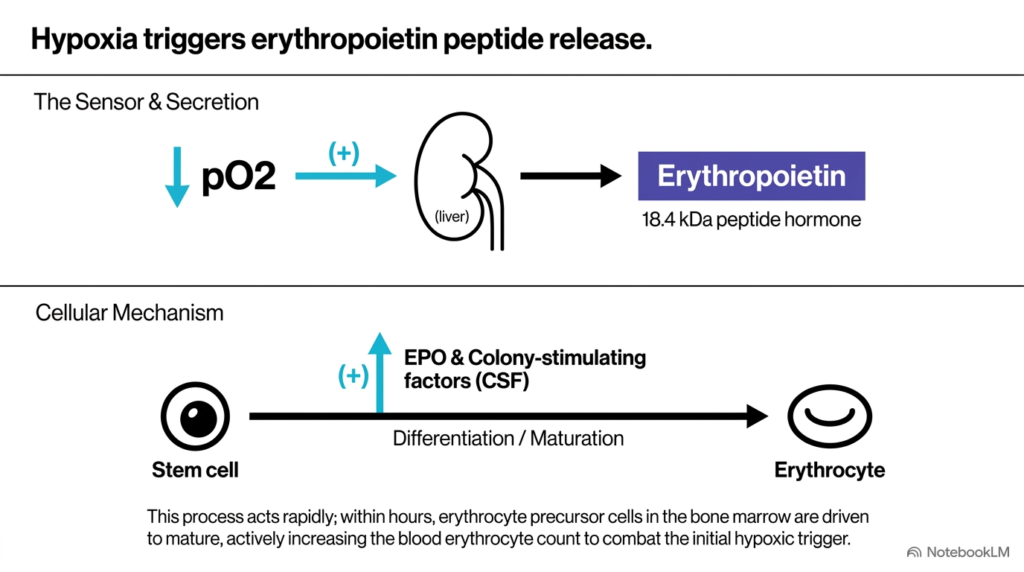
Oxygen delivery to peripheral tissues is vital for cellular survival, and the kidney serves as the body’s primary oxygen sensor. When the partial pressure of oxygen drops, renal cells respond by releasing erythropoietin, one of the most critical Renal Hormones. This glycoprotein signal initiates a complex cellular cascade that expands total red blood cell mass across circulation.
The cellular mechanism begins when specialized renal interstitial cells detect hypoxic conditions in systemic blood. In response, the kidney rapidly secretes erythropoietin, an 18.4-kilodalton peptide hormone, into the bloodstream. This signaling molecule travels directly to the bone marrow, where it collaborates with local colony-stimulating factors to direct progenitor cell maturation.
Within hours of secretion, these Renal Hormones drive bone marrow stem cells to differentiate into mature erythrocytes. This rapid cellular response increases circulating red blood cell counts, thereby restoring systemic oxygen transport capacity. The speed and precision of this endocrine pathway prevent tissue ischemia during altitude changes, blood loss, or respiratory distress.
Studying hypoxia-induced signaling illustrates how Renal Hormones maintain metabolic performance under stress. Erythropoietin connects renal oxygen sensing directly to hematopoiesis, ensuring that peripheral tissues receive adequate oxygenation. For medical students, understanding these Renal Hormones forms the foundation for analyzing respiratory adaptation and clinical blood physiology.
Slide 6: Clinical Impact of Erythropoietin and Disrupted Renal Hormones
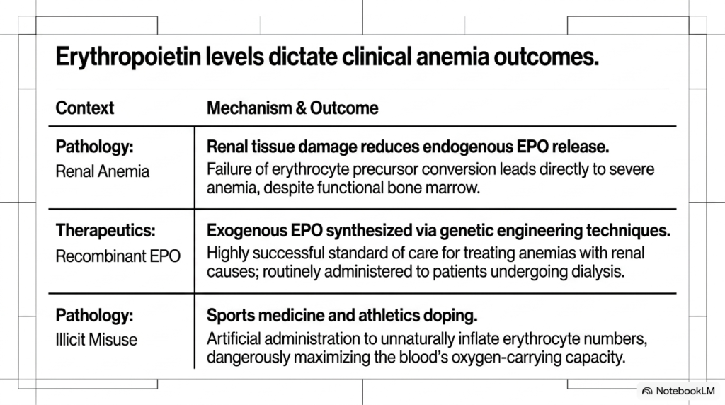
The physiological importance of erythropoietin becomes striking when renal tissue sustains chronic damage. In patients with severe kidney disease, impaired production of Renal Hormones leads directly to severe clinical complications. Examining these pathological states helps students appreciate the vital homeostatic roles of renal signaling molecules in human health.
As renal parenchyma deteriorates, endogenous erythropoietin synthesis declines significantly. Bone marrow stem cells lose their maturation signal, resulting in severe anemia despite normal iron stores. To manage this condition, modern medicine utilizes recombinant erythropoietin synthesized through genetic engineering, providing an essential therapy that replaces deficient Renal Hormones in patients undergoing regular dialysis treatment.
Conversely, the potency of these Renal Hormones has led to illegal misuse in competitive sports. Athletes sometimes administer exogenous erythropoietin to artificially elevate erythrocyte counts and enhance aerobic stamina. This illicit practice dangerously increases blood viscosity, raising the risk of stroke and severe cardiovascular events under heavy exertion.
Comparing therapeutic administration with athletic doping illustrates the broad clinical scope of Renal Hormones across medicine. Whether treating chronic kidney disease or studying sports physiology, healthcare providers must understand erythropoietin regulation. Medical students must grasp both the therapeutic benefits and potential hazards of altering Renal Hormones in clinical practice.
Slide 7: Renin Synthesis and Enzymatic Triggering of Renal Hormones
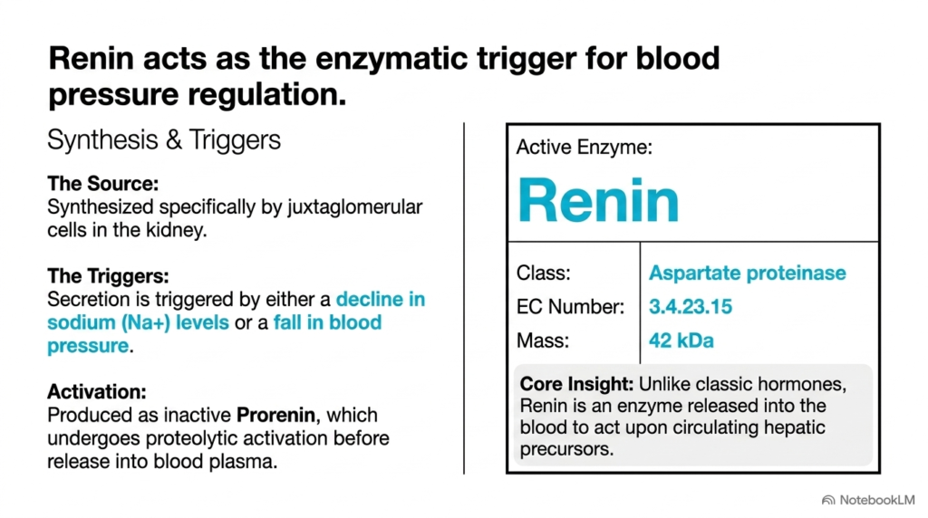
Systemic blood pressure regulation relies heavily on the enzymatic activity of renin, a unique protein produced by the kidney. Unlike typical circulating messengers, renin acts as an enzyme rather than a traditional receptor-binding factor among Renal Hormones. Its release marks the initial step in a cascade that governs arterial tone, systemic resistance, and fluid volume.
Specialized juxtaglomerular cells surrounding the renal afferent arterioles synthesize renin. The kidney releases this 42-kilodalton aspartate proteinase in response to two primary physiological triggers: a drop in renal perfusion pressure or a decline in circulating sodium levels. Before secretion, inactive prorenin is proteolytically cleaved to yield active renin.
Once secreted into blood plasma, renin initiates the renin-angiotensin cascade by acting on circulating hepatic precursors. As a circulating catalyst, renin amplification allows small renal adjustments to produce major systemic vascular responses. This enzymatic mechanism sets renin apart from other Renal Hormones that act via direct target binding.
For students exploring cardiovascular biochemistry, renin represents a master control switch among Renal Hormones. Its synthesis in juxtaglomerular cells showcases how specialized renal structures sense blood pressure and electrolyte balance. Understanding how these Renal Hormones trigger downstream cascades provides crucial context for analyzing hypertension and clinical fluid balance disorders.
Slide 8: Renin Cleavage of Angiotensinogen in Renal Hormone Pathways
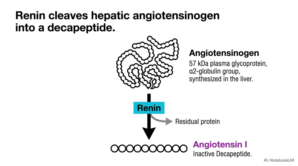
Following its release into the bloodstream, renin cleaves angiotensinogen, an abundant plasma glycoprotein produced continuously by the liver. This initial proteolytic step defines cascades driven by Renal Hormones. By cleaving a specific peptide bond, renin converts a large circulating precursor into a smaller signaling intermediate.
Angiotensinogen exists in blood plasma as a 57-kilodalton glycoprotein belonging to the alpha-2-globulin fraction. Renin selectively cleaves this precursor, releasing a residual protein fragment and generating angiotensin I. Angiotensin I is an inactive decapeptide composed of ten amino acids that serves as a necessary precursor for downstream vasoactive peptides in blood plasma.
This cleavage step represents the rate-limiting reaction in the renin-angiotensin cascade. Because angiotensinogen circulates in excess, the rate of decapeptide generation depends almost entirely on the concentration of active Renal Hormones released by the kidney. Thus, renal renin secretion directly dictates the rate of peptide activation in peripheral blood.
Analyzing this enzymatic cleavage helps biochemistry students understand precursor processing driven by Renal Hormones. The transition from a 57-kilodalton liver protein to a 10-amino-acid peptide illustrates how localized signals initiate systemic cascades. Mastering this step is key to understanding how Renal Hormones transform into widespread systemic vascular responses.
Slide 9: Angiotensin-Converting Enzyme and Cascades of Renal Hormones
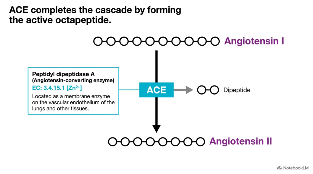
The activation cascade initiated by renal renin reaches completion through the action of angiotensin-converting enzyme, commonly abbreviated as ACE. This enzyme processes the inactive decapeptide generated by Renal Hormones into a potent vasoconstrictor. This conversion step links renal sensing to vascular smooth muscle contraction throughout the arterial tree.
Biochemically, ACE is classified as peptidyl dipeptidase A, a zinc-dependent membrane protein located primarily on the vascular endothelium of the lungs. ACE cleaves two terminal amino acids from the inactive angiotensin I decapeptide, producing the active octapeptide angiotensin II alongside a free dipeptide fragment. This enzymatic removal transforms an inactive chain into a powerful circulating hormone.
The strategic localization of ACE along pulmonary capillaries ensures rapid processing as blood flows through the lungs. Consequently, inactive peptides generated by Renal Hormones convert into active hormones before entering systemic arterial circulation. This elegant spatial arrangement allows the body to exert immediate control over arterial blood pressure, vascular resistance, and organ perfusion.
For medical students, ACE represents a pivotal junction in cardiovascular pharmacology and Renal Hormones regulation. Inhibiting this zinc-dependent enzyme prevents angiotensin II formation, lowering blood pressure. Understanding how ACE processes products of Renal Hormones provides fundamental knowledge for evaluating antihypertensive drug therapies.
Slide 10: Structure and Lifespan of Angiotensin II in Renal Hormones Signaling
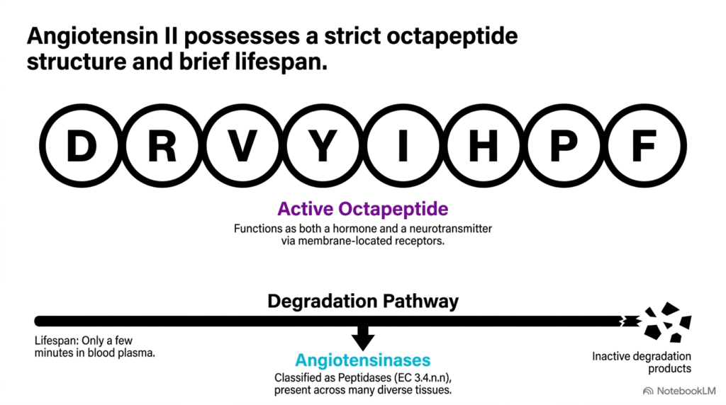
Angiotensin II is a tightly regulated octapeptide whose primary structure dictates its binding specificity and physiological lifespan. Derived from pathways initiated by Renal Hormones, this eight-amino-acid sequence exerts rapid, potent effects across multiple organ systems. Its precise amino acid arrangement determines its binding affinity for membrane-bound receptors.
The exact amino acid sequence of angiotensin II consists of Aspartate, Arginine, Valine, Tyrosine, Isoleucine, Histidine, Proline, and Phenylalanine. This peptide chain allows the molecule to act as both a systemic hormone and a central neurotransmitter. By binding specialized G-protein coupled receptors, it triggers rapid intracellular signal transduction cascades in target tissues.
To prevent excessive vasoconstriction, angiotensin II possesses an extremely brief lifespan, surviving for only a few minutes in blood plasma. Diverse tissue enzymes known as angiotensinases rapidly cleave the octapeptide into inactive fragments. This swift degradation ensures that signaling driven by Renal Hormones remains tightly controlled and responsive to real-time systemic demands.
Studying the structural biochemistry of angiotensin II highlights the balance between activation and degradation in Renal Hormones pathways. The brief half-life of this octapeptide underscores why continuous renal monitoring is necessary to sustain blood pressure. Students should note how Renal Hormones govern short-lived homeostatic signals across the circulatory network.
Slide 11: Adrenal and Renal Targets of Angiotensin II from Renal Hormones
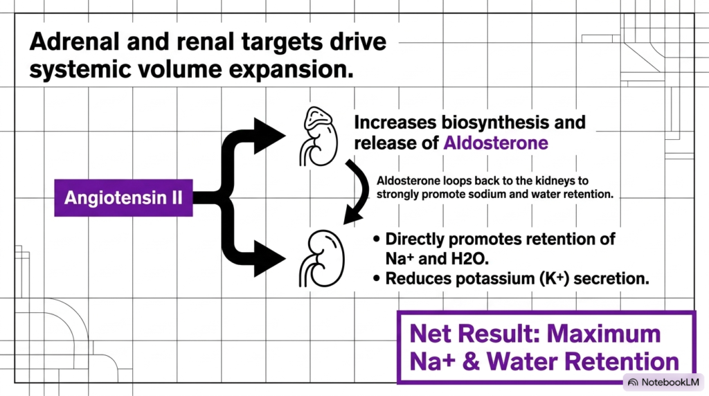
Once generated, angiotensin II acts on target tissues to expand blood volume and increase systemic pressure. The downstream effects of pathways triggered by Renal Hormones involve both direct intrarenal mechanisms and endocrine interactions with the adrenal cortex. Together, these actions maximize sodium retention and fluid conservation across the body.
In the adrenal cortex, angiotensin II stimulates aldosterone synthesis and secretion, a potent mineralocorticoid hormone. Aldosterone acts on the renal distal tubules, where it enhances sodium and water reabsorption while promoting potassium excretion. Concurrently, angiotensin II acts directly on kidney tubules to increase sodium uptake, amplifying the effects of Renal Hormones.
Together, these pathways maximize sodium and water retention within the extracellular fluid compartment. By expanding plasma volume, the body counters hypotensive states and restores effective arterial blood flow. This integrated mechanism illustrates how signals from Renal Hormones coordinate systemic volume regulation during fluid loss.
Understanding adrenal and renal targeting allows students to trace the full reach of kidney signaling. Angiotensin II does not act alone; it engages secondary endocrine organs to reinforce the homeostatic goals of Renal Hormones. Mastering these volume-expanding pathways of Renal Hormones is essential for comprehending clinical fluid management and electrolyte balance.
Slide 12: Vascular and Neural Targets in Renal Hormones Cascades
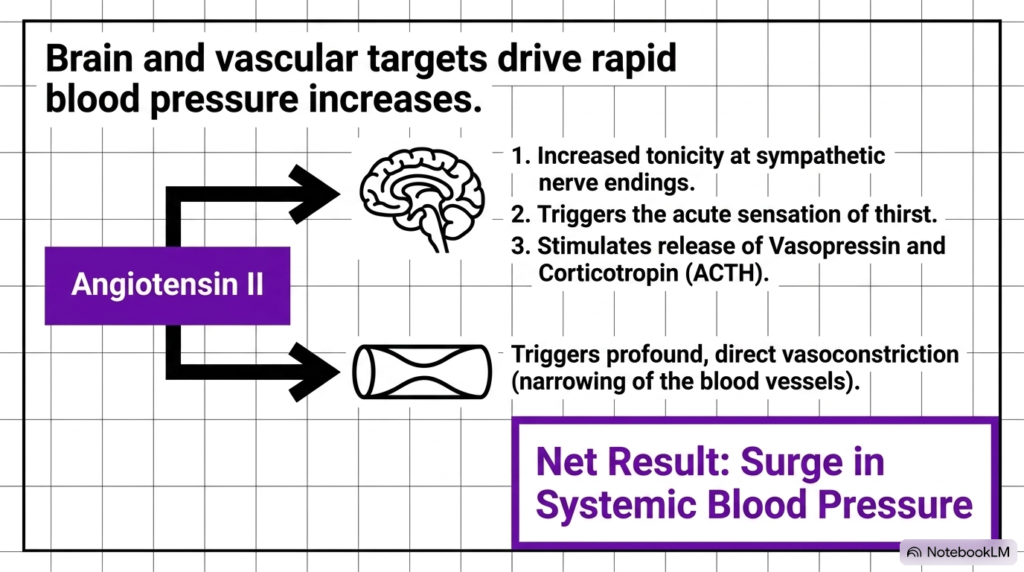
Beyond controlling fluid volume, angiotensin II rapidly increases systemic blood pressure by targeting vascular smooth muscle and the central nervous system. These swift responses showcase how pathways managed by Renal Hormones achieve both immediate and long-term hemodynamic control during severe physiological stress and circulatory challenges.
When angiotensin II binds receptors on vascular smooth muscle cells, it triggers intense, direct vasoconstriction. This narrowing rapidly increases total peripheral resistance, elevating arterial pressure within seconds. At the same time, the peptide acts on brain centers, increasing sympathetic outflow and stimulating thirst to drive fluid intake alongside renal hormone actions in peripheral blood.
Furthermore, central action of angiotensin II stimulates the pituitary gland to release vasopressin and adrenocorticotropic hormone. Vasopressin increases renal water reabsorption, while adrenocorticotropic hormone supports adrenal steroid synthesis. These neuroendocrine mechanisms reinforce the primary actions of Renal Hormones, ensuring a coordinated, multi-organ response to acute circulatory collapse.
Analyzing vascular and neural targets shows the multi-system impact of Renal Hormones across human physiology. By combining rapid vasoconstriction with neural thirst stimulation, the body effectively restores cardiovascular stability under stress. College and medical students should recognize how Renal Hormones trigger critical defense mechanisms against severe systemic hypotension.
Slide 13: Pharmacological Interventions Targeting Renal Hormone Pathways
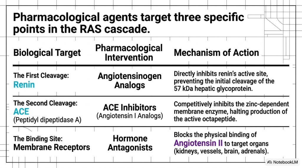
Given the renin-angiotensin system’s central role in cardiovascular disease, modern pharmacology focuses heavily on disrupting these pathways. Pharmacological agents target three specific molecular points in cascades driven by Renal Hormones. Understanding these drug classes allows medical students to connect biochemical mechanisms to clinical therapeutics.
The first intervention point involves direct renin inhibitors, such as angiotensinogen analogs, that block renin’s active site. By preventing renin from cleaving its hepatic substrate, these agents halt the cascade’s initial step. The second target is ACE, where competitive ACE inhibitors bind the zinc-dependent active site, preventing the formation of active octapeptides from precursor angiotensin I.
The third pharmacological strategy utilizes hormone receptor antagonists, commonly known as angiotensin receptor blockers. These compounds selectively block angiotensin II from binding membrane receptors on target organs, including blood vessels and the adrenal cortex. Each drug class provides a precise way to modulate blood pressure and regulate pathways governed by Renal Hormones.
Reviewing these three intervention points highlights the clinical utility of targeting endocrine cascades in cardiology. By blocking specific enzymatic cleavages or receptor sites, clinicians effectively manage hypertension and heart failure. Mastering these pharmacological targets demonstrates how understanding Renal Hormones directly informs modern medical treatments and patient care.
Slide 14: Systemic Integration of Renal Hormones and Homeostasis
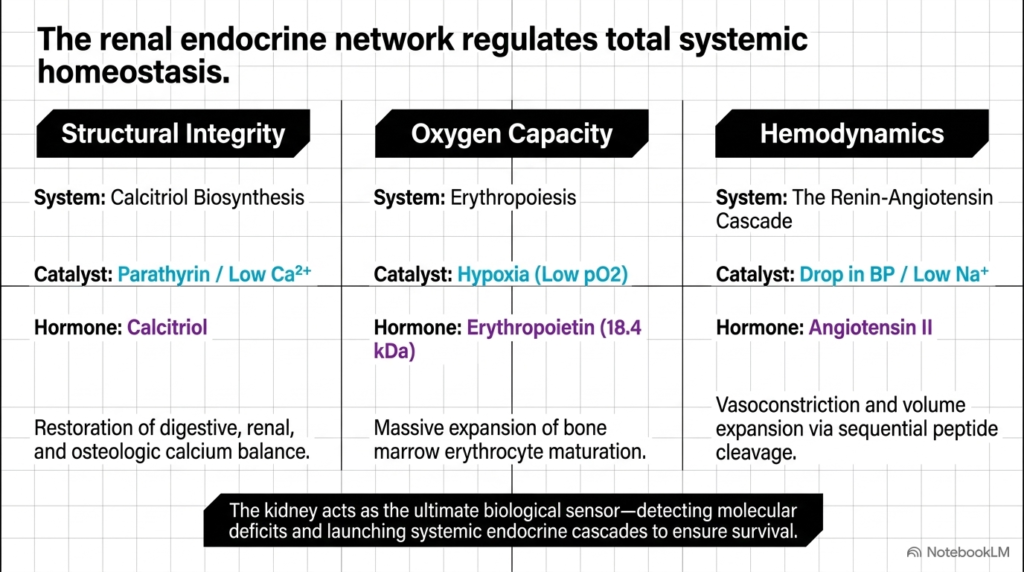
The renal endocrine network integrates three major physiological systems to maintain total body homeostasis. By evaluating structural integrity, oxygen capacity, and hemodynamics together, students can appreciate the overarching impact of Renal Hormones. The kidney serves as an ultimate biological sensor that continuously adjusts chemical output to preserve health.
First, the calcitriol pathway preserves structural integrity by regulating intestinal and osteologic calcium balance in response to parathyroid hormone signals. Second, the erythropoietin pathway maintains oxygen-carrying capacity by driving bone marrow erythrocyte maturation during hypoxia. Third, the renin-angiotensin cascade governs hemodynamics, executing sequential peptide cleavages to induce vasoconstriction and volume expansion when blood pressure drops.
Each pathway demonstrates how specialized catalysts trigger specific responses from Renal Hormones to correct physiological deficits. Whether responding to low plasma calcium, hypoxia, or hypotension, the kidney launches precise chemical signals. Thus, renal hormones form a unified defense network that protects systemic stability against environmental changes and internal stressors.
In summary, studying renal endocrine biochemistry reveals the kidney as far more than an excretory organ. Its ability to sense molecular shifts and secrete targeted signaling factors is vital for human survival. College and medical school students who master these Renal Hormones pathways will possess a deep understanding of integrative human physiology and clinical medicine.
Please read our Content Disclaimer Statement.
Check out our social media channels: