129. Biochemistry of Muscle Contraction Control
Every movement, from the blink of an eye to a sprinting stride, relies on precise cellular orchestration. At the heart of skeletal physiology lies a remarkable biochemical sequence that bridges electrical signaling and physical force. This slide deck explores the molecular blueprint governing human movement, showing how electrical impulses at the cell surface trigger mechanical work deep within muscle fibers. Designed for college and medical students, this presentation breaks down three core physiological domains, showing how calcium ions and specialized protein structures interact to maintain precise physiological regulation.
Slide 01: Excitation-Contraction Coupling and Muscle Contraction Control
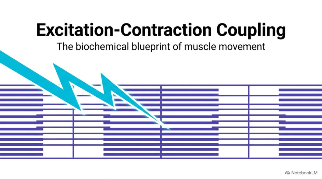
The fundamental process of physiological movement depends on a highly coordinated biological sequence known as excitation-contraction coupling. This intricate biochemical cascade drives muscle contraction control, converting a brief electrical impulse from a somatic motor neuron into sustained physical work. When an action potential stimulates a skeletal muscle fiber, an electrical wave sweeps rapidly across the outer plasma membrane, initiating a crucial intracellular signaling cascade. Biochemistry students must recognize that this fundamental event bridges cell membrane biophysics with force generation across microscopic tissue domains.
Within the broader physiological framework of Muscle Contraction Control, the individual muscle cell acts as an extraordinary biological transducer. The incoming electrical wave does not move microscopic filaments directly; instead, it activates specialized membrane proteins to release secondary chemical messengers into the interior sarcoplasm. By translating nerve excitation into rapid physical shortening, the muscle cell ensures that voluntary physical actions occur with remarkable speed, substantial force, and absolute temporal precision.
Understanding these initial electrical and chemical surface events establishes an essential foundation for studying advanced Muscle Contraction Control in clinical medicine and sports exercise physiology. Without this initial coupling process, electrical nerve signals would fail to engage the muscle fiber’s internal machinery, leaving the body unable to generate coordinated physical movements.
Slide 02: The Excitation-Contraction Relay in Muscle Contraction Control
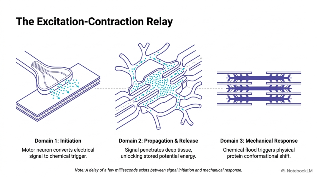
The cellular response during excitation relies on an organized three-part relay system that coordinates signal transfer across distinct anatomical regions within the muscle fiber. In the systematic study of Muscle Contraction Control, Domain 1 handles signal initiation, where the motor neuron converts an electrical nerve impulse into a chemical neurotransmitter. Domain 2 governs signal propagation and internal calcium release, directing the stimulus deep into intracellular tissue layers to unlock potential energy stored within membrane-bound organelles. Finally, Domain 3 executes the mechanical response, converting chemical shifts into physical protein movements that generate contractile force.
Physiologists emphasize that effective Muscle Contraction Control involves a crucial millisecond delay between initial surface stimulation and actual physical shortening. This brief latency period reflects the finite time required for electrical action potentials to travel down membrane invaginations, specialized channels to open, and calcium ions to diffuse across compartments. By dividing the overall signaling sequence into three distinct functional domains, Muscle Contraction Control achieves extraordinary operational speed while preventing spontaneous or unwanted muscle twitches during quiet resting states.
This multi-domain structure also ensures that Muscle Contraction Control maintains modular regulation across the entire cell volume. Each domain serves as an independent control point where metabolic conditions, ion availability, or pharmaceutical interventions can fine-tune the resulting mechanical output.
Slide 03: Neuromuscular Junction Events in Muscle Contraction Control
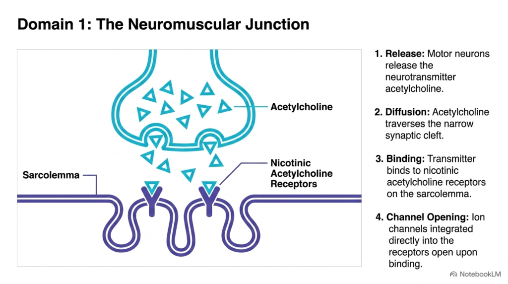
Signal initiation takes place at the neuromuscular junction, a highly specialized chemical synapse where the motor neuron terminal meets the muscle cell sarcolemma. Under normal mechanisms of Muscle Contraction Control, an arriving nerve action potential stimulates the presynaptic axon terminal to release the chemical neurotransmitter acetylcholine into the synaptic cleft. Acetylcholine diffuses rapidly across this narrow extracellular gap and binds directly to nicotinic acetylcholine receptors on the postsynaptic sarcolemma. This specific transmitter-receptor interaction represents the first essential chemical checkpoint in the broader cascade of Muscle Contraction Control.
The nicotinic acetylcholine receptor functions as a ligand-gated ion channel embedded in the folded membrane of the motor end plate. When two acetylcholine molecules attach to the receptor’s extracellular binding pockets, a structural conformational change opens the central channel pore. This opening permits rapid influx of positive sodium ions across the lipid bilayer, showing how Muscle Contraction Control uses transmitter binding to convert an electrical nerve signal into postsynaptic membrane excitation.
Medical students should note that pathological conditions or pharmacological agents directly targeting these nicotinic receptors can severely impair Muscle Contraction Control. For instance, autoimmune disorders such as myasthenia gravis reduce functional receptor density, disrupting normal neurotransmission and leading to profound skeletal muscle fatigability.
Slide 04: Sarcolemma Depolarization during Muscle Contraction Control
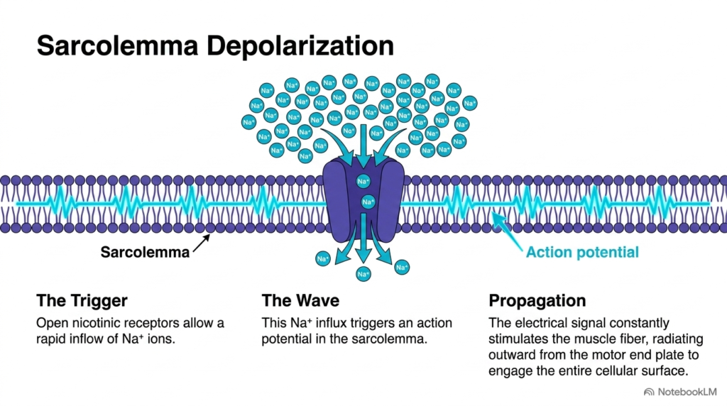
After nicotinic acetylcholine receptors open at the motor end plate, sodium ions rapidly enter the sarcoplasm down their steep electrochemical gradient. This rapid sodium ion influx plays a central role in Muscle Contraction Control by depolarizing the local resting membrane potential of the sarcolemma. The sudden shift in membrane voltage activates nearby voltage-gated sodium channels, generating a self-propagating action potential that sweeps rapidly across the entire cellular surface. Through this highly effective mechanism, Muscle Contraction Control transforms a localized chemical binding event into a widespread electrical wave.
The propagating action potential spreads outward from the neuromuscular junction across the sarcolemma like ripples spreading across a pond surface. This uniform surface conduction ensures that every individual myofibril within the large muscle fiber receives the contractile signal almost simultaneously. Students should appreciate how effective Muscle Contraction Control depends on fast, unattenuated membrane depolarization to synchronize cellular activity across extensive tissue volumes during active physical movement.
Without robust sarcolemma depolarization, voltage-dependent mechanisms deeper in the cell would remain completely inactive, halting the sequence of Muscle Contraction Control. Maintaining proper systemic electrolyte balances, particularly extracellular sodium and potassium ion concentrations, is therefore vital for sustaining healthy sarcolemma excitability and preventing muscular fatigue during physical exertion.
Slide 05: Architectural Elements of Muscle Contraction Control
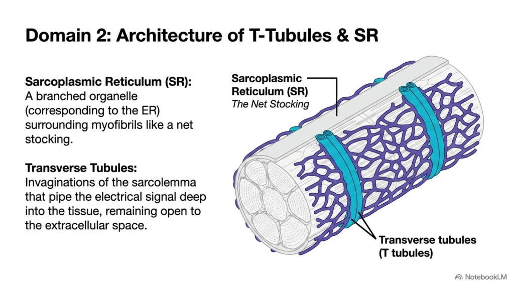
To convey the electrical signal from the outer sarcolemma deep into the cell interior, muscle fibers rely on specialized structural architecture. In muscle contraction control, transverse tubules—or T-tubules—form deep tubular invaginations of the sarcolemma that channel surface depolarization directly into the cellular core. Because T-tubules remain continuous with the extracellular fluid environment, the action potential penetrates deep into the muscle fiber without losing signal strength or propagation speed. This specialized structural adaptation is absolutely essential for uniform Muscle Contraction Control across thick muscle cells.
Closely associated with the T-tubule network is the sarcoplasmic reticulum, a highly branched organelle that wraps around individual myofibrils like a net stocking. The sarcoplasmic reticulum works with T-tubules to form specialized intracellular junctional triads throughout the muscle fiber. By positioning calcium storage compartments directly adjacent to invaginated surface membranes, Muscle Contraction Control minimizes internal diffusion distances and optimizes excitation speed across the entire contractile apparatus.
This elegant geometric arrangement lets large skeletal muscle fibers bypass the physical limits of simple cytoplasmic ion diffusion. By bringing membrane depolarization within nanometers of intracellular calcium reservoirs, Muscle Contraction Control ensures rapid, synchronous activation of every sarcomere, allowing the whole cell to shorten as a unified functional unit.
Slide 06: Sarcoplasmic Calcium Dynamics in Muscle Contraction Control
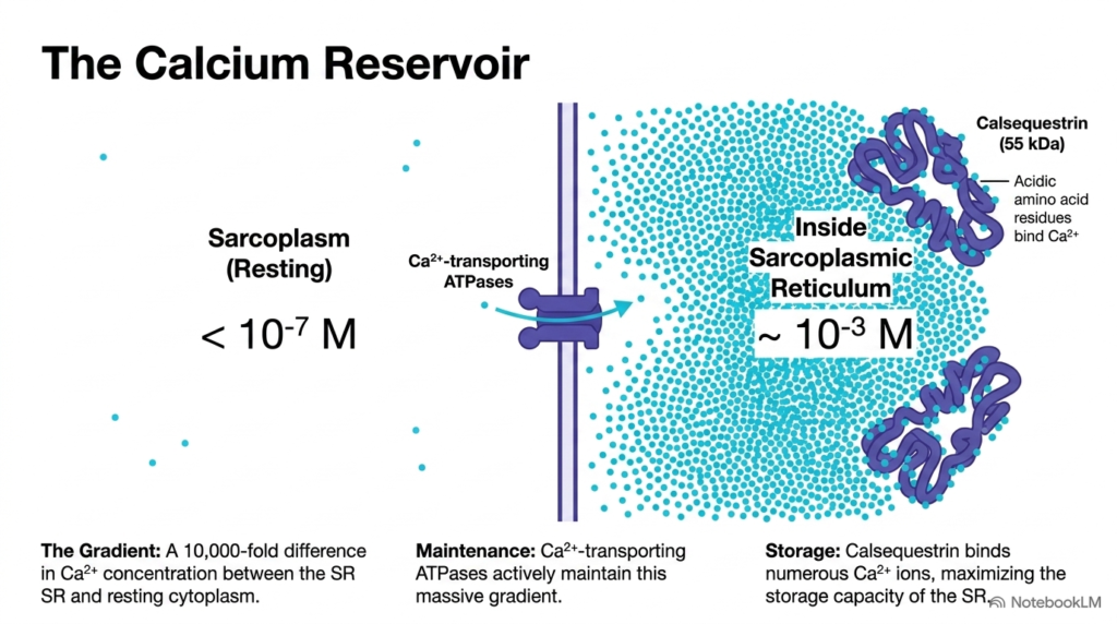
The sarcoplasmic reticulum serves as the primary intracellular calcium storage reservoir, maintaining an extraordinary concentration gradient across its membrane. In a resting muscle fiber, active ATP-dependent calcium pumps maintain sarcoplasmic calcium levels below 10^-7 M, while internal reservoir levels reach 10^-3 M. This ten-thousand-fold concentration difference represents a foundational feature of Muscle Contraction Control, storing substantial chemical potential energy ready for rapid cytosolic release during excitation.
To store large amounts of calcium without osmotic swelling or toxic precipitation, the sarcoplasmic reticulum uses a specialized luminal protein called calsequestrin. Calsequestrin is a highly acidic protein that binds numerous calcium ions with low affinity and high capacity, effectively buffering the stored pool within the terminal cisternae. Proper Muscle Contraction Control relies on both active ATP-dependent pumps and calsequestrin buffering to ensure the muscle cell can repeatedly release and re-sequester calcium ions during prolonged exercise.
The dynamic equilibrium between bound calsequestrin and free luminal calcium guarantees that an enormous pool of soluble ions is instantly available upon stimulation. Consequently, Muscle Contraction Control operates with exceptional biochemical efficiency, relying on pre-established ion gradients rather than slow de novo ion synthesis to initiate mechanical force generation.
Slide 07: SR Foot and Ryanodine Receptor in Muscle Contraction Control
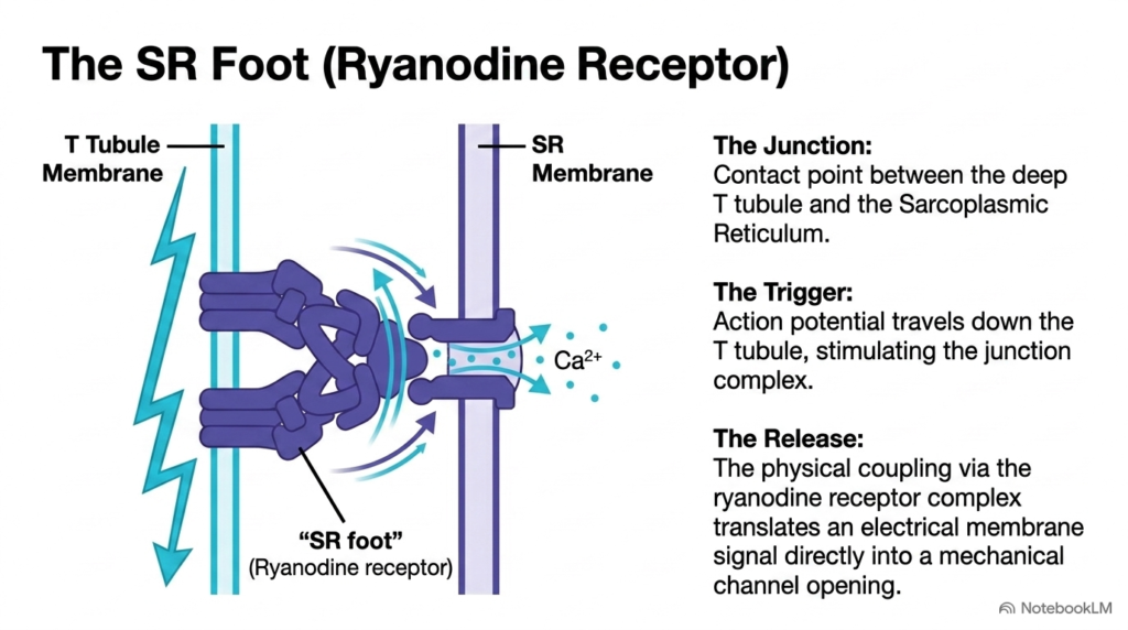
The crucial junctional interface where electrical membrane depolarization meets internal calcium storage occurs where the T-tubule contacts the sarcoplasmic reticulum membrane. In the precise execution of Muscle Contraction Control, a large tetrameric protein complex known as the ryanodine receptor—or SR foot—spans the narrow junctional gap separating these two membranes. When the action potential travels down the T-tubule membrane, voltage-sensing dihydropyridine receptors physically interact with the adjacent ryanodine receptor complex. This direct mechanical coupling plays a pivotal role in Muscle Contraction Control.
This structural interaction translates a change in membrane voltage directly into mechanical ion-channel opening within milliseconds. Rather than relying on slow chemical messenger diffusion across the gap, the physical link forces the ryanodine receptor channel pore to open almost instantaneously upon membrane depolarization. This direct electromechanical linkage ensures that Muscle Contraction Control achieves the rapid, millisecond-level response times necessary for precise motor activities in skeletal tissues.
Biochemists highlight that the ryanodine receptor functions as a giant ion channel capable of tremendous ionic flux rates during activation. Understanding this physical junction gives students clear insight into how Muscle Contraction Control seamlessly converts membrane electrical excitation into massive intracellular messenger release, bridging membrane biophysics with organelle biology to trigger movement.
Slide 08: Sarcoplasmic Calcium Flooding in Muscle Contraction Control
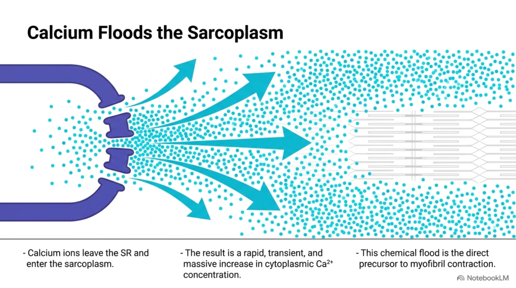
When mechanical opening of ryanodine receptor channels occurs, stored calcium ions rapidly pour out of the sarcoplasmic reticulum lumen into the surrounding cytoplasm. This massive ionic flux marks a key event in Muscle Contraction Control, raising cytosolic calcium concentration by several orders of magnitude within milliseconds. The sudden ionic surge floods the myofibrillar space, converting an electrical membrane potential into a powerful intracellular chemical messenger system. Without this dramatic calcium surge, effective Muscle Contraction Control would be entirely impossible within the cell.
The elevated cytosolic calcium acts as a transient chemical trigger that diffuses rapidly toward nearby contractile protein filaments. Because the sarcoplasmic reticulum surrounds each myofibril, released calcium ions reach their target regulatory sites nearly simultaneously throughout the cell volume. Biochemists view this localized calcium flood as the obligate molecular precursor that initiates cross-bridge formation during active Muscle Contraction Control.
The precise magnitude and spatial distribution of this calcium flood directly dictate the force and duration of the resulting muscle twitch. By tightly regulating channel opening and closing kinetics, Muscle Contraction Control modulates mechanical force output to match varying physical demands during daily motor activities, ensuring both brute strength and refined motor coordination during complex physical tasks.
Slide 09: Resting State Apparatus in Muscle Contraction Control
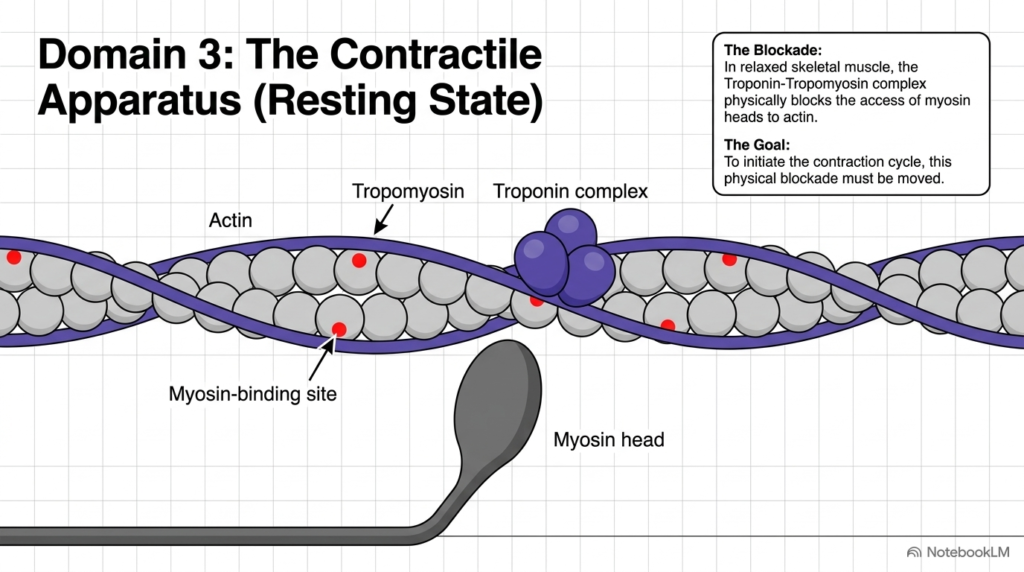
In relaxed skeletal muscle tissue, physical structural barriers prevent force generation even though cellular ATP supplies remain highly abundant. Under resting conditions governed by Muscle Contraction Control, a continuous fibrous protein strand called tropomyosin wraps around the helical actin filament. Tropomyosin physically covers the active myosin-binding sites on actin, preventing myosin cross-bridge heads from forming tight attachments with the thin filament. This physical steric hindrance ensures that Muscle Contraction Control prevents wasteful ATP hydrolysis and involuntary muscle tension during quiet resting states.
Held securely in position by the multi-protein troponin complex, tropomyosin functions as a molecular gatekeeper along the thin filament surface. Because myosin heads cannot physically access actin binding sites, the contractile apparatus remains locked in a stable off state. To initiate active muscle shortening, Muscle Contraction Control must overcome this steric blockade by shifting regulatory proteins away from the binding sites, allowing actin and myosin filaments to interact.
This resting inhibition represents a crucial energy-saving mechanism for the entire organism during routine metabolic maintenance. By maintaining a strict structural barrier during rest, Muscle Contraction Control prevents uncoordinated baseline contractions, preserving vital cellular ATP energy supplies for intended, voluntary motor activities required during demanding physiological work throughout the human body.
Slide 10: Troponin Complex Anatomy in Muscle Contraction Control
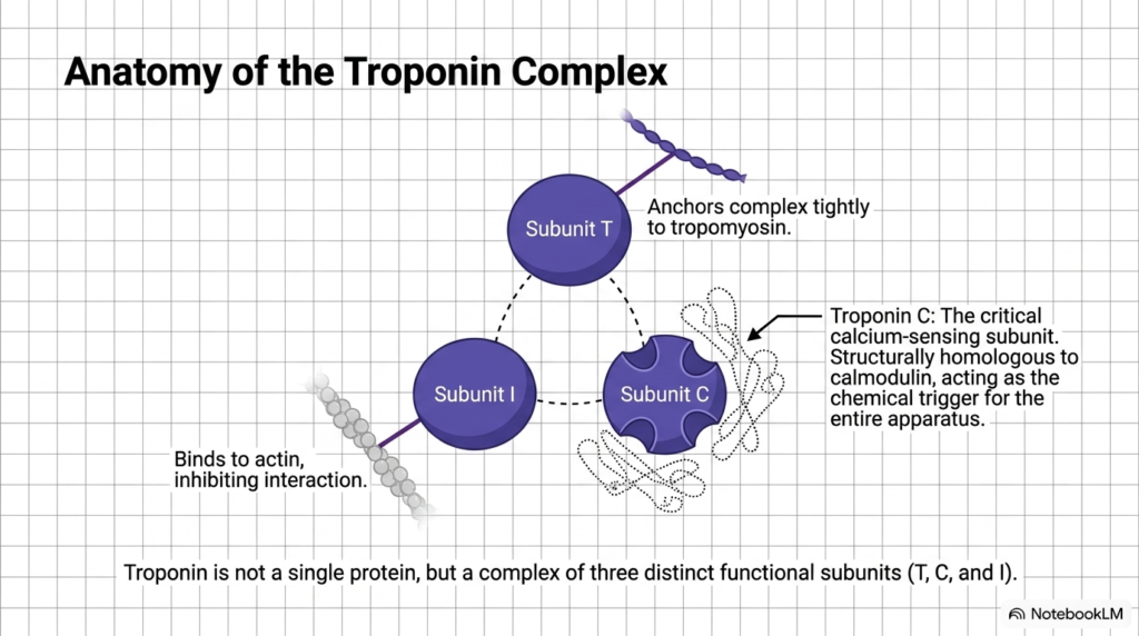
Troponin controls the regulatory blockade along the thin filament; it is a heterotrimeric protein complex composed of three distinct functional subunits. In the structural framework of Muscle Contraction Control, Subunit T binds firmly to tropomyosin, anchoring the regulatory complex tightly along the fibrous actin strand. Subunit I binds attached actin monomers, maintaining the inhibitory position that prevents myosin head attachment during resting states. Finally, Subunit C functions as the specialized calcium sensor, possessing specialized amino acid binding pockets structurally homologous to calmodulin.
Each distinct subunit performs an indispensable role that contributes directly to overall Muscle Contraction Control within the muscle fiber. Subunit C serves as the primary molecular trigger, translating calcium ion binding into physical conformational shifts across the entire regulatory unit. Understanding the specialized architecture of subunits T, I, and C provides students with a complete structural basis for how Muscle Contraction Control regulates cross-bridge activity during contraction.
In clinical diagnostics, elevated cardiac troponin isoforms in the bloodstream serve as essential biomarkers of myocardial injury. This clinical application shows how studying troponin’s molecular structure advances both basic biochemical science and emergency diagnostics under Muscle Contraction Control principles, helping clinicians assess cardiac tissue damage.
Slide 11: Calcium Trigger Mechanism in Muscle Contraction Control
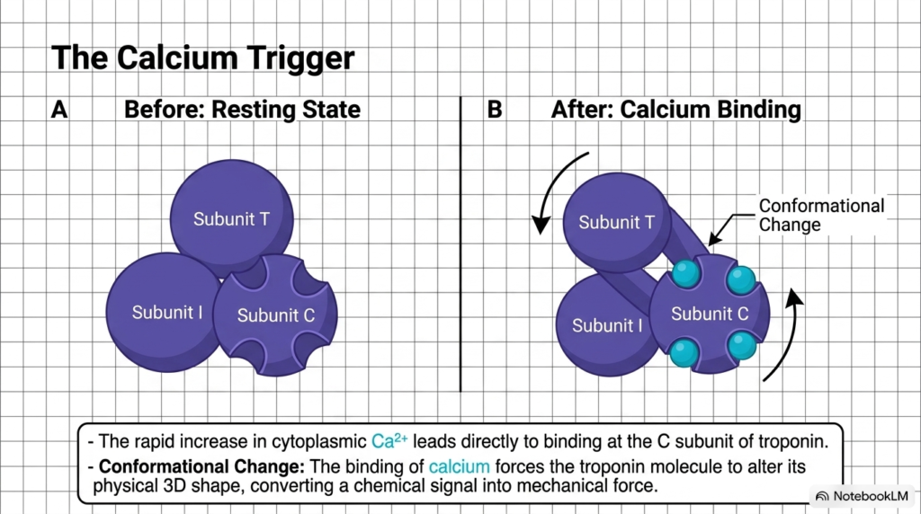
When calcium ions flood the sarcoplasm after the channel opens, they bind directly to the empty coordination pockets on Subunit C of troponin. This precise binding event drives Muscle Contraction Control by inducing a dramatic conformational shift throughout the entire troponin complex. As calcium ions occupy the binding pockets, troponin alters its three-dimensional tertiary structure, converting chemical binding energy directly into physical mechanical motion. This distinct structural reorganization represents the critical molecular switch in Muscle Contraction Control.
The conformational change in Subunit C alters its molecular interactions with Subunits I and T, relieving the inhibitory tension on the actin filament. By reorganizing its structural domain, troponin relays the calcium signal directly to the surrounding thin filament proteins. This pivotal molecular step shows how Muscle Contraction Control uses precise protein conformational changes to convert microscopic chemical signals into macroscopic biological work, enabling muscle fibers to generate active mechanical tension.
Subunit C’s high thermodynamic affinity for calcium ensures rapid, full activation even during modest ionic surges in the cell cytoplasm. Consequently, Muscle Contraction Control achieves near-instantaneous cross-bridge activation as soon as intracellular calcium levels cross the critical threshold concentration, ensuring immediate muscle responsiveness to neural stimuli during rapid movements and complex physical activities.
Slide 12: Tropomyosin Movement and Exposure in Muscle Contraction Control
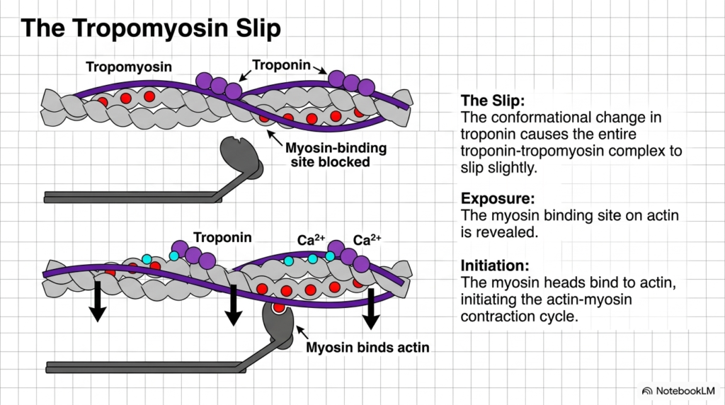
As troponin undergoes its calcium-induced conformational change, it pulls the attached tropomyosin strand. In the mechanical execution of Muscle Contraction Control, this force causes tropomyosin to slip laterally into the structural groove of the actin helix. This lateral displacement uncovers the previously hidden myosin-binding sites on the actin monomers, removing the steric block that maintained muscle relaxation. The clear exposure of these binding sites marks a decisive functional transition in Muscle Contraction Control.
With the actin binding sites fully exposed, energized myosin heads attach rapidly to actin, initiating the iterative actin-myosin cross-bridge cycle. The myosin head releases stored energy through a mechanical power stroke, sliding the thin filament toward the center of the sarcomere. Through this elegant structural movement, Muscle Contraction Control successfully translates chemical signal transduction into active mechanical force production across the entire muscle tissue.
As long as calcium ions remain bound to troponin C, tropomyosin remains in this displaced position, permitting repeated cross-bridge cycles to occur in rapid succession. This continuous cycling activity demonstrates how Muscle Contraction Control maintains sustained force generation during prolonged, heavy muscular work across all skeletal muscle groups, powering efficient human movement, physical work, and active locomotion.
Slide 13: Integrated Contraction Cascade in Muscle Contraction Control
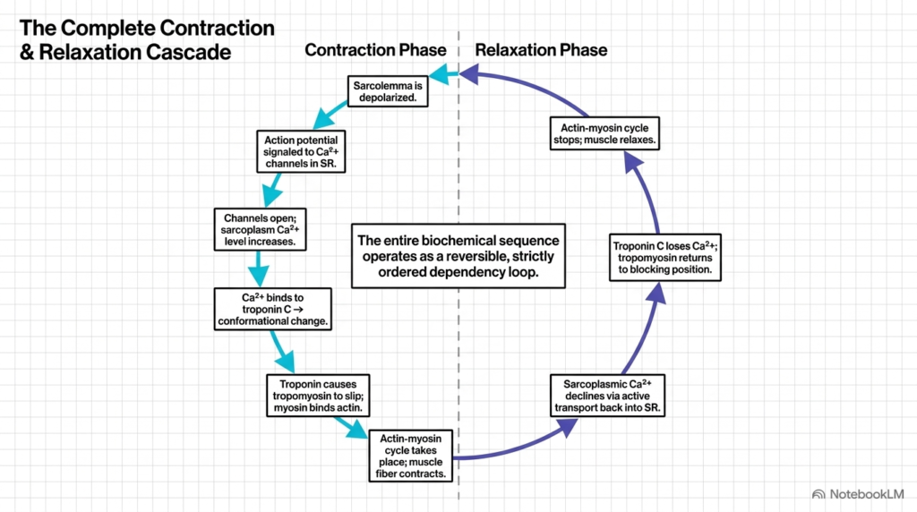
The full cycle of muscular activation and recovery operates as a strictly ordered, highly reversible biological dependency loop within the cell. During the contraction phase of Muscle Contraction Control, sarcolemma depolarization triggers sarcoplasmic calcium release, troponin binding, tropomyosin displacement, and cross-bridge cycling. To return the muscle fiber to its resting state, active transport pumps actively re-sequester calcium back into the sarcoplasmic reticulum lumen against its concentration gradient. This energy-dependent re-uptake is essential for proper Muscle Contraction Control.
As cytosolic calcium levels decline below the activation threshold, calcium ions dissociate from Subunit C of troponin. Without calcium, troponin reverts to its resting conformation, allowing tropomyosin to slide back into its original position, covering actin binding sites. This reversible regulatory mechanism stops cross-bridge cycling and permits muscle relaxation, illustrating how Muscle Contraction Control governs both active force generation and quiet recovery phases.
The precise balance between calcium release and calcium re-uptake determines the duration and intensity of muscular tension the cell develops. By tightly controlling both halves of this dependency loop, Muscle Contraction Control prevents continuous muscle tetany and enables precise movement timing during daily physical tasks, ensuring physiological flexibility, metabolic efficiency, and cellular safety during repetitive muscular actions.
Slide 14: Physiological State Matrix in Muscle Contraction Control
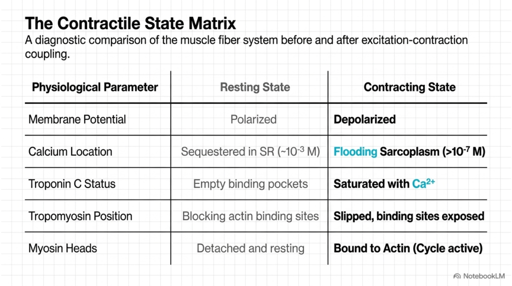
Evaluating the cellular parameters across resting and contracting states highlights how various organelle systems coordinate during physiological activation. In Muscle Contraction Control, the sarcolemma membrane potential transitions from a polarized resting state to a depolarized state during active electrical stimulation. Simultaneously, calcium location shifts from high-density sequestration within the sarcoplasmic reticulum to a high concentration flooding the sarcoplasm. These distinct physiological state changes illustrate the systemic nature of Muscle Contraction Control within the muscle cell.
At the thin-filament level, troponin C shifts from empty binding pockets to full calcium saturation during active contraction. Tropomyosin moves from a blocking position to a slipped position, enabling myosin heads to transition from detached resting units into active actin-bound cross-bridges. Analyzing this state matrix emphasizes how Muscle Contraction Control organizes discrete molecular events into a unified, highly predictable physiological response required for motor function.
Understanding these distinct physiological states provides students with a valuable diagnostic framework for evaluating overall muscle health, cellular energetic demands, and neuromuscular function. Each parameter in the matrix represents a potential therapeutic target where pharmaceutical interventions can modify Muscle Contraction Control in diseased states or metabolic disorders, offering promising avenues for clinical treatment, rehabilitation, and long-term patient recovery programs in clinical settings.
Slide 15: Structural Blueprint Summary of Muscle Contraction Control
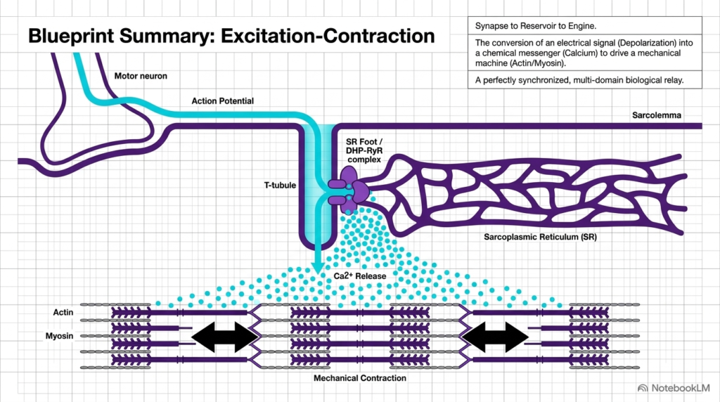
In summary, excitation-contraction coupling functions as an integrated, multi-domain biological relay that connects nervous excitation to mechanical muscle work. Throughout this entire physiological pathway, Muscle Contraction Control depends on converting an electrical surface potential into a chemical messenger that drives a molecular engine. From presynaptic acetylcholine release to the physical slipping of tropomyosin, every step is exquisitely regulated to ensure reliable movement. This comprehensive blueprint underscores why Muscle Contraction Control remains a central focus of human physiology and medical science.
For medical and graduate students, mastering these interconnected cellular domains provides essential insight into normal motor function and neuromuscular pathology. Pathological disruptions at any point—whether in channel voltage sensing, calcium storage buffering, or contractile protein binding—can severely impair motor performance. Ultimately, studying Muscle Contraction Control reveals the extraordinary biochemical precision that allows simple electrical impulses to produce life-sustaining physical action across all living organisms and complex tissue systems in nature.
By integrating membrane biophysics, organelle storage, and structural protein mechanics, the muscle cell achieves flawless operational harmony during movement. This blueprint attests to the elegant molecular design underlying human movement, governed by Muscle Contraction Control across diverse physiological environments, physical demands, and health conditions in clinical practice.
Please read our Content Disclaimer Statement.
Check out our social media channels: