133. Collagen Biochemistry: Structure and Biosynthesis Explained
Have you ever wondered why human skin stays firm, bones resist fractures, and tendons withstand massive physical strain? The secret lies in the molecular architecture of the body’s primary structural protein. This slide deck delivers a complete exploration of Collagen Biochemistry, guiding students through molecular structures, post-translational modifications, and cellular pathways. Readers will gain a clear understanding of how individual polypeptide chains assemble into resilient extracellular matrices that support human life.
Slide 1: Fundamentals of Collagen Biochemistry and Structural Overview
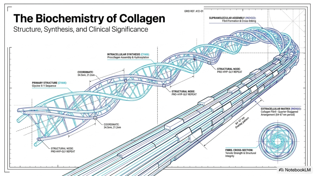
The study of Collagen Biochemistry reveals how specialized biomolecules provide essential mechanical strength to multicellular organisms. At the macroscopic level, animal tissues require rigid support and elastic tension to maintain structural integrity under constant environmental stress. This overview introduces the multi-tiered architecture that enables this physical resilience. By examining both intracellular synthesis and extracellular assembly, researchers show how genetic instructions become dense fibrous matrices that support organs, bones, and skin.
Intracellular processing begins with assembling specialized procollagen precursors within the lumen of the rough endoplasmic reticulum. Within this pathway, precise enzymatic modifications establish the biochemical foundation required for stable triple-helical folding. Understanding Collagen Biochemistry requires tracking these precursor molecules as they move through secretory vesicles, exit the cell through exocytosis, and enter the extracellular matrix. Outside the cell, individual tropocollagen units undergo highly organized self-assembly to build higher-order structural fibrils.
These extracellular fibrils assemble into a distinct quarter-staggered arrangement characterized by a repeating sixty-four to sixty-seven nanometer period. This staggered layout creates alternating gap and overlap regions that maximize tensile resistance under physical strain. Cross-sectional analysis also shows how individual fibrils pack densely to form tough structural cables with exceptional load-bearing capacity. Through this hierarchical organization, Collagen Biochemistry demonstrates how microscopic molecular geometry translates directly into macroscopic tissue durability throughout the animal kingdom.
Slide 2: Quantitative Abundance and Collagen Biochemistry Typology
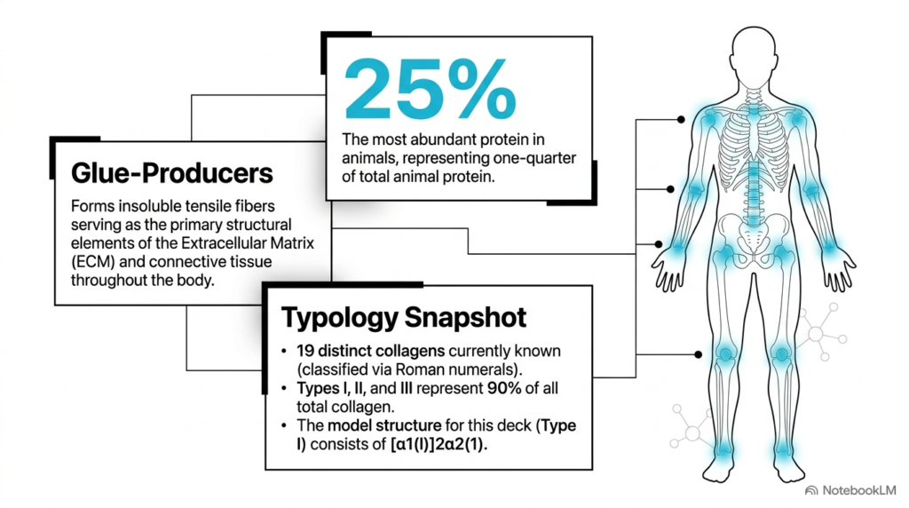
Understanding Collagen Biochemistry begins with recognizing its immense quantitative abundance throughout the animal kingdom. Representing twenty-five percent of total animal protein mass, this fibrous molecule serves as the principal structural element of the extracellular matrix. It acts as a primary biological glue, forming insoluble tensile fibers that anchor cells, support organ architecture, and reinforce connective tissues across the entire human body. Without this crucial protein network, soft tissues would lack cohesive strength.
Collagen Biochemistry classifies these structural proteins into nineteen distinct types using Roman numeral nomenclature based on discovery and chain composition. Despite this diversity, Types I, II, and III dominate biological tissues, accounting for ninety percent of all body collagen. Type I collagen serves as the primary model structure for structural analysis, consisting of a heterotrimeric combination of two alpha-one chains and one alpha-two chain designated as [α1(I)]2α2(1).
Each collagen type fulfills specialized mechanical roles based on its distinct chain composition and tissue distribution. Type I provides high tensile strength in skin, bone, and tendons, whereas Type II provides compressive resistance within articular cartilage. Type III forms flexible reticular networks within vascular walls and visceral organs. By studying Collagen Biochemistry, students appreciate how subtle variations in chain pairing create specialized scaffold structures tailored to specific physiological demands.
Slide 3: Primary Sequence Repeating Units in Collagen Biochemistry
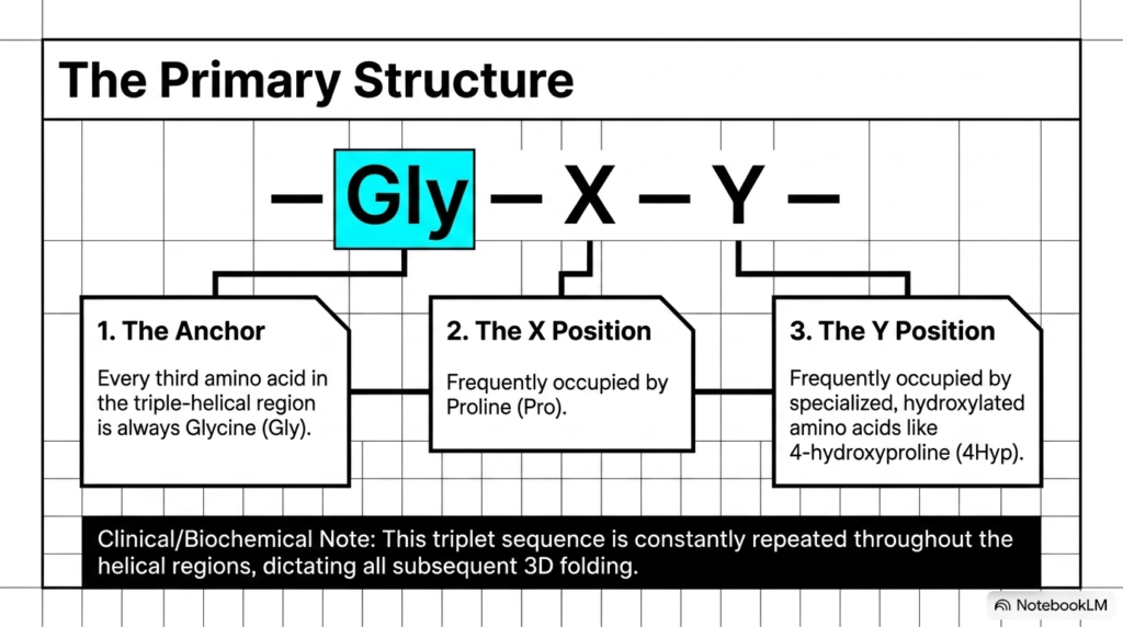
At the primary structural level, Collagen Biochemistry relies on a continuous repeating tripeptide sequence defined as Glycine-X-Y. This strict mathematical pattern dictates all subsequent three-dimensional folding and helical assembly. Every third amino acid position throughout the helical domain must contain glycine, an indispensable structural anchor that allows tight backbone packing within the molecule’s interior core.
The unique spatial constraints of Collagen Biochemistry explain why glycine is strictly required at every third position throughout the triple helix. Glycine possesses a single hydrogen atom as its side chain, making it the smallest amino acid in biological systems. Because the interior of the triple helix is extremely crowded, only glycine fits without causing severe steric hindrance that would disrupt helical stability and destabilize the protein scaffold.
The X and Y positions of the triplet sequence accommodate larger amino acid residues with specialized functional roles in structural stabilization. The X position is frequently occupied by proline, which introduces rigid ring structures that constrain polypeptide backbone flexibility. The Y position is commonly occupied by four-hydroxyproline, a modified residue crucial for interchain hydrogen bonding. Together, this repeating Glycine-X-Y motif forms the foundational sequence that governs the structural mechanics of Collagen Biochemistry.
Slide 4: Post-Translational Hydroxylation in Collagen Biochemistry
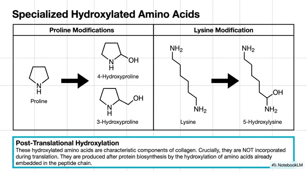
A central concept in Collagen Biochemistry is post-translational modification, specifically the enzymatic hydroxylation of key amino acid residues. These modifications occur exclusively after protein translation on ribosomes, meaning transfer RNA molecules never directly incorporate hydroxylated amino acids during polypeptide synthesis. Instead, specific oxygenase enzymes modify proline and lysine residues already embedded within the newly synthesized peptide chain inside the rough endoplasmic reticulum lumen.
In Collagen Biochemistry, proline residues undergo site-specific modification to form four-hydroxyproline and three-hydroxyproline derivatives. Four-hydroxyproline plays a paramount role in stabilizing the triple helix by forming essential interchain hydrogen-bonding networks across adjacent alpha-chains. Without sufficient proline hydroxylation, the triple helix cannot maintain its structural stability at physiological body temperature, leading to rapid thermal denaturation, structural collapse, and premature intracellular degradation before cellular secretion can occur.
Lysine residues also undergo targeted enzymatic modification to generate five-hydroxylysine within the rough endoplasmic reticulum lumen. These hydroxylated lysine residues serve as critical attachment sites for subsequent carbohydrate additions during glycosylation and participate directly in forming mature covalent cross-links later in the extracellular space. Studying these enzymatic modifications highlights how Collagen Biochemistry transforms standard amino acid chains into specialized structural building blocks that endure continuous mechanical stress across human tissues and organs.
Slide 5: The Tropocollagen Triple Helix in Collagen Biochemistry
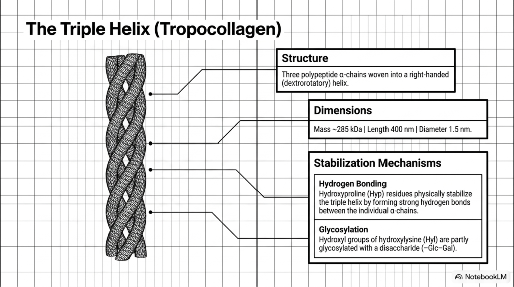
The fundamental functional unit of Collagen Biochemistry is tropocollagen, a rigid rod-like molecule measuring four hundred nanometers in length and one point five nanometers in diameter. Tropocollagen possesses a molecular mass of approximately two hundred eighty-five kilodaltons. Its structure consists of three left-handed polypeptide alpha-chains coiled together to form a tight, right-handed dextrorotatory superhelix with remarkable resistance to longitudinal pulling forces across connective tissues in human organs.
In Collagen Biochemistry, the thermal and physical stability of the tropocollagen superhelix depends on distinct chemical stabilization mechanisms operating in unison. Interchain hydrogen bonding provides primary structural reinforcement, mediated directly by the hydroxyl groups of hydroxyproline residues. These hydrogen bonds form physical bridges between adjacent alpha-chains, locking the three individual strands into a tight, cohesive rod that resists mechanical stretching, unwinding, and enzymatic degradation under physiological conditions.
Additionally, tropocollagen undergoes enzymatic glycosylation as part of its structural maturation process inside the cell. Hydroxyl groups on specific hydroxylysine residues are modified by the covalent attachment of disaccharide units consisting of glucose and galactose. This enzymatic carbohydrate attachment influences molecular packing, surface hydration, and fibril interactions during supramolecular assembly. Understanding tropocollagen structure shows how Collagen Biochemistry uses precise molecular geometry and chemical bonds to build incredibly durable extracellular scaffold units throughout animal bodies.
Slide 6: Supramolecular Fibril Assembly in Collagen Biochemistry
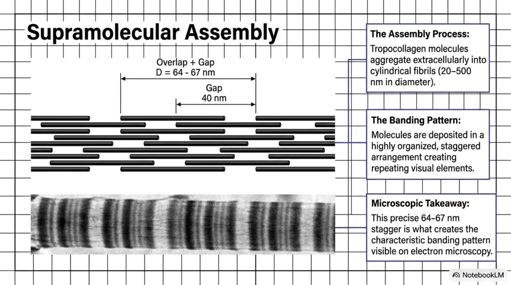
The extracellular phase of Collagen Biochemistry involves the self-assembly of individual tropocollagen rods into large cylindrical fibrils. Spanning twenty to five hundred nanometers in diameter, these supramolecular aggregates form through spontaneous, self-directed physical interactions once terminal propeptides are proteolytically removed. Tropocollagen units align in parallel rows, creating extensive structural networks that provide immense mechanical support and tensile strength to surrounding connective tissues throughout the human body.
A defining feature of Collagen Biochemistry is the highly organized staggered arrangement of tropocollagen molecules within each mature fibril. Individual tropocollagen rods line up in a quarter-staggered pattern, leaving a distinct forty-nanometer gap between successive longitudinal molecules. This precise alignment creates a repeating structural period of sixty-four to sixty-seven nanometers, defined by alternating regions of high and low electron density along the assembled fibril matrix.
Under transmission electron microscopy, this staggered molecular packing creates a characteristic cross-striated banding pattern along the fiber axis. This distinct visual signature confirms proper extracellular fibril formation, correct alignment, and structural maturity in connective tissue samples. By organizing tropocollagen into staggered supramolecular arrays, Collagen Biochemistry ensures uniform stress distribution across tissues, preventing mechanical failure, shear damage, and structural breakdown under heavy physical loads and longitudinal stretching forces.
Slide 7: Functional Typology and Networks in Collagen Biochemistry
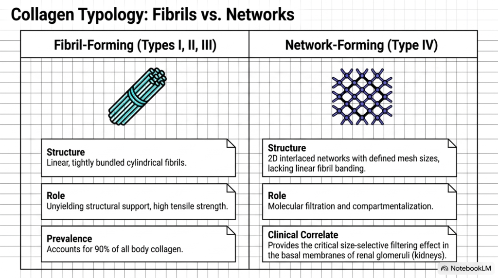
The study of Collagen Biochemistry highlights a fundamental division between fibril-forming and network-forming collagen architecture across different tissue types. Fibril-forming collagens, including Types I, II, and III, assemble into linear, tightly bundled cylindrical fibers designed for high tensile strength. These unyielding structures resist longitudinal pulling forces and make up ninety percent of all collagen in the human body, providing essential structural support to bone, cartilage, tendon, and skin matrices.
In contrast, Collagen Biochemistry identifies Type IV collagen as the premier example of network-forming structural architecture. Instead of bundling into parallel linear fibrils, Type IV molecules form two-dimensional interlaced meshworks with defined pore sizes. These flat networks lack the linear banding pattern characteristic of fibril-forming collagens and instead function as specialized planar scaffolds for cell attachment, tissue separation, and selective fluid filtration in basement membranes.
Physiologically, Type IV networks provide essential molecular filtration and compartmentalization within basement membranes throughout the human body. In renal glomeruli, this meshwork acts as a size-selective physical filter that lets small solutes pass while retaining large plasma proteins in the blood. Comparing these structural classes demonstrates how Collagen Biochemistry tailors molecular architecture to fulfill distinct mechanical support or fluid filtration roles across diverse organs.
Slide 8: Intracellular Phase of Synthesis in Collagen Biochemistry
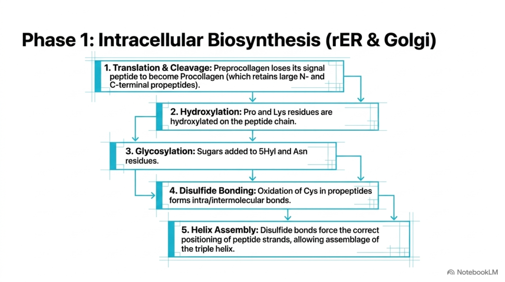
Intracellular biosynthesis in Collagen Biochemistry represents a tightly regulated multi-step pathway within the rough endoplasmic reticulum and Golgi apparatus. Synthesis begins with the translation of preprocollagen on ribosomes attached to the endoplasmic reticulum membrane. Signal peptidases rapidly cleave the N-terminal signal peptide, converting the nascent chain into procollagen containing large non-helical N-terminal and C-terminal propeptides that prevent premature intracellular fibril assembly before cellular secretion occurs into the extracellular environment.
Within the lumen, collagen undergoes vital post-translational modifications, including hydroxylation of specific proline and lysine residues by specialized oxygenase enzymes. Subsequently, glycosyltransferase enzymes attach glucose and galactose residues to selected hydroxylysine amino acids. After glycosylation, cysteine residues within the C-terminal propeptides oxidize, forming crucial intra- and intermolecular disulfide bonds that bring the three alpha-chains into close physical alignment and proper registration for triple-helix assembly.
These disulfide bonds act as molecular guides, initiating correct triple-helical folding from the C-terminus toward the N-terminus in a zipper-like manner. As the chains zip together, they form a stable procollagen triple helix flanked by bulky globular propeptide ends. By examining intracellular processing, students discover how Collagen Biochemistry ensures proper chain alignment, structural stabilization, and solubility before precursor molecules are transported through the Golgi apparatus and secreted via exocytosis into the extracellular space.
Slide 9: Micronutrient Cofactors and Scurvy in Collagen Biochemistry
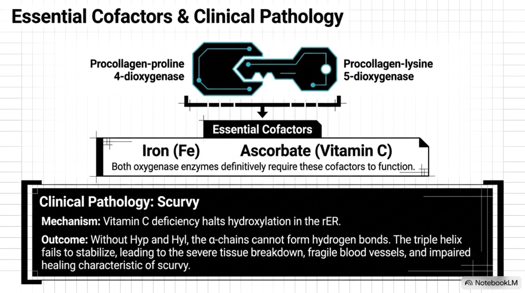
Enzymatic hydroxylation in Collagen Biochemistry requires specific micronutrient cofactors to maintain catalyst activity during protein maturation inside the cell. The enzymes procollagen-proline four-dioxygenase and procollagen-lysine five-dioxygenase depend on ferrous iron and ascorbate, commonly known as Vitamin C. Ferrous iron sits directly at the enzyme active site and participates in the oxidative decarboxylation of alpha-ketoglutarate during targeted amino acid residue modification within the endoplasmic reticulum lumen.
During catalysis, iron can oxidize to its inactive ferric state, halting enzyme function in Collagen Biochemistry. Vitamin C acts as a vital antioxidant reducing agent, transferring an electron to convert ferric iron back into functional ferrous iron. Without Vitamin C, these oxygenase enzymes remain inactive, completely preventing the hydroxylation of proline and lysine residues during intracellular procollagen processing and arresting normal collagen maturation in human connective tissue cells.
In clinical pathology, nutritional Vitamin C deficiency causes scurvy by disrupting Collagen Biochemistry at the molecular level. Lacking hydroxyproline, procollagen alpha-chains cannot form essential interchain hydrogen bonds, preventing stable triple-helix formation at physiological body temperature. Unstable precursor chains undergo intracellular degradation in lysosomes, leading to severe capillary fragility, skin lesions, bleeding gums, poor wound healing, tooth loss, and extensive structural breakdown across human connective tissues and organ systems.
Slide 10: Extracellular Maturation Steps in Collagen Biochemistry
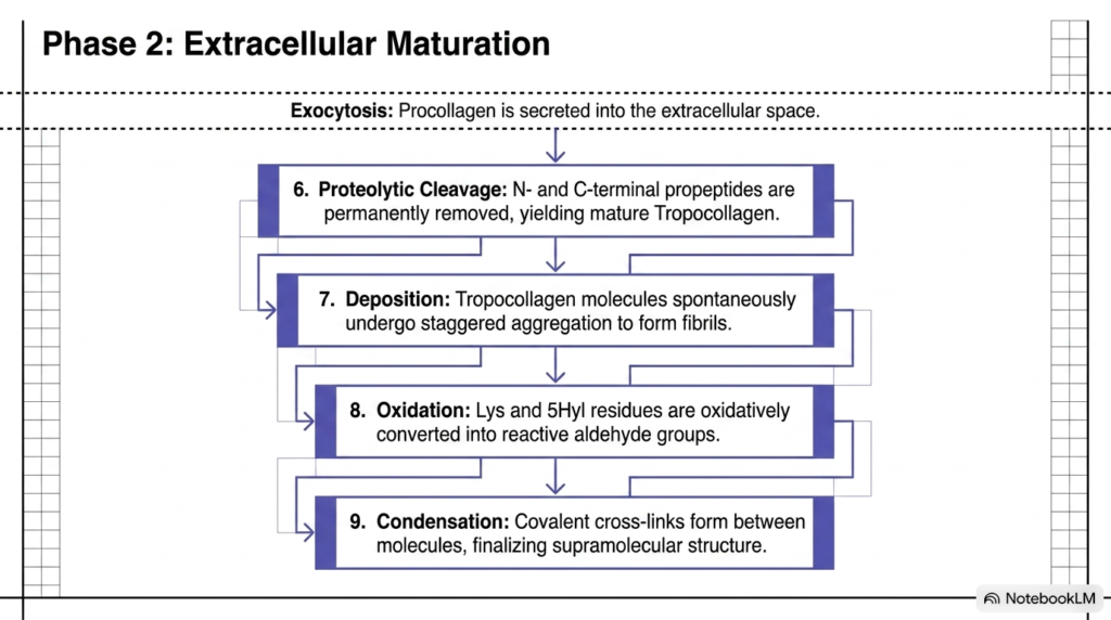
The transition from intracellular synthesis to extracellular maturation is a critical threshold in Collagen Biochemistry. Secreted procollagen molecules enter the extracellular space via exocytosis, carrying bulky globular propeptides at both ends that maintain solubility during transport through the secretory pathway. Specific extracellular peptidases then cleave the N-terminal and C-terminal propeptide domains, yielding mature, insoluble tropocollagen molecules ready for structural fibril assembly.
After propeptide removal, insoluble tropocollagen rods spontaneously undergo staggered self-assembly into linear fibrils. Once aligned in parallel, the extracellular enzyme lysyl oxidase oxidatively converts specific lysine and hydroxylysine residues into highly reactive aldehyde groups called allysine and hydroxyallysine. This enzymatic oxidation step sets the stage for final covalent cross-linking between adjacent tropocollagen molecules within the assembled fibril matrix in the extracellular space.
Finally, these reactive aldehyde groups undergo spontaneous condensation reactions with neighboring amino acid side chains on adjacent tropocollagen molecules. This process generates covalent intermolecular cross-links that permanently lock tropocollagen rods into a rigid supramolecular framework. Through these sequential maturation steps, Collagen Biochemistry converts soluble precursor proteins into insoluble, highly resistant structural fibers capable of enduring massive mechanical force and physical strain across living animal tissues and physiological organ structures.
Slide 11: Covalent Cross-linking and Aging in Collagen Biochemistry
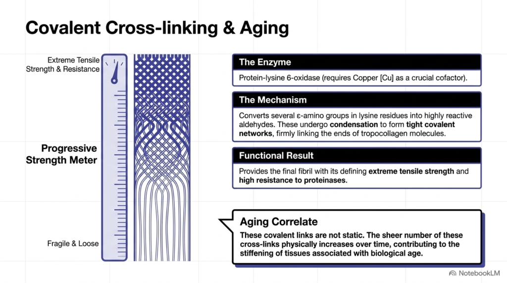
The ultimate mechanical strength in Collagen Biochemistry is achieved through covalent cross-linking catalyzed by protein-lysine six-oxidase, commonly called lysyl oxidase. This extracellular enzyme requires copper as an essential cofactor to catalyze the oxidative deamination of lysine residues. Lysyl oxidase converts epsilon-amino groups on lysine and hydroxylysine into reactive aldehyde derivatives that form highly stable, permanent covalent cross-links across adjacent polypeptide chains.
These reactive aldehydes spontaneously condense with adjacent unmodified amino acid side chains, forming a dense covalent network across tropocollagen molecules. In Collagen Biochemistry, this cross-linking process gives fibrils extreme tensile strength and high resistance to proteolytic degradation by collagenases and proteinases. Without copper-dependent lysyl oxidase activity, collagen fibers remain structurally weak, leading to severe connective tissue fragility, hyperextensible skin, joint hypermobility, skeletal deformities, and life-threatening arterial aneurysms throughout the human vascular system.
Over time, covalent cross-links progressively accumulate within connective tissues, serving as a key molecular correlate of biological aging. As cross-link density increases, collagenous tissues lose physiological elasticity and become increasingly stiff, brittle, and resistant to normal matrix turnover throughout the human body. By examining lysyl oxidase chemistry and cross-link accumulation, Collagen Biochemistry explains both the physiological maturation of young tissue and the gradual mechanical stiffening observed during organismal aging processes.
Slide 12: Integrated Synthesis Pathway in Collagen Biochemistry
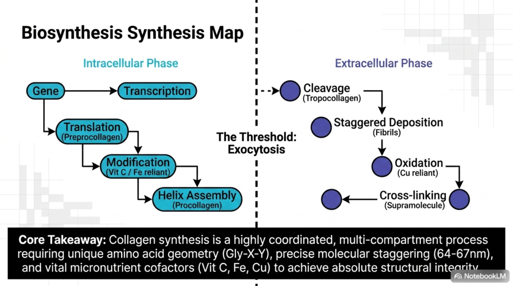
The complete biosynthetic pathway of Collagen Biochemistry operates as a highly coordinated, multi-compartment process spanning intracellular and extracellular spaces. Intracellular events begin with gene transcription and translation of preprocollagen on ribosomes attached to the endoplasmic reticulum, followed by signal peptide cleavage. Within the rough endoplasmic reticulum lumen, micronutrient-dependent modifications occur, requiring Vitamin C and iron to complete proline and lysine hydroxylation prior to procollagen triple-helix assembly and disulfide bond formation.
Crossing the cellular threshold via exocytosis marks the transition to extracellular Collagen Biochemistry. Extracellular enzymes cleave terminal propeptides to generate mature tropocollagen, which spontaneously aggregates into quarter-staggered fibrils. Next, copper-dependent lysyl oxidase oxidizes targeted residues into aldehydes, enabling condensation reactions that form covalent cross-links. This extracellular sequence transforms individual triple helices into an insoluble supramolecular scaffold with remarkable tensile durability throughout living animal tissues and physiological body systems.
In summary, collagen biochemistry shows how gene expression, post-translational modifications, and extracellular self-assembly integrate seamlessly across cellular boundaries. From the repeating Glycine-X-Y amino acid sequence to copper-mediated cross-linking, every molecular detail contributes directly to tissue architecture. Mastering these interconnected steps provides medical and biochemistry students with a foundational understanding of structural biology, matrix mechanics, and connective tissue pathology across human health and disease states.
Please read our Content Disclaimer Statement.
Check out our social media channels: