130. Muscle Metabolism: Energy Pathways, Fibers and Cycles
Skeletal muscle powers every physical action in the human body, from delicate ocular movements to maximal athletic exertion. To sustain these contractions, muscle cells must continually produce biological energy in the form of adenosine triphosphate. Because resting intracellular energy stores are extremely limited, muscle tissue relies on a coordinated network of pathways. This comprehensive post breaks down the essential biochemical mechanisms of muscle metabolism, exploring how different fiber types, energy buffers, and inter-organ cycles collaborate to maintain cellular homeostasis during rest, intense exercise, and starvation.
Slide 01: Introduction to the Biochemistry of Muscle Metabolism
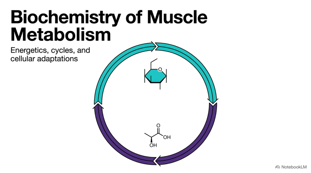
Skeletal muscle operates as a specialized biological motor that converts chemical energy into physical force. To sustain everyday movements and high-intensity exertion, muscle metabolism relies on a tightly regulated network of enzymatic reactions and bioenergetic pathways. The foundational challenge facing muscle tissue is the constant demand for adenosine triphosphate, which fuels the cyclic cross-bridge movements between actin and myosin filaments. As physical effort increases, muscle metabolism must adapt by selecting specific fuel substrates and adjusting energy-production pathways to prevent rapid cellular exhaustion.
This introductory diagram illustrates the fundamental relationship between carbohydrate fuels and metabolic end-products during intense physical activity. As glucose breaks down to provide rapid energy, key intermediates like pyruvate convert into lactate, which requires specialized handling across tissues. Understanding these initial biochemical concepts highlights how muscle metabolism connects localized cellular work with whole-body metabolic integration.
Throughout this series, students will analyze how distinct fiber profiles, high-energy phosphate buffers, and inter-organ recycling loops allow skeletal muscle to balance explosive power output with long-term survival. By studying these integrated cellular pathways, future healthcare professionals and biochemists gain crucial insights into physical performance, muscle fatigue, and metabolic disease states that impact human physiology daily. This foundational knowledge provides a clear framework for understanding complex cellular bioenergetics.
Slide 02: The ATP Deficit and Immediate Energy Needs in Muscle Metabolism
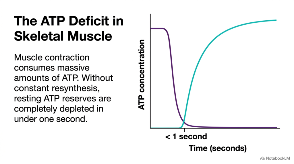
Skeletal muscle cells maintain an extremely small resting pool of adenosine triphosphate relative to their tremendous energy consumption during active work. During intense physical activity, muscle metabolism increases its energy utilization rate by several hundredfold within milliseconds. As demonstrated in the accompanying graph, the resting intracellular concentration of adenosine triphosphate drops drastically in less than one second of maximal contraction. Without rapid and continuous resynthesis, this sudden energy deficit would cause immediate muscular paralysis, structural cross-bridge locking, and cellular rigor.
To prevent complete energy exhaustion, muscle metabolism relies on immediate and delayed pathway responses to regenerate spent adenosine diphosphate back into usable fuel. The sharp initial decline in energy stores triggers instant activation of localized biochemical buffering mechanisms, followed by rapid glycolytic and oxidative pathways. This metabolic transition highlights why muscle metabolism cannot depend solely on static energy pools.
Instead, muscle tissue uses a layered network of bioenergetic systems that match energy-generation speed to the precise intensity and duration of muscular work. This immediate energy response ensures that cross-bridge cycling continues smoothly during sudden bursts of physical activity. Understanding this rapid turnover shows how cellular systems maintain energetic homeostasis under extreme functional demands and prevent early metabolic collapse.
Slide 03: Fiber Types and Metabolic Diversity in Muscle Metabolism
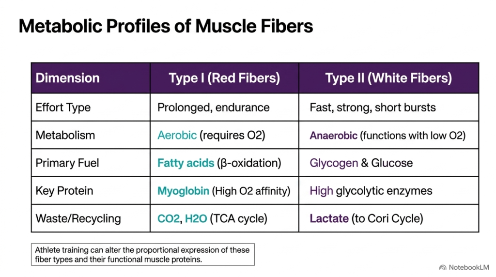
Skeletal muscle tissue contains distinct fiber types with specialized structural and biochemical characteristics. These functional differences dictate how muscle metabolism handles distinct physiological workloads, ranging from prolonged endurance activities to brief explosive sprints. Type I red fibers are optimized for slow, sustained contractions. They rely heavily on aerobic muscle metabolism, utilizing mitochondrial beta-oxidation of fatty acids to generate large quantities of adenosine triphosphate over extended periods. These red fibers contain high concentrations of myoglobin, an oxygen-binding heme protein that ensures a steady oxygen supply for electron transport chain activity.
In contrast, Type II white fibers are engineered for rapid, high-force contractions that operate largely independent of immediate oxygen availability. In these fast-twitch fibers, muscle metabolism relies predominantly on anaerobic glycolysis, burning stored glycogen and blood glucose to produce rapid energy. Because Type II fibers possess high levels of glycolytic enzymes and lower mitochondrial density, they produce substantial amounts of lactate that must be transported out of the cell.
Physical training can alter the proportional expression of specific muscle proteins within these fibers, demonstrating the remarkable adaptability of human muscle metabolism. Understanding these fiber-specific energy pathways helps explain individual athletic variations, fatigue resistance, and functional recovery after strenuous exercise in academic and clinical exercise physiology.
Slide 04: Anaerobic Glycolysis and Redox Balance in Muscle Metabolism
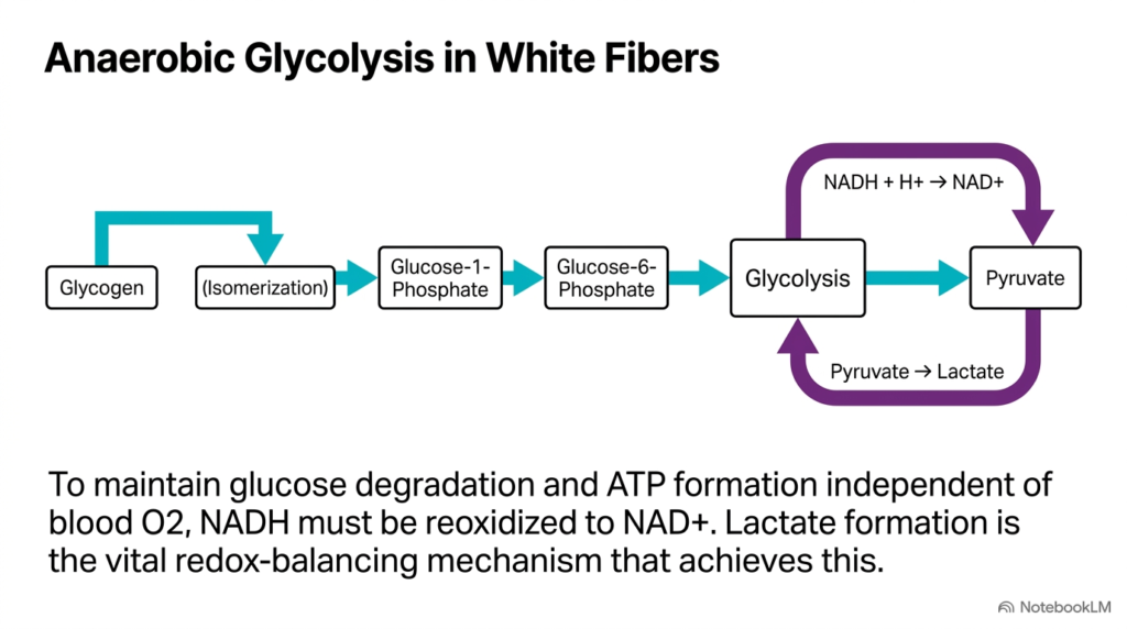
When physical exertion exceeds the vascular system’s oxygen-delivery capacity, fast-twitch muscle fibers depend heavily on anaerobic glycolysis. Under these oxygen-restricted conditions, muscle metabolism breaks down stored glycogen through glycogenolysis, yielding glucose-1-phosphate, which is converted into glucose-6-phosphate for glycolytic processing. This rapid pathway generates adenosine triphosphate quickly without entering the mitochondria. However, continuous glycolytic flux requires a steady supply of nicotinamide adenine dinucleotide in its oxidized form to sustain the glyceraldehyde 3-phosphate dehydrogenase reaction.
To maintain redox equilibrium during rapid energy production, muscle metabolism employs the enzyme lactate dehydrogenase to reduce pyruvate into lactate. This essential reaction transfers electrons from reduced nicotinamide adenine dinucleotide, regenerating the oxidized coenzyme necessary for ongoing glycolysis. By converting pyruvate to lactate, muscle metabolism avoids a metabolic bottleneck and allows high-speed energy generation to continue during anaerobic effort.
The accumulated lactate is subsequently exported into the bloodstream, where it travels to other organs for recycling, demonstrating how localized anaerobic pathways integrate with systemic physiology. Ultimately, this anaerobic pathway provides a vital energy bridge during short bursts of maximal muscular effort, ensuring that ATP resynthesis keeps pace with high contractile demands without causing immediate metabolic failure. This redox-balancing mechanism represents a classic adaptation for high-power cellular survival.
Slide 05: Aerobic Oxidation and Myoglobin Dynamics in Muscle Metabolism
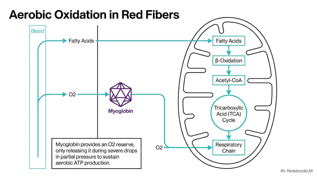
In slow-twitch red muscle fibers, energy production relies predominantly on aerobic oxidation within the mitochondrial matrix. This sustainable arm of muscle metabolism utilizes circulating fatty acids and oxygen delivered by the bloodstream. Once inside the mitochondrion, fatty acids undergo beta-oxidation to generate acetyl coenzyme A, which enters the tricarboxylic acid cycle. The resulting reduced coenzymes feed into the electron transport chain, driving oxidative phosphorylation to generate abundant adenosine triphosphate. This aerobic pathway provides high energetic efficiency, making it ideal for continuous endurance activities.
Myoglobin plays a crucial role in supporting aerobic muscle metabolism by serving as an intracellular oxygen reservoir. As shown in the slide diagram, myoglobin possesses a high affinity for oxygen, retaining bound oxygen under normal resting conditions. When local oxygen partial pressure drops sharply during vigorous contraction, myoglobin releases its stored oxygen directly to the mitochondrial respiratory chain.
This localized buffering mechanism prevents transient hypoxia and ensures that aerobic muscle metabolism continues uninterrupted during fluctuations in microvascular blood flow. By coupling intramuscular oxygen storage with efficient mitochondrial oxidation, red muscle fibers maintain long-term fatigue resistance during prolonged mechanical work. This mechanism allows endurance muscles to sustain aerobic performance without prematurely accumulating acidic metabolic byproducts or suffering from cellular energy exhaustion.
Slide 06: Adenylate Kinase and Energy Salvage in Muscle Metabolism
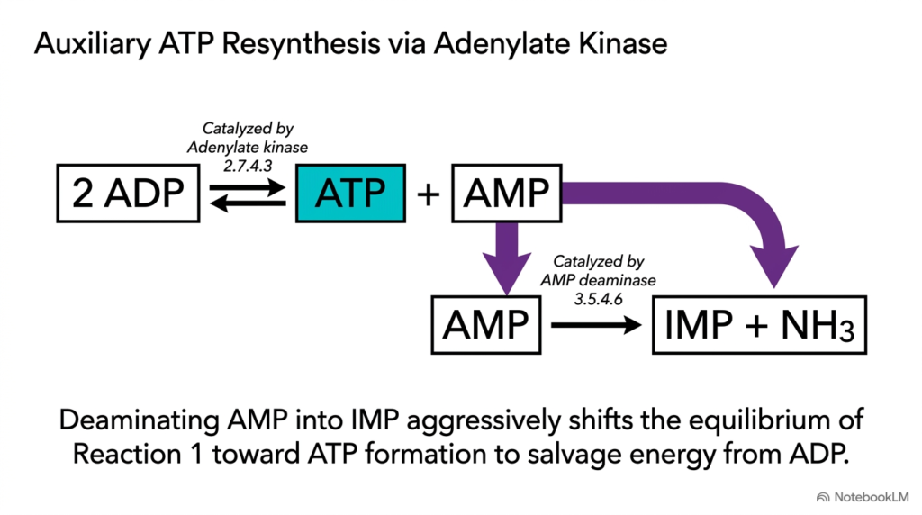
When sudden energy demands overwhelm primary metabolic pathways, muscle metabolism utilizes auxiliary enzyme systems to salvage usable energy from accumulated nucleosides. The first line of emergency energy recovery involves the enzyme adenylate kinase. During intense contraction, adenosine triphosphate breaks down, leading to a rapid accumulation of adenosine diphosphate. Adenylate kinase catalyzes a reversible reaction that transfers a phosphate group between two molecules of adenosine diphosphate, producing one molecule of adenosine triphosphate and one molecule of adenosine monophosphate.
However, because the adenylate kinase reaction is near equilibrium, accumulated adenosine monophosphate would normally limit further energy recovery. To overcome this thermodynamic constraint, muscle metabolism employs AMP deaminase to convert adenosine monophosphate into inosine monophosphate and ammonia. According to Le Chatelier’s principle, removing adenosine monophosphate strongly shifts the adenylate kinase equilibrium toward continued energy production.
Through this coordinated enzymatic mechanism, muscle metabolism salvages essential fuel from spent nucleosides during extreme metabolic stress. This emergency reaction pathway acts as a crucial energetic safety net, preserving cellular viability when high-intensity exercise rapidly consumes ATP reserves. Furthermore, the generated inosine monophosphate can later be reaminated during muscle recovery, demonstrating the remarkable efficiency of nucleoside recycling in human muscle metabolism. Understanding these salvage pathways expands our knowledge of cellular bioenergetics under stress.
Slide 07: Creatine Phosphate Buffering in Muscle Metabolism
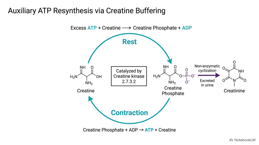
The creatine phosphate system serves as the primary high-energy phosphate buffer in skeletal muscle cells. During periods of rest, when energy production exceeds immediate consumption, muscle metabolism stores excess energy by transferring a phosphate group from adenosine triphosphate to creatine. This reversible reaction, catalyzed by creatine kinase, creates a substantial reservoir of intracellular creatine phosphate. When sudden physical effort begins, this stored phosphate is immediately transferred back to spent adenosine diphosphate, regenerating usable fuel within milliseconds without requiring oxygen or complex metabolic pathways.
Beyond its central role in rapid energy transfer, the creatine buffering system undergoes slow, continuous non-enzymatic degradation. A small fraction of intracellular creatine phosphate spontaneously cyclizes into creatinine, an inactive waste product that enters the circulation and is excreted by the kidneys. Because creatinine production correlates directly with total functional tissue mass, clinical measurements of serum creatinine provide valuable insights into renal clearance and human muscle metabolism.
Understanding these chemical pathways emphasizes how creatine buffering maintains stability in human muscle metabolism during explosive contractions. This rapid buffering network represents the fastest mechanism available for ATP resynthesis, bridging the critical temporal gap before glycolysis accelerates. By preventing rapid energy depletion, creatine phosphate allows muscles to generate immediate maximal power output safely.
Slide 08: Multi-Organ Biosynthesis of Creatine for Muscle Metabolism
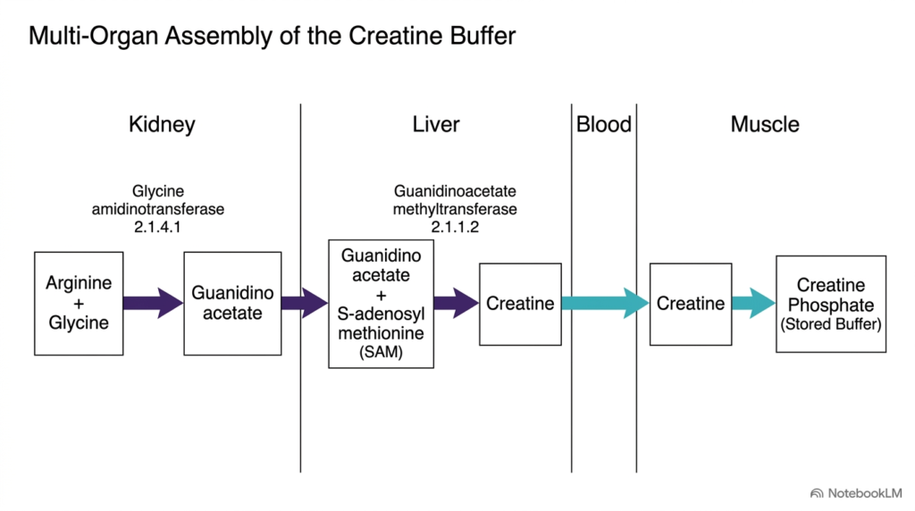
Although skeletal muscle relies heavily on creatine phosphate for rapid energy buffering, muscle tissue lacks the necessary enzymes to synthesize creatine from scratch. Instead, muscle metabolism depends on a multi-organ biosynthetic pathway involving the kidneys and liver. As detailed in the slide flow chart, the initial step occurs in the kidneys, where glycine amidinotransferase combines arginine and glycine to form guanidinoacetate. This intermediate compound is released into the bloodstream and transported to the liver for final chemical assembly.
Inside the liver, the enzyme guanidinoacetate methyltransferase transfers a methyl group from S-adenosylmethionine to guanidinoacetate, producing mature creatine. The synthesized creatine enters the systemic circulation and is actively taken up by skeletal muscle cells via specialized transporters. Once inside the muscle compartment, creatine kinase phosphorylates creatine to build the high-energy pool required for active muscle metabolism.
This multi-organ assembly line demonstrates how muscle metabolism relies on inter-organ metabolic cooperation to maintain its localized energy reserves. Without this inter-organ support system, skeletal muscle cells would quickly deplete their high-energy phosphate stores during high-intensity physical performance. This pathway underscores the vital biochemical interdependency between renal, hepatic, and muscular tissues in human bioenergetics and systemic health, illustrating the interconnected nature of human organ systems during sustained metabolic activity.
Slide 09: Inter-Organ Lactate Recycling via the Cori Cycle in Muscle Metabolism
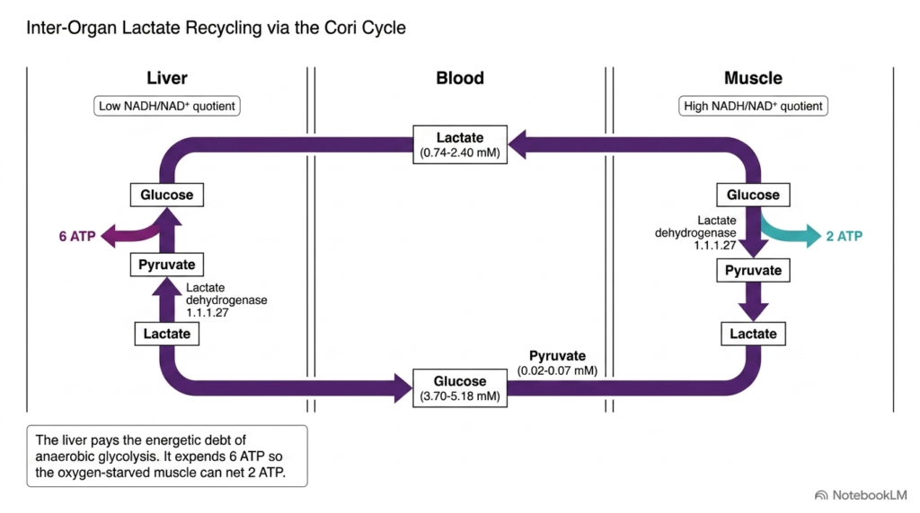
During intense physical activity, anaerobic glycolysis in fast-twitch fibers produces substantial amounts of lactate. To prevent severe intracellular acidosis and recycle metabolic waste, muscle metabolism relies on the Cori cycle, an essential inter-organ pathway connecting skeletal muscle and the liver. In contracting muscle cells, where the ratio of reduced to oxidized nicotinamide adenine dinucleotide is high, pyruvate is reduced to lactate while generating two net molecules of adenosine triphosphate. The accumulated lactate diffuses into the bloodstream and travels directly to the liver.
In hepatic tissue, where the redox environment favors oxidation, lactate dehydrogenase converts lactate back into pyruvate. The liver then uses gluconeogenesis to transform pyruvate into fresh glucose, expending six molecules of adenosine triphosphate in the process. The liver then uses gluconeogenesis to transform pyruvate into fresh glucose, expending six molecules of adenosine triphosphate in the process.
By expending energy in the liver to pay the energetic debt incurred by working muscle, the Cori cycle demonstrates how inter-organ cooperation supports sustainable muscle metabolism. This elegant metabolic exchange protects muscle cells from toxic lactate accumulation while maintaining systemic blood glucose levels during prolonged strenuous effort. Consequently, hepatic gluconeogenesis plays an indispensable role in supporting continuous exercise capacity across demanding workloads.
Slide 10: The Glucose-Alanine Cycle and Nitrogen Disposal in Muscle Metabolism
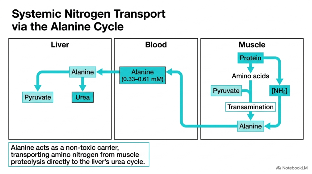
During periods of prolonged exertion or fasting, skeletal muscle degrades internal structural proteins to supply carbon skeletons for energy production. However, amino acid catabolism releases toxic ammonium ions that the cell must safely remove. To solve this nitrogen-disposal problem, muscle metabolism uses the glucose-alanine cycle. Inside contracting muscle tissue, transaminase enzymes transfer amino groups from degraded amino acids onto pyruvate, forming the non-toxic amino acid alanine.
Alanine is secreted into the blood and transported directly to the liver. Hepatic enzymes then remove the nitrogen group from alanine, transferring it into the urea cycle for safe excretion in urine. The remaining pyruvate carbon skeleton is converted into glucose via gluconeogenesis and released back into circulation to support ongoing muscle metabolism.
By acting as a non-toxic carrier of amino nitrogen, alanine lets skeletal muscle generate needed energy from protein breakdown without risking ammonia toxicity. This dual transport system highlights how muscle metabolism integrates nitrogen clearance with carbohydrate resynthesis to protect cellular integrity during severe energy deficits. As a result, muscle protein catabolism contributes safely to whole-body glucose balance without compromising systemic pH or neural function during prolonged physical stress and catabolic states in clinical metabolic physiology and cellular bioenergetics.
Slide 11: Branched-Chain Amino Acid Catabolism in Muscle Metabolism
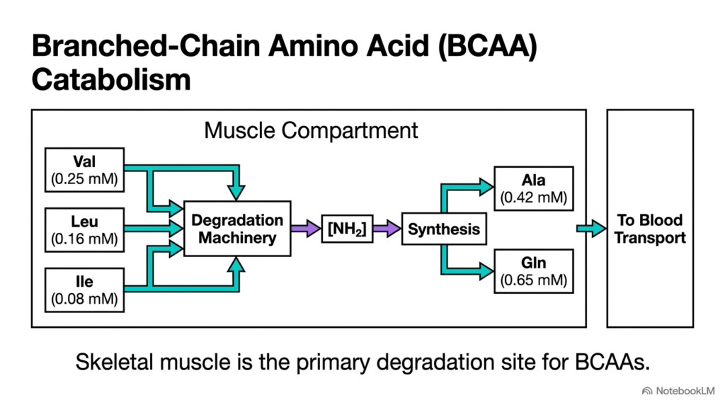
Skeletal muscle plays a unique, central role in whole-body protein turnover as the primary site of branched-chain amino acid catabolism. Unlike most other amino acids, which the liver processes predominantly, valine, leucine, and isoleucine bypass hepatic breakdown and enter skeletal muscle cells directly. Here, specialized transaminases and dehydrogenase complexes initiate the degradation of these essential amino acids, providing energy substrates directly to active muscle metabolism.
During branched-chain amino acid catabolism, liberated nitrogen groups are incorporated into pyruvate and glutamate to synthesize alanine and glutamine. These two non-toxic amino acids are exported into the blood to transport amino nitrogen safely to the liver and kidneys. Leucine catabolism is particularly important because its oxidative intermediates yield acetyl-CoA, feeding directly into the tricarboxylic acid cycle during periods of metabolic stress in muscle.
This specialized enzymatic machinery highlights how muscle metabolism actively participates in systemic amino acid homeostasis and energy production. By utilizing branched-chain amino acids during exercise, skeletal muscle generates extra ATP while generating essential nitrogen carriers for systemic metabolic health. This mechanism proves vital during endurance activities when carbohydrate availability becomes limited, and additional fuel sources are needed to sustain performance and prevent cellular energy collapse.
Slide 12: Hormonal Regulation and Starvation Adaptation in Muscle Metabolism
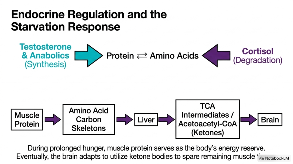
Circulating endocrine hormones tightly regulate the metabolic fate of skeletal muscle protein by balancing synthesis and degradation. Anabolic hormones such as testosterone promote amino acid uptake and structural protein synthesis, expanding functional tissue mass. Conversely, catabolic hormones like cortisol stimulate proteolysis during physical stress or extended fasting. Under prolonged starvation, muscle metabolism shifts dramatically, transforming structural muscle proteins into an essential energy reserve for the rest of the body.
During extended food deprivation, amino acid carbon skeletons derived from muscle breakdown travel to the liver, where they are converted into glucose and ketone bodies. As starvation progresses, the brain adapts to utilize ketone bodies as its primary fuel source. This endocrine adaptation significantly reduces systemic demand for glucose, sparing remaining muscle tissue from further destruction and stabilizing overall muscle metabolism.
Understanding these hormonal signals clarifies how muscle metabolism adapts dynamically to preserve whole-body survival during severe metabolic hardship. This complex endocrine crosstalk ensures that protein reserves are mobilized when needed while preventing fatal muscle wasting during starvation. These regulatory mechanisms highlight the vital balance between anabolic growth and catabolic survival signaling in human physiology and metabolic medicine during extended fasting.
Slide 13: Comparative Analysis of Smooth Muscle vs Skeletal Muscle Metabolism
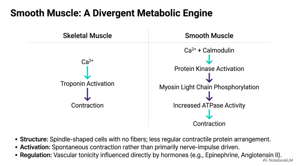
Smooth muscle tissue exhibits a divergent metabolic and structural engine compared to skeletal muscle. While skeletal muscle relies on organized sarcomeres and troponin complexes for rapid contractions, smooth muscle consists of spindle-shaped cells that lack striated banding. In smooth muscle, calcium entry triggers contraction by binding to calmodulin, which activates myosin light chain kinase. This enzyme phosphorylates myosin light chains, stimulating adenosine triphosphatase activity and cross-bridge cycling. Consequently, smooth muscle metabolism operates at a much lower rate of energy turnover, making it ideal for sustained tonic contractions in vascular and visceral organs.
Unlike skeletal muscle, which depends heavily on somatic nerve impulses and large glycogen reserves, smooth muscle contraction is frequently spontaneous or regulated by circulating hormones such as epinephrine and angiotensin II. Because smooth muscle maintains low-energy latch states, its overall energy requirement is significantly less than the high energy consumption characteristic of skeletal muscle metabolism.
Comparing these two tissue types illustrates how evolution has tailored muscle metabolism to meet specific mechanical and physiological demands across human anatomy. This functional divergence ensures that internal organs maintain continuous vascular tone and motility with minimal energetic cost. Ultimately, smooth muscle bioenergetics emphasizes efficiency and sustainability over raw mechanical power in everyday physiology.
Slide 14: Systemic Integration and the Muscle-Liver Axis in Muscle Metabolism
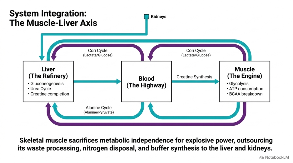
This final integrative slide illustrates how skeletal muscle functions as part of a highly coordinated systemic network rather than an isolated organ. To maximize explosive mechanical power and rapid force generation, skeletal muscle sacrifices complete metabolic independence. As the diagram shows, muscle tissue acts as the primary mechanical engine, consuming vast amounts of fuel through glycolysis and energy turnover while producing metabolic byproducts like lactate, alanine, and creatinine during active muscle metabolism.
To maintain continuous function without cellular damage, muscle metabolism outsources waste management, nitrogen disposal, and buffer synthesis to secondary organs. The liver acts as a central refinery, processing lactate via the Cori cycle, converting alanine nitrogen into urea, and completing creatine synthesis. Meanwhile, the kidneys assist with initial creatine assembly and waste clearance, supporting the ongoing demands of muscle metabolism.
By integrating these specialized inter-organ pathways, human muscle metabolism balances massive localized power output with long-term physiological stability. This systemic architecture ensures that active muscle tissue operates in complete harmony with the rest of the body, enabling both intense athletic performance and metabolic survival. Mastering these inter-organ relationships gives students a foundational framework for advanced biochemistry and physiology studies across clinical medical science and human exercise performance.
Please read our Content Disclaimer Statement.
Check out our social media channels: