121. Cytochrome P450 Systems: Biochemical Mechanisms of Biotransformation
Have you ever wondered how your body safely processes daily medications or clears toxic chemicals after a meal? The liver relies on a remarkable family of specialized enzymes to transform water-insoluble molecules into easily excretable compounds. This slide deck provides a comprehensive walk-through of these vital biochemical systems. Students will explore the structural framework, electron transport pathways, and step-by-step catalytic cycle that allow these specialized hemeproteins to perform difficult chemical transformations, offering essential insights for pharmacology, toxicology, and clinical medicine.
Slide 1: Introduction to Cytochrome P450 and Phase I Biotransformation
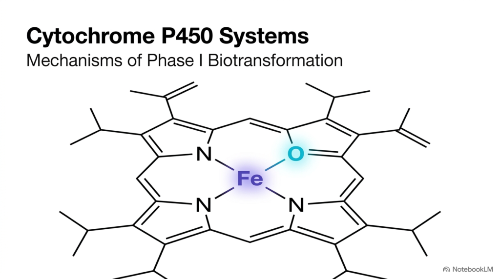
The human body encounters thousands of foreign compounds daily, including medications, environmental pollutants, and dietary chemicals. To prevent lipophilic toxins from accumulating in fatty tissues, the liver utilizes Phase I biotransformation reactions. At the center of this protective mechanism is the Cytochrome P450 enzyme superfamily, a group of heme-containing monooxygenases found across all domains of life. These enzymes convert nonpolar, water-insoluble molecules into more polar compounds that can be eliminated or further modified.
The structural hallmark of every Cytochrome P450 protein is an iron-protoporphyrin IX prosthetic group, visible in the central diagram. The iron atom at the core of the heme ring alternates between oxidation states during catalysis, allowing the enzyme to activate molecular oxygen. By inserting one oxygen atom into an unreactive carbon-hydrogen bond, Cytochrome P450 increases the solubility of its substrate.
Understanding Phase I biotransformation is fundamental for medical and pharmacy students. These enzymatic pathways dictate drug half-lives, determine therapeutic dosages, and influence how the body handles potential toxins. Without Cytochrome P450 activity, many essential modern pharmaceuticals would remain trapped in lipid membranes, leading to severe cellular toxicity. As students explore this slide deck, they will examine how structural architecture, electron flow, and precise catalytic steps enable these versatile enzymes to safeguard human health.
Slide 2: The Physiological Goals of Cytochrome P450 Enzymes
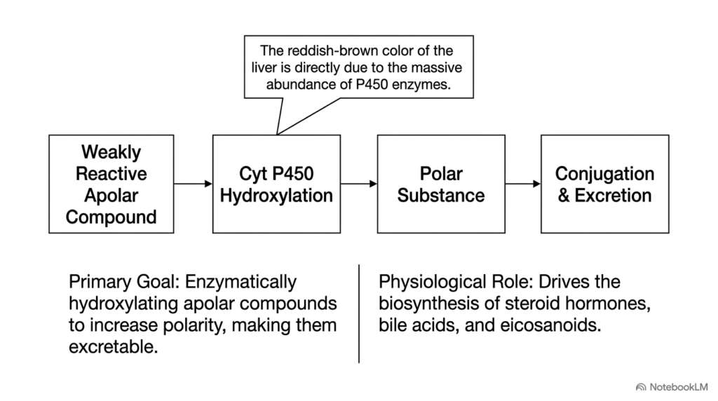
The primary goal of hepatic biotransformation is converting weakly reactive apolar compounds into soluble, excretable metabolites. Lipophilic molecules pass easily through cell membranes but resist renal excretion, making them dangerous if left unprocessed. As shown in the slide diagram, Cytochrome P450 hydroxylation adds polar hydroxyl groups to nonpolar molecules, transforming them into polar substances. These polar metabolites can then undergo Phase II conjugation and be rapidly excreted in urine or bile.
Beyond clearing foreign drugs, Cytochrome P450 systems play an indispensable role in normal human physiology. These enzymes drive the biosynthesis of essential endogenous compounds, including steroid hormones, bile acids, and inflammatory mediators like eicosanoids. In organs like the adrenal cortex and gonads, specialized family members catalyze key steps in converting cholesterol into cortisol, aldosterone, and sex steroids.
An interesting anatomical observation highlighted on this slide is the characteristic reddish-brown color of human liver tissue. This distinct coloration stems directly from the massive abundance of heme-containing Cytochrome P450 enzymes embedded within the endoplasmic reticulum of hepatocytes. Because hepatocytes process vast amounts of blood-borne substances, they maintain extraordinarily high concentrations of these hemeproteins. Through dual roles in metabolism and hormone synthesis, Cytochrome P450 serves as a cornerstone of metabolic homeostasis.
Slide 3: Structural Architecture of the Cytochrome P450 Enzyme
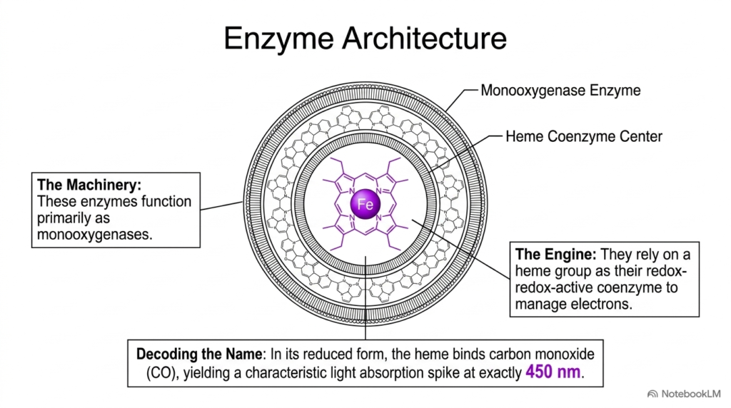
To understand how biotransformation occurs, students must examine the enzyme’s unique structural architecture. As the diagram shows, Cytochrome P450 functions as a specialized monooxygenase built around a central heme coenzyme center. The heme group features an iron protoporphyrin ring that serves as the redox-active engine, managing electron transfers necessary for oxygen activation and chemical catalysis.
The unusual name of this enzyme family originates from a distinctive spectroscopic property discovered during early biochemical studies. In its reduced ferrous state, the heme iron binds carbon monoxide, producing an intense light absorption peak at exactly 450 nanometers. This spectroscopic signature gave rise to the term Cytochrome P450, where ‘P’ stands for pigment and ‘450’ represents the characteristic absorption wavelength.
The protein shell surrounding the heme creates an active site pocket that accommodates diverse substrate molecules. A conserved cysteine residue provides an axial thiolate ligand to the heme iron, imparting unique electronic properties that facilitate oxygen splitting. By pairing a versatile protein cavity with a powerful iron-based coenzyme, Cytochrome P450 can catalyze difficult chemical reactions on remarkably diverse substrates. Medical students should appreciate how this specialized heme architecture allows Cytochrome P450 to perform oxidative chemistry with extraordinary precision inside human hepatocytes.
Slide 4: Electron Transport Components in Cytochrome P450 Reactions
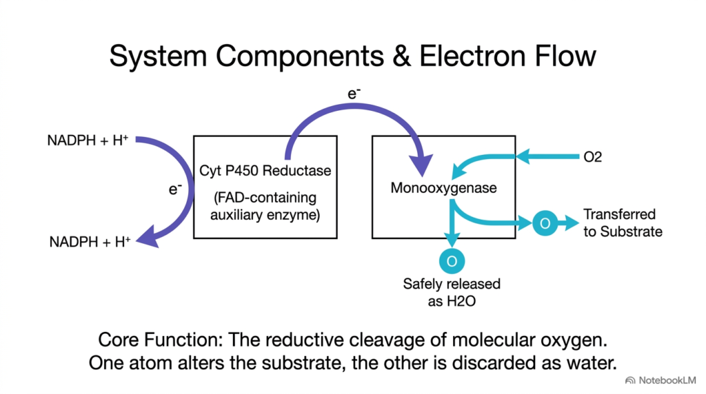
Monooxygenases require a continuous supply of electrons and molecular oxygen for catalytic activity. This slide outlines the two-component system and electron transport pathway that drive biotransformation. The principal enzyme, Cytochrome P450, relies on a membrane-bound auxiliary partner called Cytochrome P450 reductase. This flavoprotein contains FAD and FMN coenzymes, allowing it to extract two electrons from reduced NADPH and deliver them sequentially to the main catalytic site.
The diagram depicts the core biochemical function: reductive cleavage of molecular oxygen. During each catalytic cycle, one molecule of diatomic oxygen enters the active site. The enzyme utilizes two electrons transferred from NADPH via the reductase to split the oxygen-oxygen bond. One oxygen atom is inserted into the hydrophobic substrate, while the second oxygen atom is reduced and released as water.
This elegant division of oxygen atoms defines the monooxygenase class of enzymes. By coupling NADPH oxidation with molecular oxygen splitting, Cytochrome P450 ensures that reactive oxygen species are not released into the surrounding cellular environment. Students should note that without the auxiliary reductase enzyme delivering electrons, Cytochrome P450 remains completely inactive, underscoring the absolute necessity of coordinated electron transport in human cellular detoxification pathways. Consequently, understanding electron transfer components is vital for mastering monooxygenase chemistry.
Slide 5: Substrate Promiscuity and Versatility of Cytochrome P450
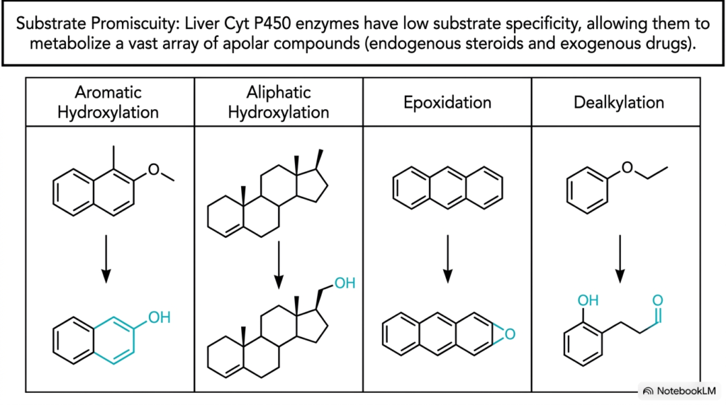
Unlike most enzymes that target a single specific substrate, hepatic Cytochrome P450 enzymes possess remarkable substrate promiscuity. This low substrate specificity allows a relatively small set of enzymes to recognize and metabolize thousands of structurally distinct apolar compounds. As the slide shows, this structural flexibility enables processing of both endogenous steroids and exogenous pharmaceuticals through several distinct chemical pathways.
The slide highlights four primary reaction types catalyzed by these versatile hemeprotein catalysts: aromatic hydroxylation, aliphatic hydroxylation, epoxidation, and dealkylation. In aromatic hydroxylation, a hydroxyl group attaches to a benzene ring system. Aliphatic hydroxylation targets hydrocarbon chains, converting hydrophobic alkyl groups into polar alcohols. Epoxidation converts double bonds into three-membered oxygen rings, whereas dealkylation removes methyl or ethyl groups from heteroatoms like oxygen or nitrogen.
This broad catalytic versatility is essential for human survival and xenobiotic metabolism. Because humans consume unpredictable combinations of natural and synthetic chemicals daily, Cytochrome P450 enzymes must adapt to varied chemical structures without requiring a unique enzyme for every molecule. Understanding substrate promiscuity helps pharmacology students predict drug-drug interactions, as multiple medications frequently compete for the same binding cavity within a single Cytochrome P450 isoform in liver microsomes. This broad binding capacity represents a primary evolutionary defense against environmental toxins.
Slide 6: Hydroxylation Pathways Catalyzed by Cytochrome P450
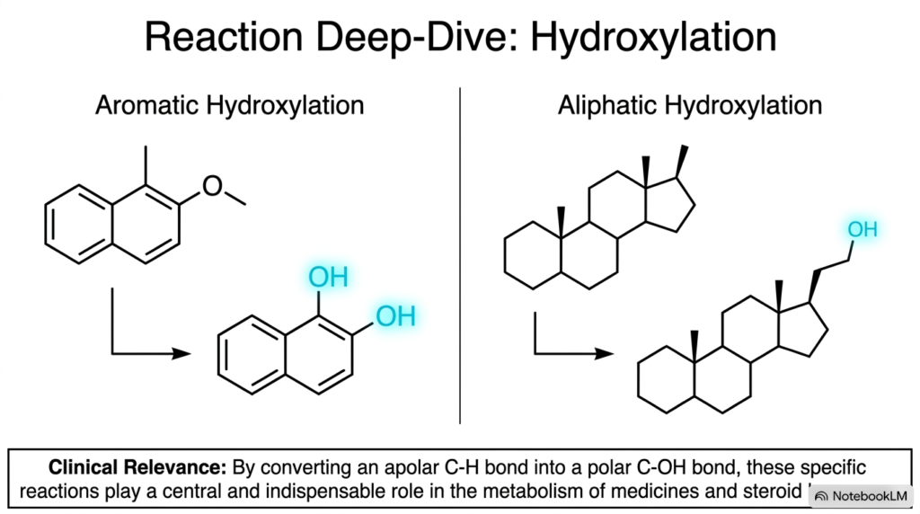
Hydroxylation is the most common chemical modification in Phase I drug metabolism. This slide provides a detailed look at aromatic and aliphatic hydroxylation pathways. In both cases, Cytochrome P450 converts an unreactive, nonpolar carbon-hydrogen bond into a polar carbon-hydroxyl group. This single-atom addition dramatically increases the target compound’s water solubility, facilitating its eventual clearance from the body.
Aromatic hydroxylation targets ring structures, converting compounds like naphthalene or pharmaceutical aromatics into phenolic derivatives. Aliphatic hydroxylation targets saturated carbon chains, such as the hydrocarbon side chains found on steroid molecules. In steroidogenesis, Cytochrome P450 hydroxylation of cholesterol derivatives is a mandatory step in synthesizing corticosteroid hormones and bile acids within adrenal and hepatic tissues.
The clinical relevance of these hydroxylation reactions cannot be overstated. By transforming apolar drugs into polar hydroxylated metabolites, Cytochrome P450 creates functional handles that Phase II conjugation enzymes can easily recognize. Furthermore, hydroxylated metabolites are often more water-soluble, allowing renal tubules to excrete them directly into urine. Students should recognize that Cytochrome P450 hydroxylation is the fundamental chemical bridge connecting lipophilic drug intake to successful metabolic clearance in human biochemistry. These crucial reactions directly safeguard human tissues and vital organs from dangerous drug accumulation and systemic toxicity.
Slide 7: Epoxidation and Toxic Activation by Cytochrome P450
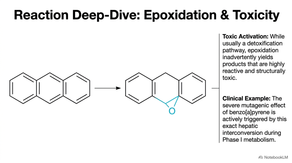
Although Phase I biotransformation is primarily a detoxification pathway, it can occasionally produce hazardous outcomes. This slide illustrates epoxidation, where Cytochrome P450 inserts an oxygen atom across a carbon-carbon double bond to form a reactive three-membered oxirane ring. While epoxide hydrolase can neutralize epoxides, highly reactive epoxides sometimes escape and cause severe cellular damage.
This phenomenon is called toxic activation, or bioactivation. Instead of inactivating a compound, Cytochrome P450 inadvertently transforms an inert pro-carcinogen into a potent electrophilic toxin. A classic clinical example shown on the slide is benzo[a]pyrene, a polycyclic aromatic hydrocarbon found in tobacco smoke and industrial exhaust. During hepatic metabolism, Cytochrome P450 converts benzo[a]pyrene into a reactive diol epoxide that covalently binds to DNA, initiating mutations and carcinogenesis.
Medical and toxicology students must appreciate this dual nature of drug metabolism. While Cytochrome P450 evolved to protect tissues from lipophilic poisons, the chemical intermediates generated during oxygen activation can occasionally react with cellular macromolecules. Understanding which chemical structures undergo toxic epoxidation allows medicinal chemists to design safer pharmaceuticals that avoid harmful bioactivation pathways during human drug metabolism. Thus, studying epoxidation helps scientists prevent adverse therapeutic outcomes, cellular injury, and organ toxicity in clinical practice.
Slide 8: Clinical Drug and Alcohol Interactions with Cytochrome P450
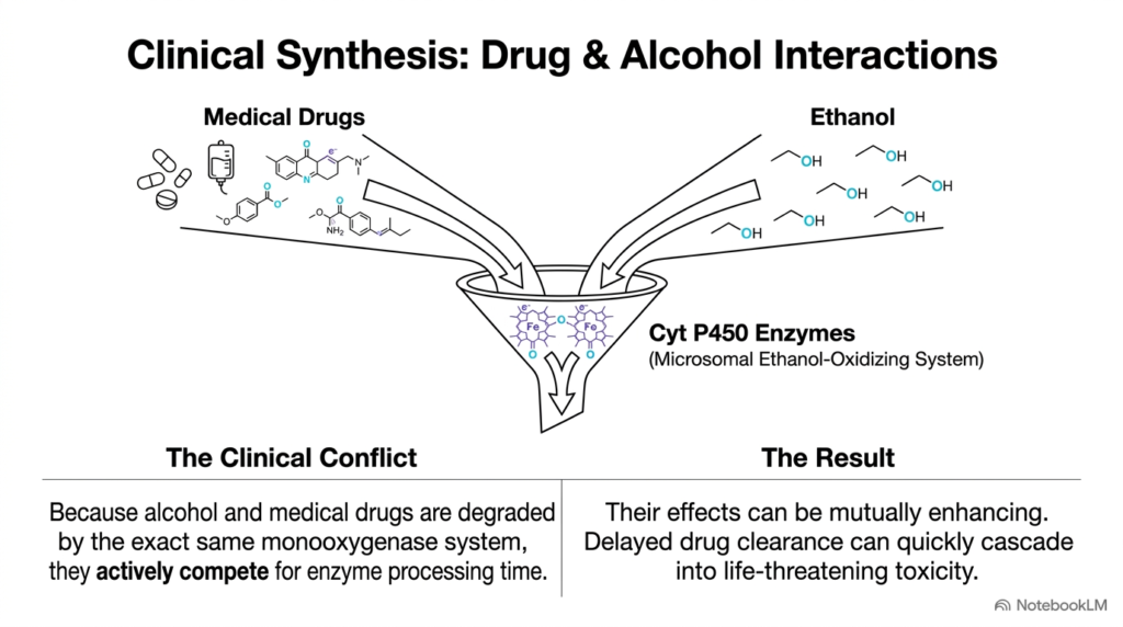
The broad substrate promiscuity of liver enzymes creates significant clinical challenges when patients consume multiple substances simultaneously. This slide illustrates the metabolic conflict between medical drugs and ethanol. Both classes of compounds are processed in hepatocytes by the same monooxygenase system, specifically the Microsomal Ethanol-Oxidizing System involving Cytochrome P450 isoforms such as CYP2E1.
When a patient consumes alcohol alongside prescription medications, ethanol and drug molecules compete directly for binding time within the Cytochrome P450 active site. This competitive inhibition reduces the clearance rate of prescription drugs, causing them to accumulate in the plasma to dangerous levels. Delayed drug clearance can quickly elevate therapeutic drug concentrations into toxic ranges, risking severe adverse drug events or organ damage.
Conversely, chronic alcohol intake increases expression of Cytochrome P450 enzymes. When sober, chronic drinkers metabolize certain medications unusually fast, rendering standard drug doses therapeutic failures. Understanding these competitive and inductive mechanisms is crucial for healthcare providers prescribing sedatives, anticoagulants, or analgesics. Pharmacy and medical students must recognize that Cytochrome P450 competition underlies many life-threatening clinical drug-alcohol interactions in emergency medicine and clinical pharmacology. Managing these interactions requires careful dosage adjustments, thorough drug screening, detailed patient histories, and close patient monitoring.
Slide 9: Overview of the Cytochrome P450 Catalytic Cycle
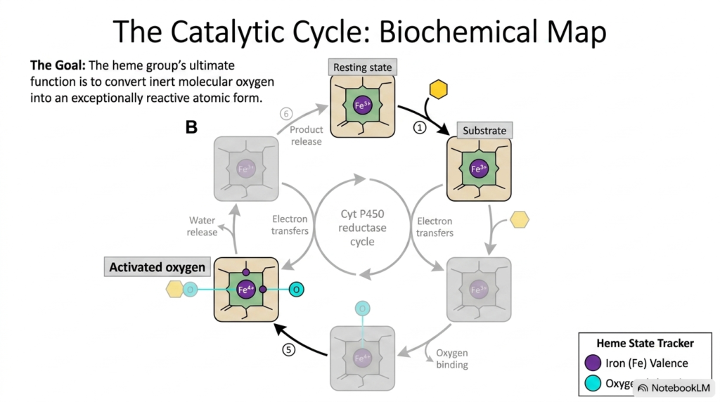
To perform difficult oxidation reactions on unreactive hydrocarbons, the enzyme follows a tightly regulated multi-step catalytic cycle. This slide provides a comprehensive biochemical map of the six-step reaction sequence executed by Cytochrome P450. The overarching goal of this complex cycle is converting stable molecular oxygen into an exceptionally reactive atomic oxygen species capable of modifying apolar substrates.
The cycle begins with the substrate entering the active site pocket near the resting ferric iron atom. Subsequent steps involve electron transfers from the Cytochrome P450 reductase partner, binding of diatomic oxygen, protonation, and water splitting to generate a high-valent iron-oxo intermediate. Once this activated oxygen inserts into the substrate, the oxygenated product dissociates, allowing the Cytochrome P450 protein to return to its original resting state.
Students should trace the iron valence changes tracked in the diagram, observing how the heme iron cycles between ferric, ferrous, and ferryl states. Precise coordination between electron transfer and oxygen binding prevents the premature release of harmful reactive oxygen species like superoxide or peroxide. Mastering this overarching Cytochrome P450 catalytic map gives students the conceptual foundation needed to examine each reaction step in detail on subsequent slides. This comprehensive cycle represents an essential landmark in biochemical education.
Slide 10: Substrate Binding and First Reduction in Cytochrome P450
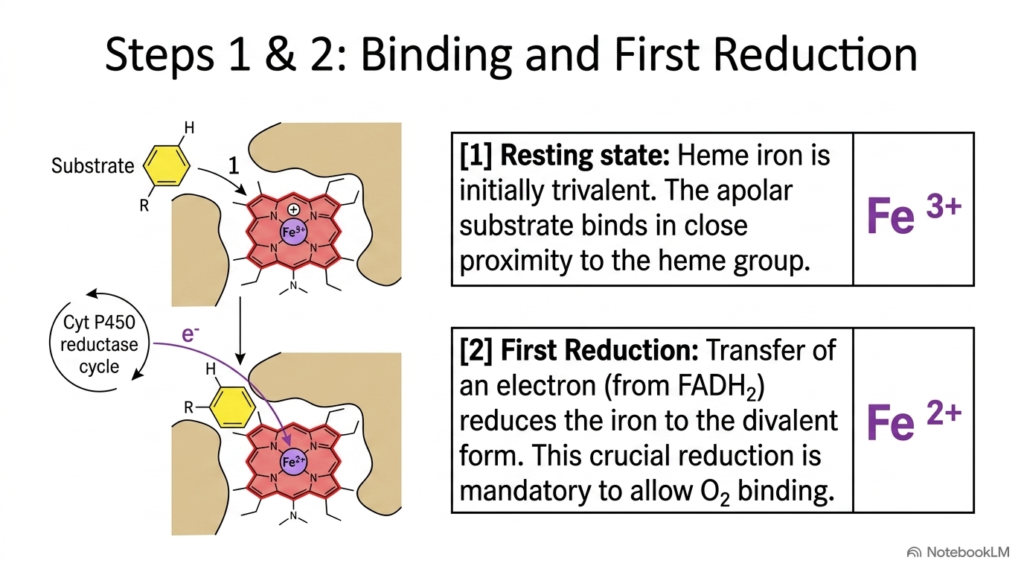
The first two steps of the catalytic cycle prepare the heme iron for oxygen binding. As depicted in Step 1, the enzyme begins in its resting state with the heme iron in the trivalent ferric valence state. The apolar substrate enters the hydrophobic active-site pocket of Cytochrome P450, positioning itself close to the heme ring. Substrate binding displaces a water molecule coordinated to the iron, inducing a spin-state transition that raises the heme group’s redox potential.
Step 2 represents the first reduction step of the cycle. An electron originates from NADPH and is delivered by the Cytochrome P450 reductase system. This electron transfer reduces the ferric iron to the divalent ferrous valence state. This initial reduction is a mandatory gatekeeping step; resting ferric heme cannot bind diatomic oxygen, whereas reduced ferrous iron has the electronic configuration required to coordinate molecular oxygen.
Biochemistry students should note how substrate binding logically precedes electron transfer. By making electron entry dependent on substrate presence, Cytochrome P450 avoids wasting reducing equivalents from NADPH when no target substrate is bound. This tight control highlights the efficiency of Cytochrome P450 catalysis, ensuring electron flow occurs only when a substrate is properly positioned for biotransformation.
Slide 11: Oxygen Binding and Peroxide Intermediate Formation in Cytochrome P450
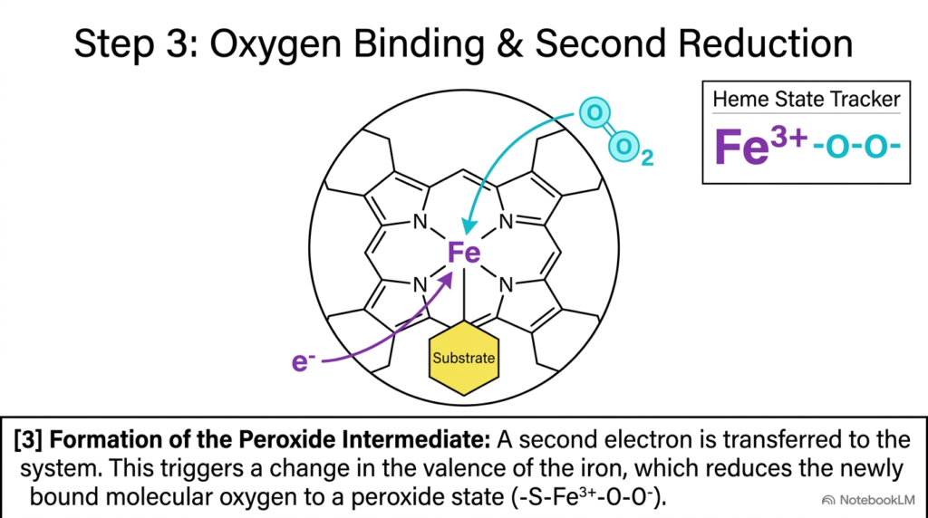
Once the heme iron reaches the reduced ferrous state, it can engage diatomic oxygen. In Step 3, a molecule of atmospheric oxygen enters the active site and coordinates to the ferrous iron of Cytochrome P450, forming a ferrous-dioxygen complex. Almost immediately, a second electron is transferred into the system from the Cytochrome P450 reductase pathway, driving a crucial electronic rearrangement.
This second electron transfer reduces the bound oxygen molecule to a peroxide state, creating a ferric-peroxo intermediate represented as an iron-peroxide species within the Cytochrome P450 active site. The iron atom returns to the trivalent ferric state while holding the reduced oxygen molecule tightly within the catalytic pocket. This intermediate is highly unstable and must be processed rapidly to prevent dangerous reactive oxygen species from leaking into the hepatocyte cytoplasm.
Students analyzing this step should recognize the crucial role of exact timing in enzyme catalysis. The Cytochrome P450 system must manage two sequential single-electron transfers while keeping atmospheric oxygen bound to the heme center. This peroxide intermediate is the immediate chemical precursor to oxygen-oxygen bond cleavage, setting the stage for generating the active oxygen species needed for substrate hydroxylation. Understanding this step illuminates how enzymes manage reactive intermediates safely.
Slide 12: Ferryl Radical Generation and Oxygen Activation in Cytochrome P450
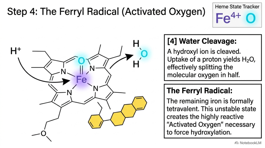
The fourth step of the catalytic cycle is the most energetically demanding. As shown in the diagram, two protons enter the active site pocket of Cytochrome P450 from the surrounding aqueous environment. Proton uptake causes heterolytic cleavage of the peroxide oxygen-oxygen bond. One oxygen atom combines with the two protons, is cleaved, and is safely released as a molecule of water.
The remaining oxygen atom remains double-bonded to the heme iron inside Cytochrome P450, forming an activated oxygen species known as the ferryl radical. In this high-valent state, the iron atom is formally tetravalent, paired with an oxo ligand and a porphyrin cation radical. This unstable, highly reactive intermediate provides the chemical motive force needed to break aliphatic carbon-hydrogen bonds that are otherwise chemically inert.
Understanding oxygen activation is essential for advanced biochemistry students. Unassisted carbon-hydrogen bonds are notoriously difficult to oxidize at body temperature. By channeling electron energy and protonation steps to generate a ferryl species, Cytochrome P450 creates one of the most powerful oxidizing agents in biological systems. This extraordinary activation mechanism allows Cytochrome P450 to hydroxylate recalcitrant drugs and steroids with remarkable speed and specificity. Thus, generating the ferryl radical represents the chemical core of biotransformation.
Slide 13: Substrate Oxygenation and Enzyme Reset in Cytochrome P450
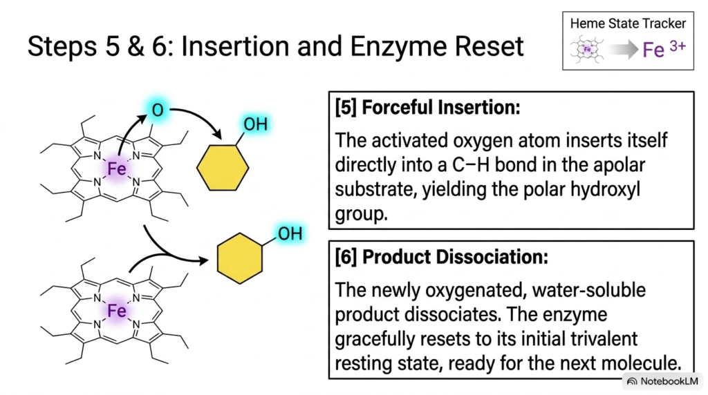
With the reactive ferryl radical established, the catalytic cycle reaches its chemical climax in Steps 5 and 6. Step 5 involves the forceful insertion of the activated oxygen atom directly into a carbon-hydrogen bond of the bound apolar substrate inside Cytochrome P450. Through a radical rebound mechanism, the ferryl oxygen abstracts a hydrogen atom from the substrate, forming a transient substrate radical that rapidly recombines with the iron-bound hydroxyl radical.
This oxygen insertion transforms the nonpolar substrate into a polar hydroxylated product. In Step 6, the newly oxygenated product dissociates from the active site pocket of Cytochrome P450. Because the product is now hydrophilic, its affinity for the hydrophobic binding pocket drops sharply, driving rapid dissociation into the surrounding aqueous cytosol.
Following product release, a water molecule re-enters the active site, and the heme iron inside Cytochrome P450 gracefully resets to its initial trivalent ferric resting state. The enzyme is now fully restored and ready to bind another substrate molecule. Students should appreciate the structural elegance of this complete cycle: Cytochrome P450 executes a dangerous oxidation reaction, releases a functional polar product, and resets itself without sustaining self-inflicted oxidative damage. This elegant reset completes the cycle smoothly and efficiently.
Slide 14: Complete Reaction Cycle and Synthesis of Cytochrome P450 Action
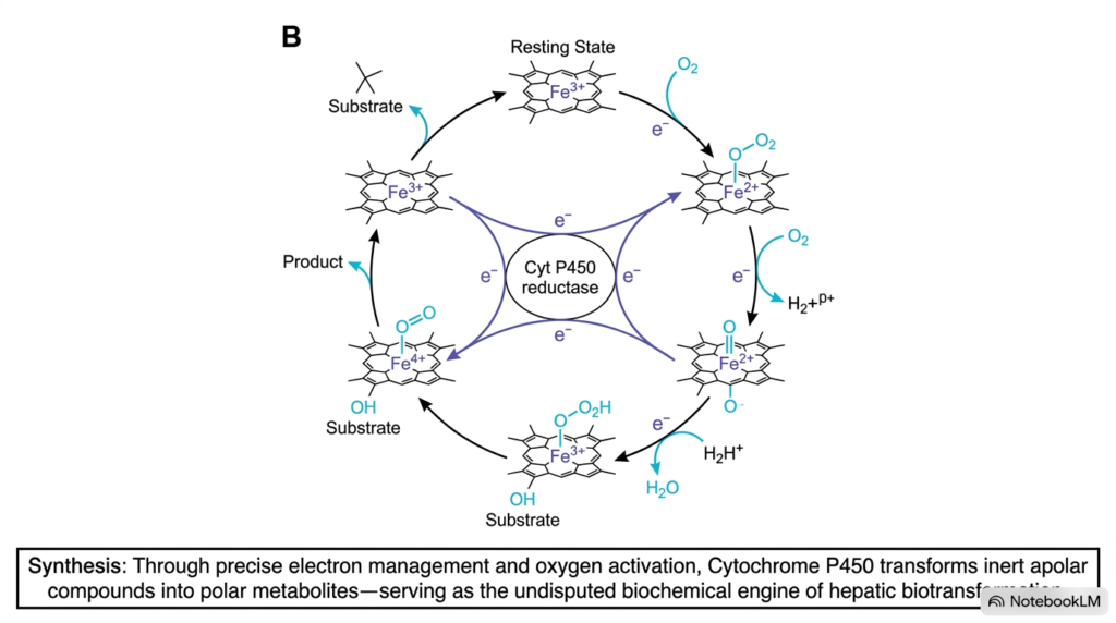
This final slide synthesizes the entire reaction cycle into an integrated biochemical framework. As shown in the comprehensive diagram, the Cytochrome P450 catalytic sequence coordinates electron supply from NADPH, precise iron redox transitions, and proton uptake to split oxygen. By managing these energetic inputs, the enzyme converts inert molecular oxygen into a powerful oxo-ferryl intermediate that readily oxidizes nonpolar target molecules.
The core physiological synthesis is straightforward: through precise electron management and oxygen activation, Cytochrome P450 transforms inert apolar compounds into polar metabolites. These Phase I modifications serve as the prerequisite for Phase II conjugation, rendering insoluble drugs, environmental toxins, and metabolic waste products water-soluble for effective excretion through urine or bile.
As the undisputed biochemical engine of hepatic biotransformation, Cytochrome P450 bridges fundamental chemistry and clinical medicine. Mastery of this system helps students understand drug pharmacokinetics, toxicological mechanisms, steroidogenesis, and dangerous drug interactions. Whether evaluating patient drug clearance or designing novel therapeutics, medical and science students will repeatedly encounter the vital impact of Cytochrome P450 enzymes throughout their clinical and scientific careers. Comprehensive understanding of this remarkable hepatic enzyme system is therefore essential for future healthcare professionals, practicing clinicians, biomedical researchers, and educators in modern medical practice.
Please read our Content Disclaimer Statement.
Check out our social media channels: