123. Renal Biochemistry: Mechanisms of Kidney Function and Urine Formation
Every second, human kidneys process an extraordinary volume of fluid to keep the body in chemical equilibrium. Without this continuous filtration, toxic metabolic waste would accumulate rapidly, leading to fatal systemic collapse. The provided slide deck explores the foundational molecular principles of renal function, breaking down complex physiological mechanisms into clear biochemical steps. For medical and university students, mastering these pathways is crucial for understanding systemic metabolic control, electrolyte balance, and kidney pathology. This comprehensive guide walks through each slide in sequence to illuminate the structural and functional marvels of the renal system.
Slide 01: Introduction to Renal Biochemistry and Molecular Kidney Function
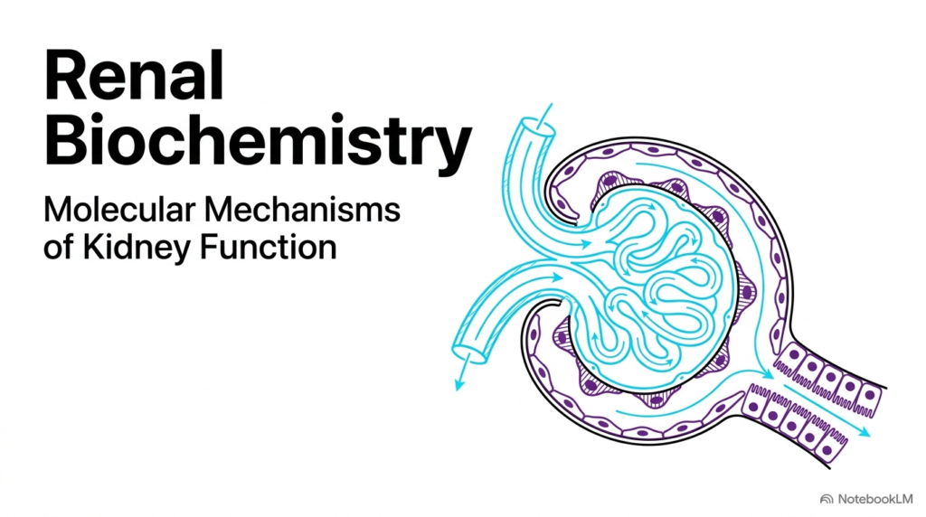
The opening slide introduces the foundational framework of Renal Biochemistry, highlighting the intricate molecular structures and vascular arrangements that drive human kidney function. At the heart of this system is the renal corpuscle, which contains the glomerulus enclosed by Bowman’s capsule. Blood enters through the afferent arteriole and circulates through specialized capillary networks, where hydrostatic pressure pushes fluid into the urinary space. This initial structural arrangement establishes the foundational physical architecture required for high-capacity blood filtration.
In the broader context of Renal Biochemistry, understanding this microvascular network reveals how structural adaptations enable rapid ultrafiltration without damaging delicate cellular components. Specialized fenestrated endothelial cells, the glomerular basement membrane, and podocyte foot processes work together to form a highly selective filtration barrier. As plasma flows through the glomerular tuft, essential plasma proteins remain safely inside the bloodstream while water, small solutes, and metabolic waste products enter the nephron tubule for further processing.
Ultimately, renal biochemistry shows how physical hemodynamic forces and specialized cellular anatomy work together to initiate urine formation. The initial fluid collected in Bowman’s space, known as primary filtrate, serves as the essential raw material for all downstream reabsorptive and secretory processes. Scholars studying renal function appreciate that this opening stage sets the baseline for systemic fluid balance, blood pressure regulation, and metabolic waste clearance across the human body.
Slide 02: The Four Pillars of Renal Biochemistry and Systemic Function
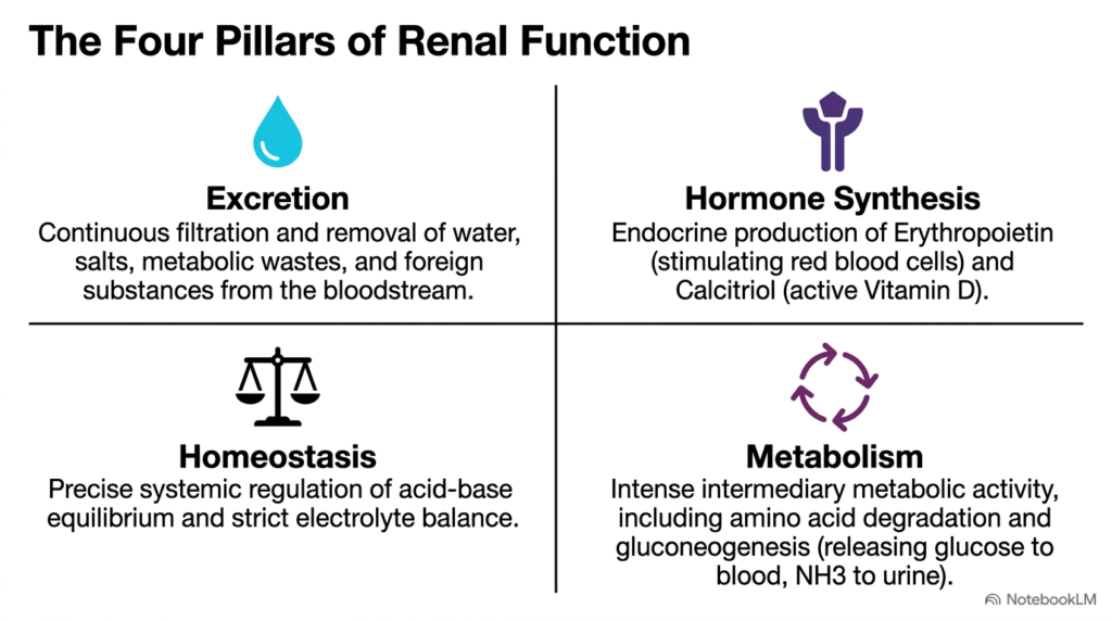
The second slide outlines the four fundamental pillars that define the operational scope of Renal Biochemistry across human physiology. Beyond simple filtration, the kidney functions as a dynamic metabolic organ with widespread endocrine, homeostatic, and excretory responsibilities. Excretion represents the most familiar pillar, involving the continuous filtration and elimination of metabolic nitrogenous wastes, excess salts, water, and exogenous foreign compounds. Through constant blood processing, the organ maintains a clean internal chemical environment.
A second critical pillar of Renal Biochemistry involves endocrine hormone synthesis, specifically the production of erythropoietin and calcitriol. Erythropoietin directly stimulates bone marrow stem cells to generate red blood cells, ensuring adequate tissue oxygenation throughout the body. At the same time, renal conversion of vitamin D precursors into active calcitriol regulates systemic calcium and phosphate metabolism, highlighting how renal tissues communicate with distal skeletal and endocrine systems.
The remaining pillars highlighted in Renal Biochemistry are systemic homeostasis and intermediary metabolism. Kidneys maintain precise acid-base equilibrium and strict electrolyte balance through controlled ion transport. Renal cells also carry out intense metabolic activity, including amino acid degradation and renal gluconeogenesis. During fasting, renal gluconeogenesis releases glucose into the bloodstream while diverting ammonia into urine, effectively linking energy production with systemic acid buffering.
Slide 03: The Hemodynamic Paradox in Renal Biochemistry
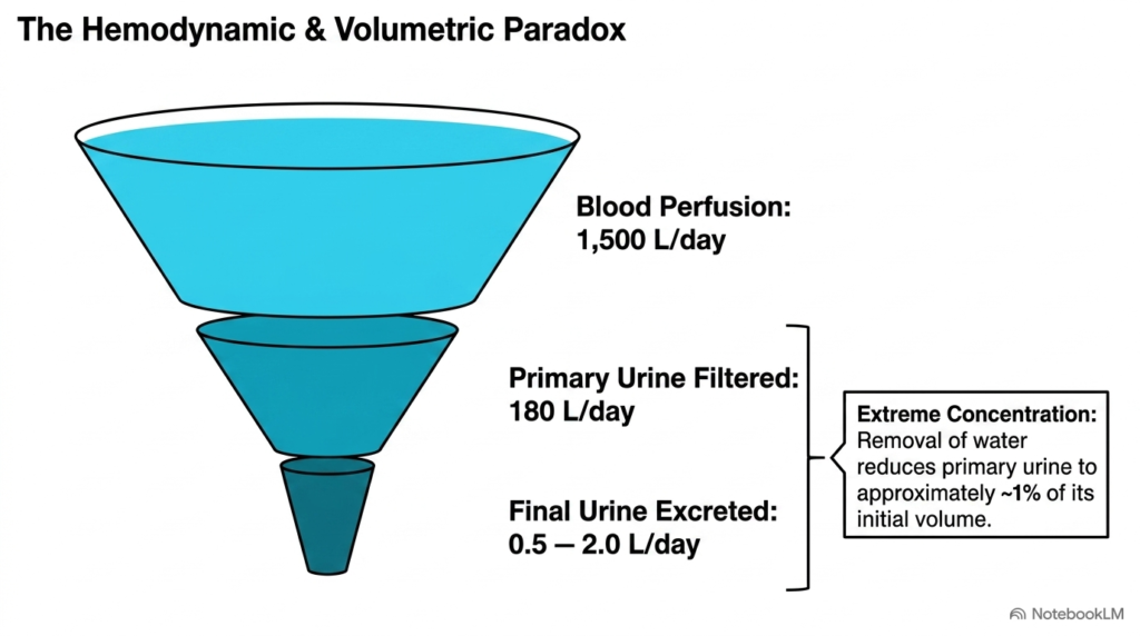
The third slide illustrates the remarkable volumetric funnel known as the hemodynamic paradox in renal biochemistry. Every single day, the human kidneys receive an astonishing blood perfusion volume of approximately 1,500 liters. From this massive cardiac throughput, the glomerular capillaries filter roughly 180 liters of primary urine into Bowman’s capsules. This immense volumetric throughput underscores the extraordinary physiological workload required to continuously screen circulating blood plasma for metabolic waste products and chemical toxins.
Despite generating 180 liters of primary filtrate daily, Renal Biochemistry reveals that the body excretes only 0.5 to 2.0 liters of final urine. This drastic volumetric reduction highlights the renal tubular system’s extreme concentrating capacity. Over 99% of filtered water, electrolytes, and essential plasma solutes are systematically recovered back into the surrounding peritubular capillaries. Without this intensive retrieval process, rapid dehydration and lethal nutrient depletion would occur within hours.
Understanding this volumetric paradox allows students of Renal Biochemistry to appreciate the high-efficiency architecture of the functional nephron. The massive initial filtration rate guarantees thorough clearance of small metabolic byproducts, while extensive tubular reabsorption selectively reclaims critical organic resources. This delicate balance between high-volume ultrafiltration and precise tubular recovery represents one of the most elegant examples of physiological bioenergetics and fluid conservation in human biology.
Slide 04: Anatomical Blueprint and Stages of Renal Biochemistry
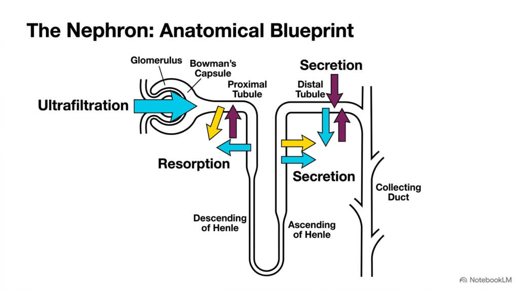
The fourth slide maps the nephron’s structural blueprint, detailing the sequential processing zones essential to Renal Biochemistry. The nephron is the kidney’s primary functional unit, divided into distinct anatomical segments with specialized transport capabilities. Processing begins at the renal corpuscle, comprising the glomerulus and Bowman’s capsule, where blood undergoes initial ultrafiltration. This non-selective fluid movement pushes water and small molecules out of capillaries into the nephron lumen.
Following ultrafiltration, Renal Biochemistry highlights the sequential roles of the proximal convoluted tubule, the loop of Henle, the distal tubule, and the collecting duct. The proximal tubule performs bulk reabsorption, reclaiming most filtered solutes and water back into the bloodstream. As filtrate flows through the descending and ascending limbs of the loop of Henle, countercurrent mechanisms concentrate the tubular fluid, establishing a steep osmotic gradient across the renal medulla.
The final stages of Renal Biochemistry occur within the distal convoluted tubule and collecting duct, where targeted secretion and regulated reabsorption take place. Tubule cells actively secrete waste ions and foreign molecules directly into the lumen while fine-tuning water and salt recovery. This spatial organization ensures that filtrate undergoes progressive chemical refinement, transforming raw primary filtrate into highly concentrated final urine ready for excretion.
Slide 05: Molecular Biophysics of Ultrafiltration in Renal Biochemistry
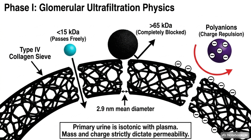
The fifth slide delves into Phase I of kidney function, examining the biophysical mechanisms of glomerular ultrafiltration in renal biochemistry. Filtration across the glomerular membrane acts as a molecular sieve governed by molecular mass, effective pore diameter, and electrical charge. This barrier’s structural foundation is a Type IV collagen meshwork with a mean pore diameter of about 2.9 nanometers. This tight physical matrix allows water and small solutes to pass freely while restricting larger structures.
A central rule in Renal Biochemistry is that molecules smaller than 15 kilodaltons pass effortlessly into Bowman’s space, whereas plasma proteins larger than 65 kilodaltons are completely blocked. Beyond simple size exclusion, electrostatic charge repulsion plays an equally vital role. The glomerular basement membrane and podocytes are densely coated with negatively charged polyanions. These fixed negative charges actively repel negatively charged plasma proteins, such as albumin, preventing them from entering the tubule filtrate.
Through these combined physical and electrostatic constraints, Renal Biochemistry explains why primary urine remains virtually protein-free yet isotonic with blood plasma. Small uncharged or positively charged molecules cross the filter effortlessly along pressure gradients. Understanding these filtration mechanics gives medical students a clear foundation for diagnosing clinical pathologies such as glomerulonephritis or nephrotic syndrome, where barrier damage causes abnormal protein leakage into urine.
Slide 06: Proximal Resorption Pathways and Pathology in Renal Biochemistry
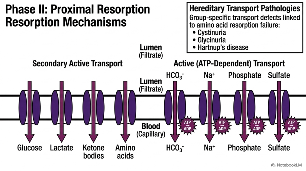
The sixth slide explores Phase II of renal processing, focusing on the extensive nutrient recovery mechanisms that define proximal tubule Renal Biochemistry. The proximal convoluted tubule reclaims most filtered organic nutrients, water, and electrolytes before they are lost in urine. Transport occurs across the luminal membrane through two primary modalities: secondary active transport driven by electrochemical sodium gradients, and primary active transport utilizing direct adenosine triphosphate hydrolysis.
For substrate recovery, Renal Biochemistry shows that glucose, lactate, ketone bodies, and amino acids are reabsorbed via specialized secondary active co-transporters. Sodium ions flowing down their concentration gradient power the entry of these vital organic nutrients back into tubule cells. At the same time, primary active ATP-dependent transporters directly drive reabsorption of inorganic ions, including bicarbonate, sodium, phosphate, and sulfate, ensuring complete recovery of essential systemic buffers and minerals into peritubular capillaries.
The clinical significance of Renal Biochemistry becomes strikingly evident when hereditary transport pathologies disrupt these membrane carrier proteins. Genetic defects in group-specific amino acid transporters lead to well-characterized metabolic disorders such as cystinuria, glycinuria, and Hartnup’s disease. In these conditions, unabsorbed amino acids accumulate in the tubular lumen, highlighting how localized transporter failures directly cause systemic metabolic dysfunction and urinary solute accumulation.
Slide 07: Hormonal Control of Distal Resorption in Renal Biochemistry
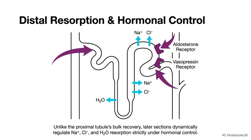
The seventh slide examines distal tubular resorption and endocrine regulation, detailing a highly dynamic phase of Renal Biochemistry. Unlike the proximal tubule, which conducts non-discriminatory bulk recovery of fluid and solutes, the distal convoluted tubule and collecting duct fine-tune electrolyte and water retention according to physiological demand. This precise regulatory control relies on specific endocrine hormone receptors located along the basolateral membranes of tubular epithelial cells.
A major focus in distal Renal Biochemistry is hormonal signaling mediated by aldosterone and vasopressin receptors. Aldosterone binds to intracellular mineralocorticoid receptors, upregulating sodium-potassium ATPase pumps and epithelial sodium channels to promote sodium and chloride reabsorption. Concurrently, vasopressin, also known as antidiuretic hormone, binds to basolateral membrane receptors to trigger aquaporin channel insertion, dramatically increasing water permeability and driving concentrated water recovery back into systemic blood circulation.
Through these endocrine feedback loops, Renal Biochemistry coordinates systemic blood volume, arterial blood pressure, and plasma osmolality. When dehydration, hemorrhage, or hypovolemia occurs, elevated circulating hormone levels maximize water and salt retention to restore cardiovascular stability. Medical students gain key insights into clinical pharmacology through these mechanisms, as diuretic medications target these specific distal transport proteins to manage hypertension, heart failure, and fluid overload disorders.
Slide 08: Mechanisms of Active Tubular Secretion in Renal Biochemistry
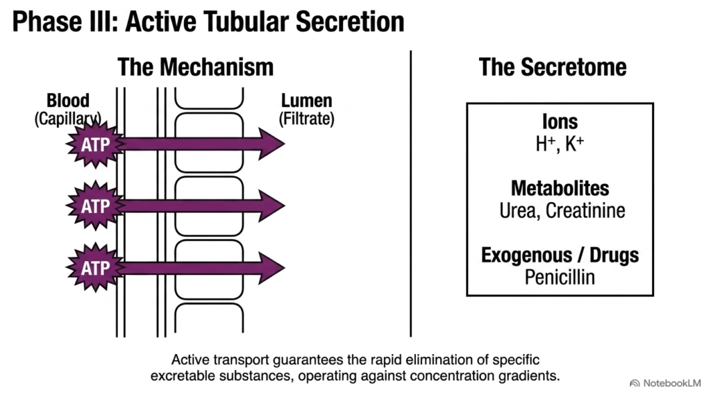
The eighth slide highlights Phase III of kidney function by analyzing the molecular mechanisms of active tubular secretion in renal biochemistry. While ultrafiltration and bulk reabsorption handle large fluid volumes, active tubular secretion provides a highly targeted elimination pathway. Specialized epithelial cells utilize primary and secondary active transport pumps, powered directly or indirectly by ATP hydrolysis, to transfer specific waste substances from peritubular capillary blood across the tubular epithelium into the lumen.
The wide diversity of secreted compounds underscores the vital homeostatic scope of Renal Biochemistry. Secreted substances, collectively referred to as the renal secretome, include essential inorganic ions such as hydrogen and potassium, which are moved against steep concentration gradients to maintain systemic acid-base balance and neuromuscular stability. Additionally, endogenous metabolic byproducts like urea and creatinine are actively transferred into the tubular fluid to guarantee complete waste clearance.
Furthermore, Renal Biochemistry plays a vital role in clinical pharmacology through the active secretion of exogenous drugs and environmental xenobiotics. Membrane-bound organic anion and organic cation transporters actively pump foreign chemical structures, including beta-lactam antibiotics like penicillin, from plasma into urine. Understanding these ATP-dependent secretory pathways allows researchers and clinicians to accurately predict drug clearance rates, optimize therapeutic drug dosing schedules, and prevent toxic accumulation.
Slide 09: Quantifying Function through Renal Clearance in Renal Biochemistry
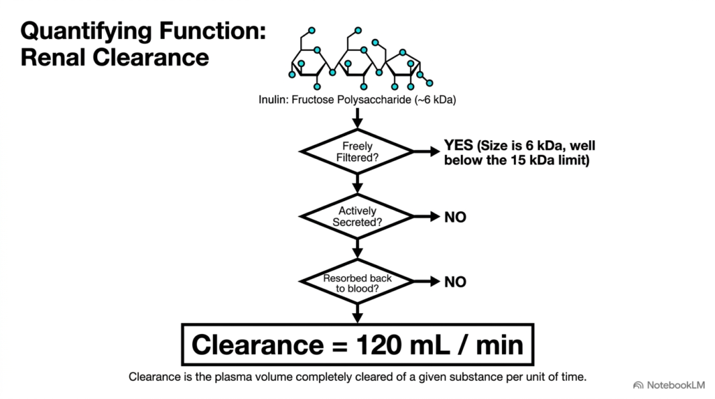
The ninth slide introduces renal clearance, a fundamental quantitative analytical tool in clinical Renal Biochemistry. Renal clearance is defined as the specific volume of blood plasma completely cleared of a given substance by the kidneys per unit of time, typically expressed in milliliters per minute. This relationship provides clinicians and medical researchers with an accurate quantitative measure of glomerular filtration rate and overall functional renal tissue mass.
In renal biochemistry, the fructose polysaccharide inulin is the gold-standard clinical marker for measuring true glomerular filtration rate. Inulin possesses an ideal molecular weight of approximately 6 kilodaltons, allowing it to be freely filtered across the glomerular barrier without structural restriction. Crucially, as inulin flows through the nephron tubules, it undergoes neither active tubular secretion nor reabsorption back into the surrounding blood capillary network.
Because inulin passes through the nephron entirely unmolested by tubular transport processes, its calculated clearance rate precisely equals the true filtration rate of approximately 120 milliliters per minute. Comparing the clearance of other compounds against inulin allows students of Renal Biochemistry to deduce their net nephron handling. A clearance below 120 milliliters per minute indicates net tubular reabsorption, whereas a clearance exceeding this baseline confirms net active tubular secretion.
Slide 10: Compartmentalized Energetics and Substrate Preferences in Renal Biochemistry
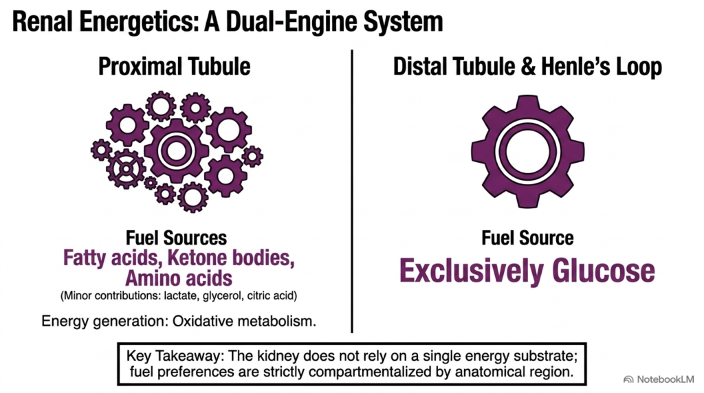
The tenth slide explores renal bioenergetics, presenting the dual-engine metabolic organization that uniquely characterizes Renal Biochemistry. To power continuous active ion transport against steep electrochemical gradients, the kidney consumes massive quantities of cellular energy. However, metabolic substrate utilization is not uniform across the nephron; rather, fuel preferences are strictly compartmentalized between distinct anatomical regions based on local tissue oxygen availability and specific enzymatic machinery.
In the proximal tubule, Renal Biochemistry relies almost exclusively on aerobic oxidative metabolism fueled by non-glucose substrates. Epithelial cells in this cortical region preferentially metabolize long-chain fatty acids, ketone bodies, and amino acids, with minor contributions from lactate, glycerol, and citric acid cycle intermediates. Because proximal tubules carry out intense bulk reabsorption, their high mitochondrial density requires abundant oxygen to sustain oxidative phosphorylation and maintain robust cellular ATP pools.
Conversely, the distal tubule and loop of Henle exhibit a strikingly different energy profile. These distal and medullary nephron segments rely exclusively on glucose as their primary metabolic fuel source, utilizing glycolytic pathways that function effectively under lower medullary oxygen tensions. This strict metabolic compartmentalization ensures that different nephron segments maintain optimal ATP production without competing for identical metabolic substrates under changing physiological conditions.
Slide 11: Intermediary Metabolism and Urinary Buffering in Renal Biochemistry
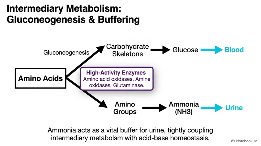
The eleventh slide details intermediary metabolic pathways, highlighting renal gluconeogenesis and urinary acid-base buffering in Renal Biochemistry. Specialized renal cortical epithelial cells possess remarkable enzymatic machinery that enables them to process organic amino acids during metabolic stress or extended physiological fasting. Through high-activity enzymes, including amino acid oxidases, amine oxidases, and glutaminase, tubular cells cleave amino acids into carbohydrate carbon skeletons and nitrogenous amino groups.
The carbon skeletons derived from deamination enter renal gluconeogenesis, an important metabolic pathway in Renal Biochemistry. During prolonged fasting, starvation, or severe metabolic acidosis, renal gluconeogenesis actively synthesizes new glucose molecules and releases them directly into systemic circulation to maintain plasma glucose levels. This crucial metabolic function positions the kidney alongside the liver as a primary human organ capable of endogenous glucose synthesis for systemic bioenergetic needs.
Simultaneously, Renal Biochemistry links this metabolic turnover to urinary acid-base buffering. The removed amino groups are converted into ammonia gas, which is actively secreted into the tubular lumen. Ammonia combines with filtered hydrogen ions to form ammonium, trapping excess protons in urine and preventing severe urinary acidification. This dual process elegantly couples nitrogen metabolism with systemic acid-base homeostasis, protecting the body against severe metabolic acidosis.
Slide 12: The Integrated Biochemical Matrix of Nephron Function in Renal Biochemistry
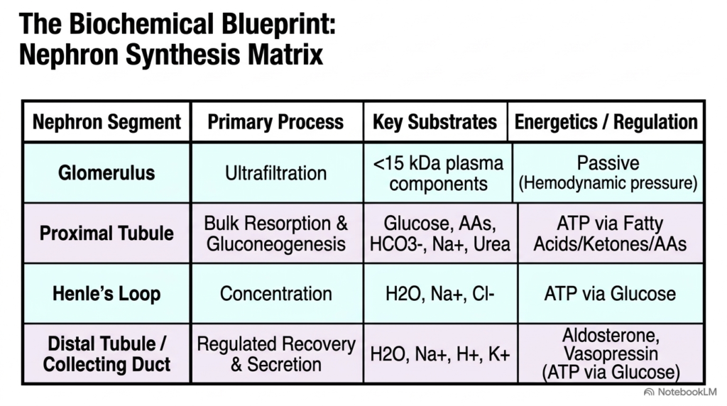
The final slide integrates all preceding physiological concepts into a comprehensive synthesis matrix defining Renal Biochemistry. This structural blueprint organizes nephron function across four distinct anatomical zones, outlining the primary physiological processes, essential transport substrates, metabolic fuel sources, and hormonal regulatory mechanisms operating in each region. This synthesis gives students a holistic view of how these compartmentalized molecular processes work in unison.
Starting at the glomerulus, Renal Biochemistry establishes passive ultrafiltration driven by hemodynamic pressure, allowing small solutes under 15 kilodaltons to enter the nephron. Fluid then enters the proximal tubule for bulk reabsorption and gluconeogenesis, where glucose, amino acids, bicarbonate, and sodium are recovered using ATP generated from fatty acid and ketone oxidation. Next, the loop of Henle concentrates filtrate by reabsorbing water, sodium, and chloride using ATP derived from glucose glycolysis.
Finally, Renal Biochemistry concludes in the distal tubule and collecting duct, where regulated recovery of water and sodium occurs alongside active hydrogen and potassium secretion. Controlled strictly by aldosterone and vasopressin signaling, these terminal segments fine-tune final urine composition. By mastering this integrated matrix, medical and graduate students gain a complete molecular understanding of how kidney physiology preserves human health, acid-base equilibrium, and metabolic homeostasis.
Please read our Content Disclaimer Statement.
Check out our social media channels: