151. Hydrophilic Hormone Mechanism of Action: Receptors and Pathways
Imagine delivering an urgent message inside a heavily fortified castle without ever stepping through the front gates. That is the exact challenge water-soluble hormones face every second in the human body. Because these chemical messengers cannot pass through lipid membranes, cells rely on complex relay systems to pass signals inward. This slide deck breaks down the hydrophilic hormone mechanism of action, exploring how cell-surface receptors, G proteins, and secondary messengers translate external signals into rapid cellular responses that control human metabolism and homeostasis.
Slide 1: Introduction to the Hydrophilic Hormone Mechanism of Action
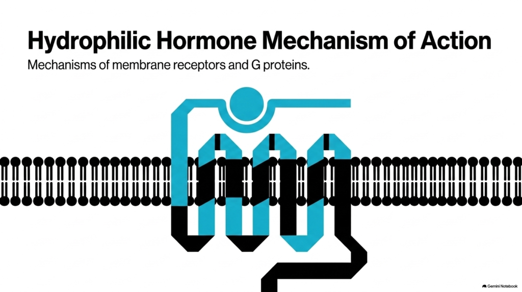
Cells depend on molecular signals to coordinate vital physiological tasks across different organ systems. The hydrophilic hormone mechanism of action explains how peptide and amine hormones communicate with target tissues without physically entering the cytoplasm. Because water-soluble molecules cannot cross the hydrophobic lipid bilayer, they must bind to specialized receptor domains exposed on the outer surface of the plasma membrane. This binding interaction initiates the hydrophilic hormone mechanism of action, converting an extracellular chemical message into an immediate internal conformational change. Biochemistry students must appreciate that these membrane receptors act as translators, bridging the extracellular matrix with the cell interior.
At its core, the hydrophilic hormone mechanism of action ensures that vital physiological instructions, such as insulin-mediated glucose regulation or epinephrine-driven stress responses, occur rapidly and reversibly. Membrane-bound receptors recognize specific ligands with high affinity, preventing accidental cellular activation by non-target molecules. Once a hormone binds its receptor, the receptor’s structural change triggers downstream cascades that modify enzyme activity or ion fluxes within milliseconds. Understanding the hydrophilic hormone mechanism of action provides the foundational framework for studying human endocrinology, pharmacology, and cellular signaling networks in medical education.
Slide 2: The Biochemical Barrier in the Hydrophilic Hormone Mechanism of Action
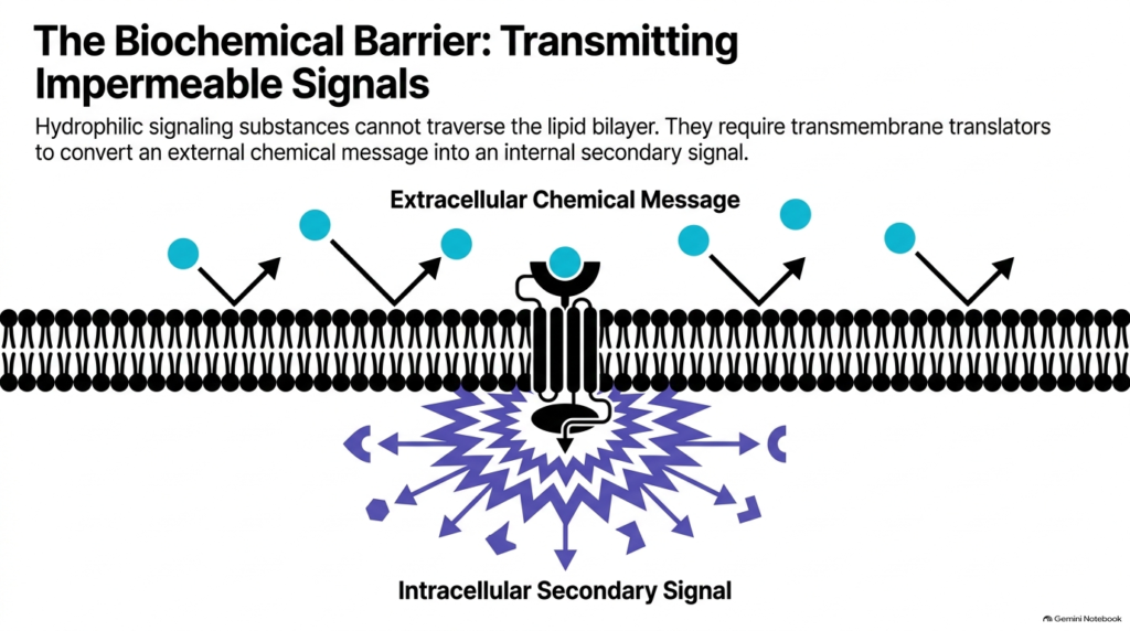
The plasma membrane presents a formidable biochemical barrier to circulating signaling molecules due to its hydrophobic phospholipid core. The hydrophilic hormone mechanism of action begins when water-soluble signaling substances, such as catecholamines, peptide hormones, and local mediators, encounter this impermeable lipid bilayer. Unlike lipophilic steroid hormones that diffuse freely into the cytoplasm, hydrophilic molecules bounce off the nonpolar fatty acid tails. Consequently, hydrophilic hormone action relies entirely on integral transmembrane proteins to convert an external chemical message into a robust intracellular secondary signal without physically transferring the hormone across the cell border.
Transmembrane receptor proteins span both leaflets of the lipid bilayer, positioning a specialized ligand-binding domain externally and a functional signaling domain in the cytoplasm. In this hydrophilic hormone mechanism of action, this key spatial arrangement lets cells detect environmental chemical signals instantly while keeping their internal metabolic contents isolated and protected. When a hormone attaches to its receptor, the induced structural shift propagates directly through the membrane helix, activating intracellular messenger systems. This physical separation shows why the hydrophilic hormone mechanism of action enables rapid cellular responses, as target cells do not need to wait for internal hormone transport, nuclear entry, or slow gene transcription.
Slide 3: Taxonomy of Membrane Receptors in the Hydrophilic Hormone Mechanism of Action
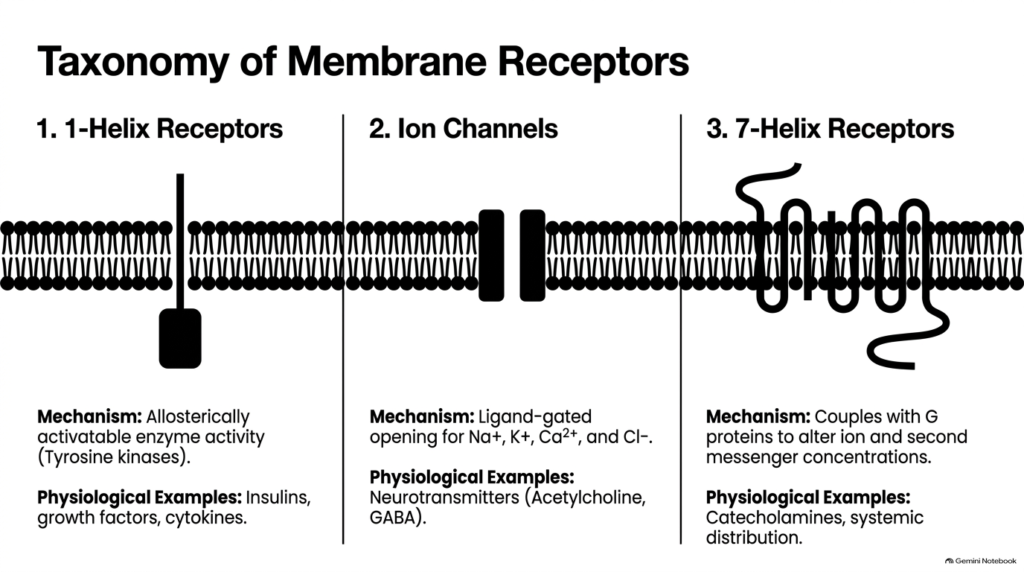
To meet diverse physiological signaling demands, target cells use three distinct classes of cell-surface receptors. In the hydrophilic hormone mechanism of action, these receptor classes include single-transmembrane 1-helix receptors, multimeric ligand-gated ion channels, and 7-helix serpentine receptors. Each structural class uses a distinct molecular method to span the plasma membrane and transmit external chemical messages inward. Single-helix receptors possess intrinsic enzymatic activity, whereas ion channels regulate direct electrochemical fluxes across membranes. Meanwhile, 7-helix receptors partner with intermediary regulatory proteins. Analyzing this structural diversity reveals how the hydrophilic hormone mechanism of action yields customized signaling kinetics for distinct biological tissues.
Specific signaling molecules select receptor classes based on the required response speeds and metabolic outcomes. For instance, neurotransmitters like acetylcholine use ion channels to produce millisecond electrical changes, while systemic endocrine signals like epinephrine use 7-helix receptors to trigger broad enzymatic cascades. In the hydrophilic hormone mechanism of action, 1-helix receptors primarily process metabolic growth factors like insulin, driving nutrient storage and cell proliferation. By organizing membrane receptors into these three functional families, the hydrophilic hormone mechanism of action provides cells with versatile molecular toolkits for regulating both rapid physiological adjustments and long-term developmental processes.
Slide 4: 1-Helix Receptors and Kinase Pathways in the Hydrophilic Hormone Mechanism of Action
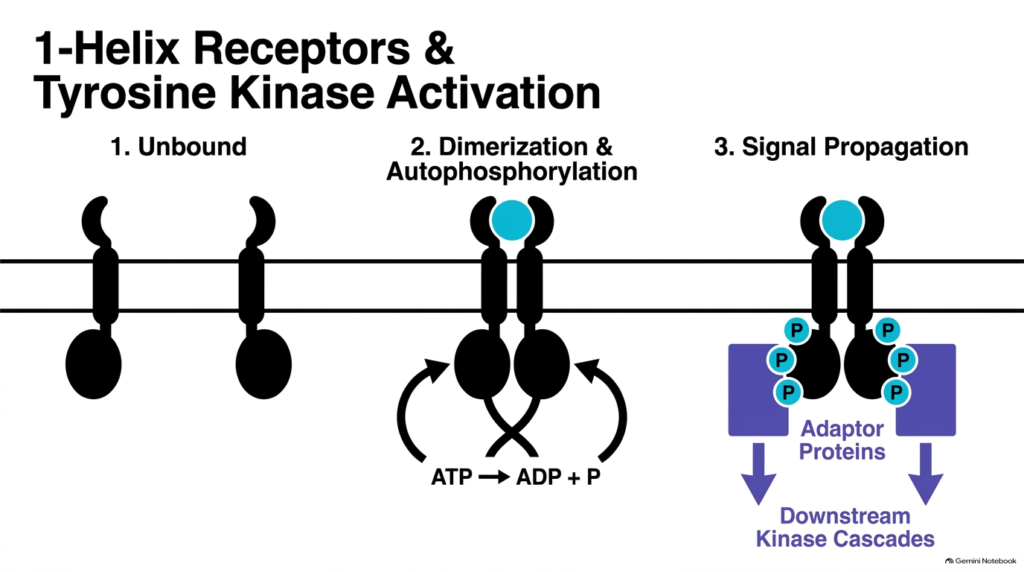
Single-pass transmembrane proteins, known as 1-helix receptors, represent a vital structural class of cell-surface transducers. In the hydrophilic hormone mechanism of action, these receptors feature an external ligand-binding domain, a single membrane-spanning alpha helix, and an internal enzymatic domain that typically functions as a tyrosine kinase. In the resting unbound state, these monomeric receptors float independently within the fluid lipid bilayer. However, when insulin or growth factors initiate the hydrophilic hormone mechanism of action, hormone binding induces two adjacent receptor monomers to dimerize, bringing their internal enzymatic kinase domains into close physical contact inside the cell.
This close physical proximity triggers autophosphorylation, where each active kinase domain uses ATP to cross-phosphorylate specific tyrosine residues on its partner receptor chain. In this hydrophilic hormone mechanism of action, these newly phosphorylated tyrosine sites serve as high-affinity docking anchors for specialized cytosolic adaptor proteins. Once docked, adaptor proteins recruit additional enzymes to activate downstream protein kinase cascades, relaying biochemical signals deep into the cytoplasm. Through this organized phosphorylation cascade, the hydrophilic hormone mechanism of action enables insulin to govern glucose storage and growth factors to stimulate cell proliferation, showing how structural dimerization yields metabolic control.
Slide 5: Ligand-Gated Ion Channels in the Hydrophilic Hormone Mechanism of Action
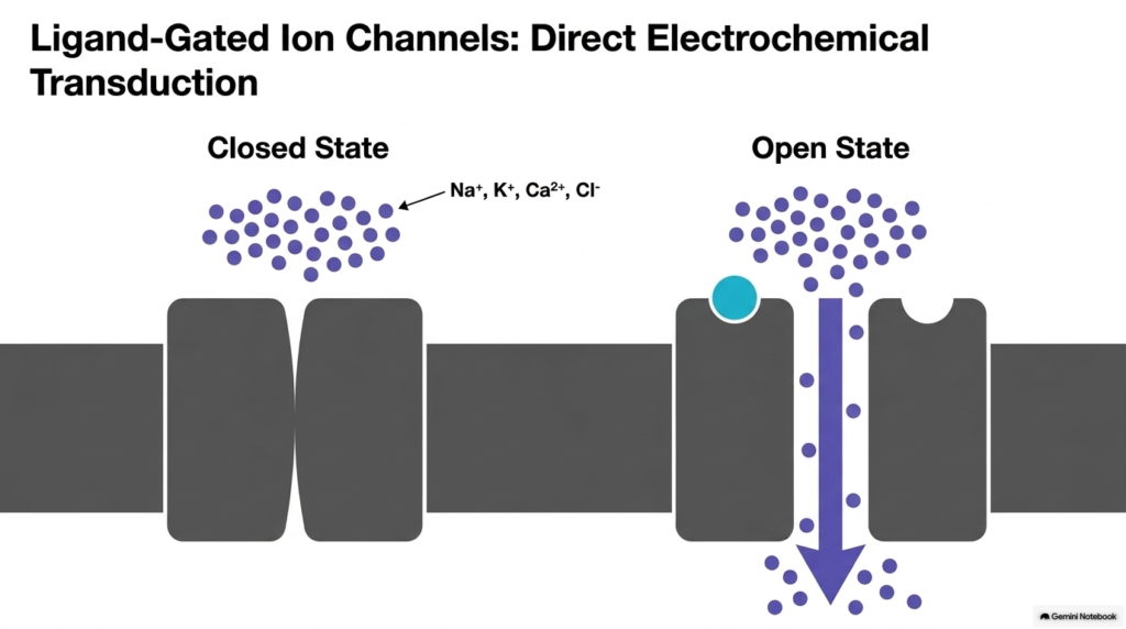
When target cells require instant physiological communication, ligand-gated ion channels serve as the primary membrane transducers. In the hydrophilic hormone mechanism of action, these multimeric protein complexes form a central transmembrane pore that remains tightly closed without signaling ligands. Hydrophilic neurotransmitters, such as acetylcholine and gamma-aminobutyric acid, bind directly to specialized extracellular receptor sites on the channel complex. This binding event triggers the hydrophilic hormone mechanism of action, inducing an immediate conformational shift that rotates the protein subunits and opens the central pore to specific inorganic ions within microseconds.
Once opened, inorganic ions like sodium, potassium, calcium, or chloride flow rapidly down their respective concentration and electrical gradients. Within the hydrophilic hormone mechanism of action, this massive ion movement alters the membrane potential across the plasma membrane, generating immediate cellular responses such as skeletal muscle contraction or nerve impulse propagation. Unlike enzyme-linked pathways that rely on multi-step intracellular biochemical cascades, the hydrophilic hormone mechanism of action operating through ion channels provides direct electrochemical transduction. This direct physical mechanism enables central nervous systems and neuromuscular junctions to process vital information and control physical movements at extraordinary physiological speeds.
Slide 6: Structural Anatomy of 7-Helix Receptors in the Hydrophilic Hormone Mechanism of Action
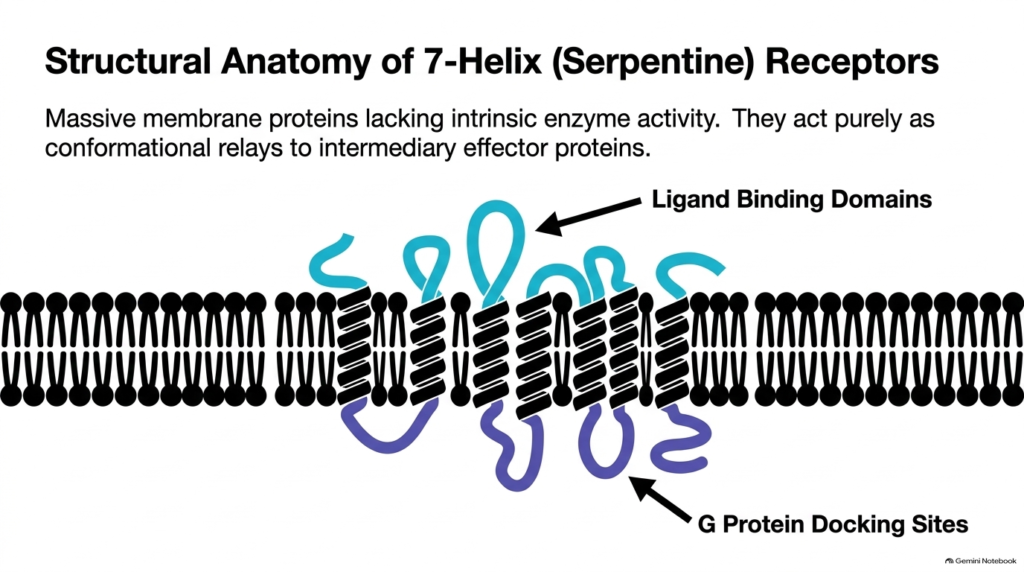
The most abundant and versatile cell-surface receptors in human physiology are 7-helix serpentine receptors, also known as G protein-coupled receptors. In the hydrophilic hormone mechanism of action, these massive membrane proteins consist of a single polypeptide chain that threads back and forth across the lipid bilayer seven times, forming seven hydrophobic alpha-helical segments. The extracellular loops fold into specialized ligand-binding domains designed to recognize specific hormones. Meanwhile, the intracellular loops form functional docking sites for intracellular partner proteins. The hydrophilic hormone mechanism of action relies on this serpentine architecture to transmit structural changes across the membrane barrier.
Unlike 1-helix receptors, serpentine receptors possess no intrinsic enzymatic catalytic activity on their cytoplasmic tails. Instead, the hydrophilic hormone mechanism of action uses these receptors strictly as conformational relays. When a signaling molecule like epinephrine binds the extracellular pocket, the seven transmembrane helices shift relative to one another. This helical movement alters the shape of the cytoplasmic loops, exposing a high-affinity docking surface for heterotrimeric G proteins. Through this mechanism, the hydrophilic hormone mechanism of action connects external ligand recognition to internal signal transduction without requiring the receptor itself to catalyze chemical reactions.
Slide 7: Anatomy and Classification of G Proteins in the Hydrophilic Hormone Mechanism of Action
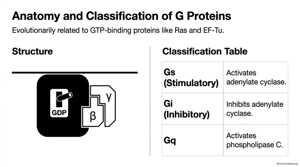
Heterotrimeric G proteins serve as the essential middle managers that link 7-helix receptors to internal effector enzymes. In the hydrophilic hormone mechanism of action, a G protein complex consists of three distinct polypeptide subunits: alpha, beta, and gamma. The alpha subunit contains a nucleotide-binding pocket that holds guanosine diphosphate in the resting state, along with intrinsic enzymatic activity to hydrolyze nucleotides. The beta and gamma subunits form a tightly associated dimer that anchors the complex to the inner membrane surface. In the hydrophilic hormone mechanism of action, these G protein switches share evolutionary ancestry with cellular proteins like Ras and EF-Tu.
Cells express multiple families of G protein alpha subunits to produce distinct physiological outcomes upon receptor activation. In the hydrophilic hormone mechanism of action, stimulatory G proteins activate adenylate cyclase to increase second-messenger levels, while inhibitory G proteins suppress adenylate cyclase activity. Additionally, the Gq family activates phospholipase C to trigger calcium release and lipid signaling. By coupling specific 7-helix receptors to particular G protein classes, the hydrophilic hormone mechanism of action lets hormones fine-tune intracellular biochemistry, enabling tissues to accelerate or slow specific metabolic pathways depending on bodily demands.
Slide 8: G Protein Activation and Nucleotide Exchange in the Hydrophilic Hormone Mechanism of Action
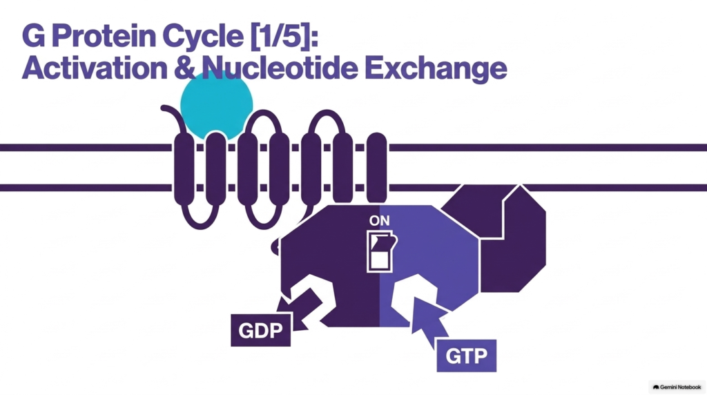
The transformation of an inactive G protein into an active signal transducer represents a central regulatory step in cellular communication. In the hydrophilic hormone mechanism of action, an inactive G protein heterotrimer carries a bound guanosine diphosphate molecule on its alpha subunit. When a hormone binds to a neighboring 7-helix receptor, the receptor undergoes a structural shift that allows it to bind the inactive G protein complex in the cytoplasm. This docking interaction initiates the hydrophilic hormone mechanism of action by forcing the alpha subunit to open its nucleotide pocket and release bound guanosine diphosphate into the surrounding cytoplasm.
Because intracellular concentrations of guanosine triphosphate are significantly higher than guanosine diphosphate, a new guanosine triphosphate molecule quickly enters the empty binding pocket. In the hydrophilic hormone mechanism of action, guanosine triphosphate binding acts as a molecular “on switch,” inducing a conformational change throughout the alpha subunit. This structural change dramatically reduces the alpha subunit’s affinity for both the receptor and the beta-gamma dimer. Consequently, the hydrophilic hormone mechanism of action converts an external hormone docking event into an active, energy-loaded protein subunit ready to travel along the membrane and activate effector target enzymes.
Slide 9: Dissociation and Effector Binding in the Hydrophilic Hormone Mechanism of Action
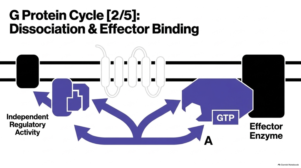
After nucleotide exchange, the G protein heterotrimer splits into two distinct functional signaling units. In the hydrophilic hormone mechanism of action, the guanosine triphosphate-bound alpha subunit detaches from the beta-gamma subunit dimer and disengages from the 7-helix receptor. Both liberated components remain firmly anchored to the plasma membrane by lipid tails, allowing them to diffuse laterally within the fluid lipid plane. In the hydrophilic hormone mechanism of action, these separate subunits engage distinct downstream effector target proteins simultaneously, amplifying the original hormonal message and diversifying the cellular response across multiple intracellular pathways.
The activated alpha subunit binds directly to primary effector enzymes, such as adenylate cyclase or phospholipase C, dramatically altering their catalytic speed. Simultaneously, in the hydrophilic hormone mechanism of action, the freed beta-gamma dimer exhibits independent regulatory activity, modulating specific ion channels or recruiting specialized receptor kinases, such as beta-adrenergic receptor kinase, to the membrane. This dual-pathway signaling clearly demonstrates how the hydrophilic hormone mechanism of action splits a single receptor activation event into complementary regulatory streams. By targeting multiple membrane effectors at once, the cell ensures a highly coordinated biological response.
Slide 10: The cAMP Amplification Cascade in the Hydrophilic Hormone Mechanism of Action
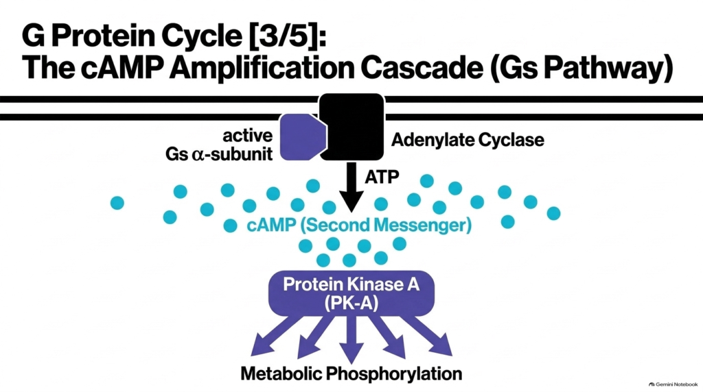
When an activated stimulatory G protein alpha subunit binds to adenylate cyclase, it triggers a massive second messenger amplification cascade. In the hydrophilic hormone mechanism of action, adenylate cyclase acts as an effector enzyme that converts cellular adenosine triphosphate into cyclic adenosine monophosphate while releasing pyrophosphate. A single active enzyme can synthesize thousands of cyclic adenosine monophosphate molecules every second. The hydrophilic hormone mechanism of action relies on this rapid enzymatic production to flood the cytoplasm with secondary signals, broadcasting the message throughout the cell far beyond the membrane boundary where the hormone docked.
Accumulated cyclic adenosine monophosphate binds to the regulatory subunits of protein kinase A, releasing its active catalytic subunits. In this hydrophilic mechanism of action, activated protein kinase A phosphorylates specific serine and threonine residues on target metabolic enzymes, switching cellular pathways on or off. For example, during epinephrine signaling, protein kinase A stimulates glycogen breakdown while inhibiting glycogen synthesis. Through this enzymatic amplification, the hydrophilic hormone mechanism of action allows tiny nanomolar concentrations of circulating hormones to produce massive millimolar shifts in intracellular metabolites, ensuring rapid physiological responses.
Slide 11: Alternative Second Messenger Pathways in the Hydrophilic Hormone Mechanism of Action
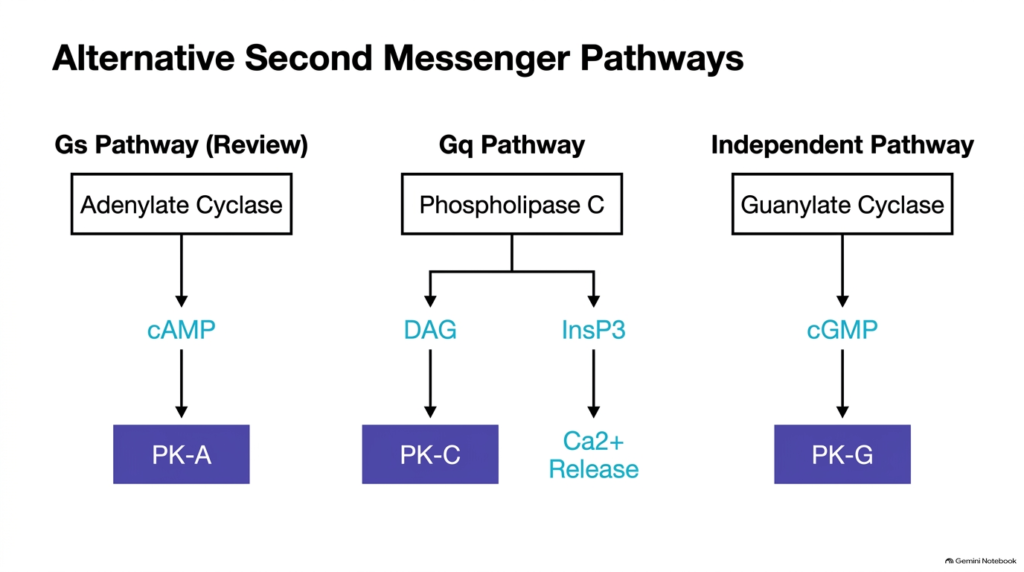
Beyond the classical cyclic adenosine monophosphate pathway, cells utilize alternative second messenger systems to execute complex regulatory functions. In the hydrophilic hormone mechanism of action, Gq activation targets the effector enzyme phospholipase C instead of adenylate cyclase. Phospholipase C cleaves membrane phosphatidylinositol bisphosphate into two distinct secondary messengers: membrane-bound diacylglycerol and soluble inositol trisphosphate. In the hydrophilic hormone mechanism of action, inositol trisphosphate opens calcium channels on the endoplasmic reticulum, releasing calcium ions into the cytoplasm to activate calcium-dependent enzymes, while membrane diacylglycerol activates protein kinase C.
Additionally, certain signaling pathways operate through guanylate cyclase to generate cyclic guanosine monophosphate, which activates protein kinase G to regulate vascular tone and fluid balance. In the hydrophilic hormone mechanism of action, these parallel second messenger networks demonstrate remarkable signaling diversity. Cells can integrate inputs from multiple hormone receptors simultaneously, balancing phosphorylation events across protein kinase A, protein kinase C, and protein kinase G. Understanding these alternative pathways within the hydrophilic hormone mechanism of action explains how different tissues produce customized physiological responses, such as smooth muscle contraction, vasodilation, or specialized glandular secretion.
Slide 12: Receptor Desensitization via Arrestin in the Hydrophilic Hormone Mechanism of Action
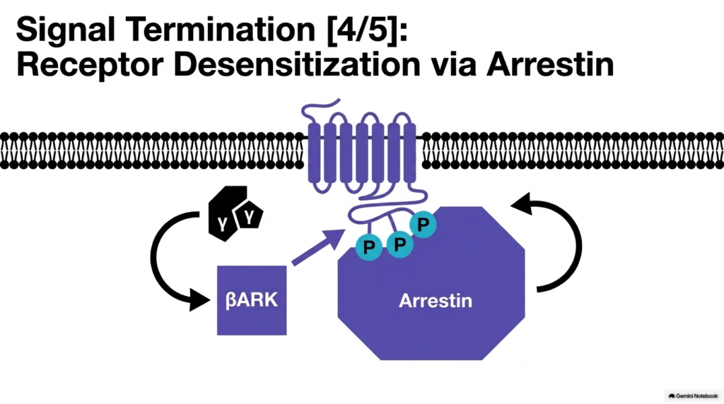
Without robust desensitization mechanisms, continuous hormone exposure would lead to dangerous cellular overstimulation. In the hydrophilic hormone mechanism of action, signal termination begins at the receptor level shortly after activation. When the beta-gamma subunit dimer releases from the G protein complex, it recruits a specialized enzyme called beta-adrenergic receptor kinase to the plasma membrane. This kinase phosphorylates specific serine and threonine residues on the cytoplasmic tail of the active 7-helix receptor. In the hydrophilic hormone mechanism of action, this receptor phosphorylation serves as a distinct chemical tag signaling that the receptor has completed its transmission duty.
Phosphorylation reduces the receptor’s affinity for G proteins and attracts a blocking protein named arrestin. Within the hydrophilic hormone mechanism of action, arrestin binds tightly to the phosphorylated cytoplasmic loops, physically preventing further G protein coupling. This steric hindrance halts secondary messenger generation even if the hormone remains attached externally. Furthermore, arrestin targets the receptor for endocytosis, pulling it into internal vesicles for degradation or recycling. Through desensitization and receptor internalization, the hydrophilic hormone mechanism of action protects target cells from chronic overstimulation, maintaining physiological homeostatic sensitivity to fluctuating blood hormone levels.
Slide 13: Intrinsic GTPase Activity in the Hydrophilic Hormone Mechanism of Action
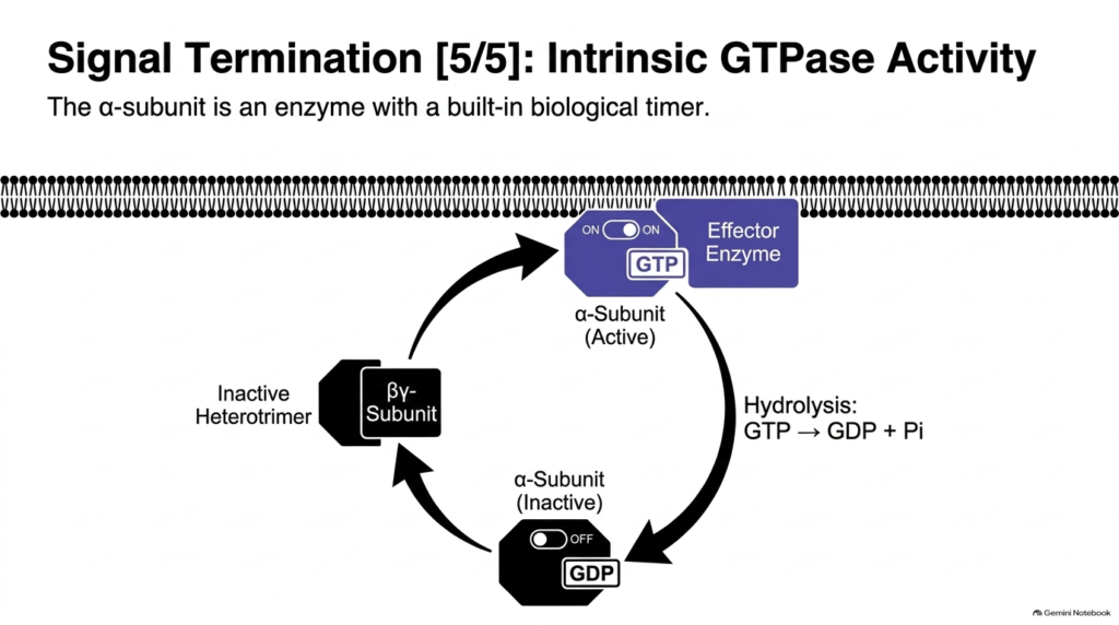
To ensure that cellular activation remains strictly temporary, G protein alpha subunits feature a built-in biological timer. In the hydrophilic hormone mechanism of action, the alpha subunit functions as a GTPase enzyme that hydrolyzes its bound guanosine triphosphate into guanosine diphosphate and inorganic phosphate. This intrinsic enzymatic reaction occurs automatically within seconds or minutes of activation. The hydrophilic hormone mechanism of action relies on this self-cleaving timer to ensure signal transmission shuts off, preventing runaway enzymatic activity inside the cell even if upstream receptor desensitization is delayed or absent.
Once guanosine triphosphate is cleaved into guanosine diphosphate, the alpha subunit shifts back to its inactive conformation. In the hydrophilic hormone mechanism of action, this structural change causes the alpha subunit to release the effector enzyme and reassociate with the beta-gamma dimer. Rejoining the beta-gamma subunits restores the inactive heterotrimeric G protein complex, preparing it for future signaling events. Through this self-limiting GTPase cycle, the hydrophilic hormone mechanism of action provides precise temporal control over signal duration, ensuring that intracellular metabolic shifts correspond accurately to the real-time presence of circulating hormones.
Slide 14: Downstream Cellular Consequences in the Hydrophilic Hormone Mechanism of Action
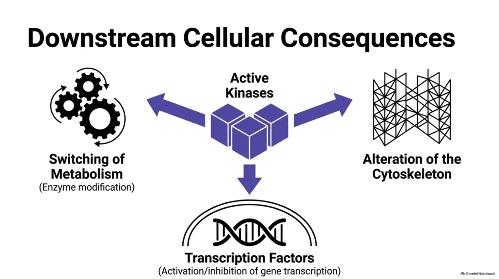
The ultimate objective of signal transduction is altering cell physiology to meet systemic biological needs. In the hydrophilic hormone mechanism of action, second messengers activate specific protein kinases that phosphorylate target proteins throughout the cytoplasm. These phosphorylation events cause immediate metabolic switching, rapidly activating catabolic pathways like glycogenolysis while inactivating anabolic pathways like glycogen synthesis. Simultaneously, the hydrophilic hormone mechanism of action impacts the cytoskeleton by modifying actin and tubulin dynamics, altering cell shape, mobility, or vesicle transport. These rapid cytoplasmic modifications allow cells to adapt their physical structure and metabolic output within moments.
In addition to fast cytoplasmic changes, activated protein kinases translocate into the nucleus to phosphorylate specific transcription factors. Within the hydrophilic hormone mechanism of action, activated transcription factors bind nuclear response elements on DNA, promoting or repressing gene transcription. This nuclear regulation alters protein synthesis over longer timeframes, reinforcing the immediate metabolic adjustments with long-term structural changes. By coordinating metabolic switching, cytoskeletal remodeling, and gene expression, the hydrophilic hormone mechanism of action converts an initial membrane binding event into a comprehensive, multi-tiered cellular response that reshapes cell biology to preserve human health.
Slide 15: Synthesis of the Hydrophilic Hormone Mechanism of Action Network
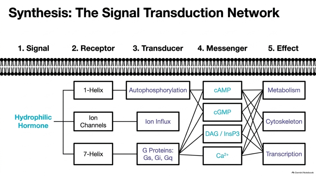
Synthesizing the entire signal transduction pathway highlights the logical, modular organization of cellular communication. In the hydrophilic hormone mechanism of action, the signaling sequence progresses systematically through five distinct stages: extracellular signal, membrane receptor, G protein transducer, second messenger, and final physiological effect. Whether operating through 1-helix receptors, ion channels, or 7-helix receptors, the hydrophilic hormone mechanism of action relies on precise structural transitions to pass information across the lipid bilayer. Each stage acts as a controlled checkpoint, allowing cells to integrate multiple hormonal inputs while maintaining tight signal fidelity across complex cellular environments.
Furthermore, this network organization provides remarkable versatility, enabling a single hormone to trigger diverse cellular outcomes depending on receptor subtypes and second-messenger machinery present in different tissues. Within the hydrophilic hormone mechanism of action, cross-talk between cAMP, calcium, and kinase cascades allows fine-tuned physiological regulation across the entire human body. From immediate neuromuscular activation to sustained metabolic modulation, hydrophilic hormone action exemplifies nature’s ability to achieve complex control through elegant molecular mechanisms. Mastering these interconnected pathways empowers students to understand disease states and pharmacological targets in modern medicine.
Please read our Content Disclaimer Statement.
Check out our social media channels: