149. Lipophilic Hormones Mechanism of Action: Molecular Pathways, and Gene Regulation
How do hormones floating in the bloodstream alter gene expression inside a cell’s nucleus? Unlike water-soluble signals that knock on the cell’s outer door, lipid-soluble messengers cross the plasma membrane to direct cellular activity from within. This slide deck breaks down the entire pathway of nuclear signaling. By exploring receptor structures, chaperone protein interactions, and DNA binding, students will gain a clear overview of this core endocrine process.
Slide 1: Lipophilic Hormones Mechanism of Action — Molecular Overview
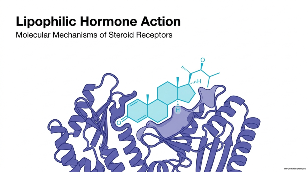
Endocrine communication relies heavily on hydrophobic messengers that can diffuse directly across cellular boundaries. The Mechanism of Action of Lipophilic Hormones begins when small, nonpolar molecules, such as steroids, bypass cell-surface receptors and enter target cells. Unlike peptide hormones that bind cell-membrane receptors to trigger rapid second-messenger cascades, lipid-soluble hormones target specialized intracellular proteins. These receptor proteins function as ligand-activated transcription factors that directly regulate nuclear gene expression.
At the structural level, the Lipophilic Hormones Mechanism of Action involves a precise lock-and-key interaction between the ligand and its receptor. As depicted in the opening diagram, the hormone fits into a designated hydrophobic binding pocket within the receptor’s ligand-binding domain. This insertion drives a crucial conformational change in the protein framework, altering the receptor from an inactive state into an active, functional complex capable of nuclear interaction.
Studying the Mechanism of Action of Lipophilic Hormones provides foundational insight into human physiology, development, and metabolic control. By converting extracellular endocrine signals directly into transcriptional changes, these molecules exert profound, long-lasting biological effects. Understanding this primary structural engagement sets the stage for examining how specific receptor domains, chaperone proteins, and DNA control elements coordinate gene expression within target cell nuclei.
Slide 2: Lipophilic Hormones Mechanism of Action — Defining Key Messengers
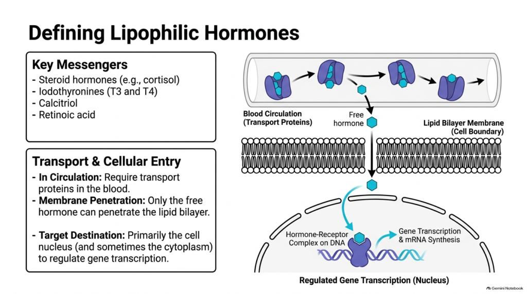
A diverse group of chemical messengers utilizes the Lipophilic Hormones Mechanism of Action to regulate physiological functions. Major examples include steroid hormones like cortisol, iodothyronines such as thyroid hormones T3 and T4, calcitriol, and retinoic acid. Because these compounds are hydrophobic, they cannot dissolve freely in aqueous blood plasma in large quantities. Consequently, they require specialized transport proteins to travel safely through the circulatory system to reach target tissues.
Circulating transport proteins maintain a dynamic equilibrium between bound and free hormone fractions. According to the Lipophilic Hormones Mechanism of Action, only the free, unbound hormone can diffuse across the lipid bilayer membrane of a target cell. Once inside, the hormone navigates through the aqueous cytoplasm toward its designated intracellular receptor. While most lipophilic receptors reside directly in the cell nucleus, some receptors act in the cytoplasm before nuclear migration.
The entry of free hormones into target cells highlights a fundamental principle of the Lipophilic Hormones Mechanism of Action. Simple diffusion lets these hydrophobic signals bypass the plasma membrane barrier without active transport channels or surface receptors. This direct access ensures that the hormone reaches its target transcription factor, initiating a signaling cascade that culminates in altered mRNA synthesis and protein production inside the nucleus.
Slide 3: Lipophilic Hormones Mechanism of Action — The Nuclear Receptor Profile
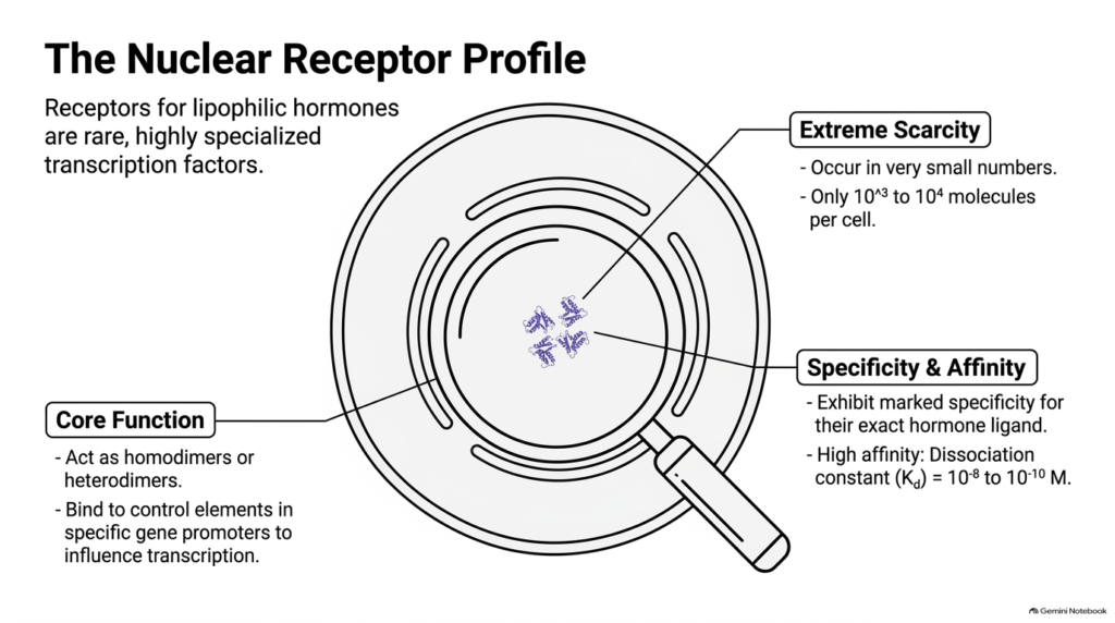
Intracellular receptors that mediate the mechanism of action of lipophilic hormones have distinct biochemical properties that set them apart from general cellular proteins. These nuclear receptors are remarkably scarce within the cell, typically occurring in small numbers ranging from only 1,000 to 10,000 molecules per target cell. Despite their low abundance, they execute critical regulatory functions by acting as highly selective transcription factors when bound to their specific ligand molecules.
To function effectively at low concentrations, receptors involved in the Lipophilic Hormones Mechanism of Action exhibit exceptional affinity and specificity for their cognate hormones. The dissociation constant, or Kd value, for these receptor-ligand interactions falls between 10^-8 M and 10^-10 M. This high-affinity binding ensures that even minute physiological concentrations of free hormone in the tissue fluid produce significant receptor activation and downstream genetic responses.
Furthermore, the Lipophilic Hormones Mechanism of Action relies on these activated receptors operating as functional dimers. Receptors bind to promoter regions on DNA as either homodimers—composed of two identical receptor units—or heterodimers partnered with distinct nuclear receptors. By binding to specific regulatory DNA sequences known as control elements, these dimeric complexes modulate gene transcription rates, orchestrating precise cellular adaptations to hormonal stimulation.
Slide 4: Lipophilic Hormones Mechanism of Action — Modular Architecture of Steroid Receptors
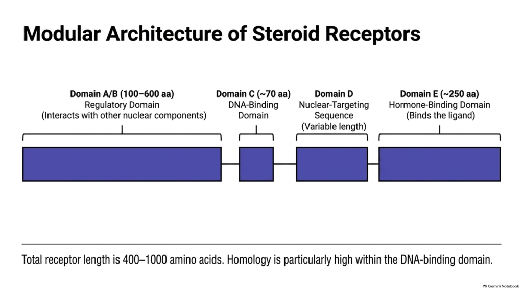
Members of the nuclear receptor superfamily that carry out the Lipophilic Hormones Mechanism of Action share a conserved modular organization. These receptor proteins range from 400 to 1,000 amino acids and consist of distinct functional domains labeled A through E. Each domain performs a specialized biochemical task necessary to receive hormonal signals and transmit them directly to the cellular genome.
Starting from the N-terminus, Domain A/B functions as the regulatory domain, spanning 100 to 600 amino acids and interacting with various nuclear transcription factors. Next lies Domain C, a highly conserved DNA-binding domain of roughly 70 amino acids that anchors the receptor to specific genetic sequences. Domain D contains a variable nuclear-targeting sequence, while Domain E at the C-terminus spans approximately 250 amino acids and houses the hormone-binding domain.
Understanding domain modularity clarifies how the Lipophilic Hormones Mechanism of Action integrates multiple biological inputs. Domain E selectively binds the hormone ligand, while Domain C engages DNA, and Domain A/B recruits transcriptional machinery. Sequence alignment reveals that amino acid homology across different steroid receptors is highest within Domain C. This structural conservation emphasizes the universal evolutionary design underpinning hormone-driven gene regulation across eukaryotic organisms.
Slide 5: Lipophilic Hormones Mechanism of Action — Structural Stabilization via Zinc Clusters
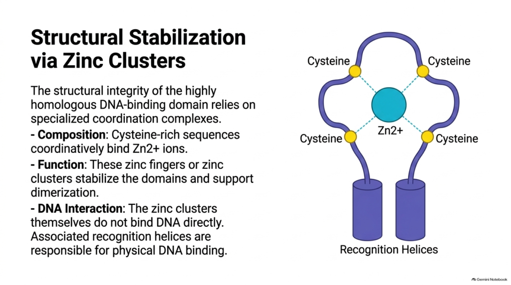
The structural integrity of the DNA-binding domain is vital to the Mechanism of Action of Lipophilic Hormones. Within Domain C, cysteine-rich amino acid sequences coordinately bind divalent zinc ions (Zn2+) to form specialized structural motifs known as zinc fingers or zinc clusters. Each zinc ion is stabilized by four surrounding cysteine residues, folding the peptide chain into a rigid secondary structure required for proper receptor function.
In the context of the Lipophilic Hormones Mechanism of Action, zinc clusters serve essential structural roles rather than directly contacting DNA. These zinc-coordinated centers stabilize the overall architecture of Domain C and promote the receptor dimerization required for activation. Adjacent peptide structures called recognition helices make physical contact with the DNA double helix and insert directly into the major groove of target DNA sequences.
Without functional zinc clusters, the Lipophilic Hormones Mechanism of Action fails because the receptor cannot maintain its tertiary fold or form stable dimers. Mutational disruption of the coordinating cysteine residues completely abolishes DNA binding. Thus, zinc coordination serves as an indispensable structural scaffold that enables recognition helices to accurately read specific base pair sequences within gene promoters during hormone signaling.
Slide 6: Lipophilic Hormones Mechanism of Action — The Cortisol Pathway: Cytoplasmic Resting State
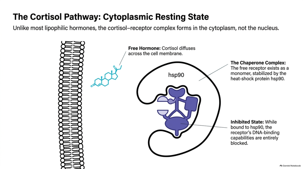
Glucocorticoid signaling provides a classic model illustrating the Lipophilic Hormones Mechanism of Action. Unlike many nuclear receptors that reside permanently inside the nucleus, the unliganded cortisol receptor resides primarily within the cytoplasm. In this resting state, free cortisol diffuses across the plasma membrane and moves toward the inactive receptor complex in the cytosolic compartment of the target cell.
In the absence of hormone, the Lipophilic Hormones Mechanism of Action keeps the receptor in an inactive conformation using molecular chaperones. The monomeric receptor protein forms a stable complex with heat-shock protein 90 (hsp90). This chaperone complex binds to the receptor, stabilizing its structure while physically masking its nuclear localization signal and DNA-binding domain, thereby preventing premature entry into the cell nucleus.
Maintaining this inhibited state is a hallmark of the Lipophilic Hormones Mechanism of Action for glucocorticoid pathways. By sequestering the unliganded receptor in the cytoplasm, the cell prevents unauthorized background transcription of target genes. The Hsp90 chaperone holds the receptor in a high-affinity conformation ready to accept incoming cortisol molecules, ensuring a rapid genetic response upon hormonal stimulation.
Slide 7: Lipophilic Hormones Mechanism of Action — The Cortisol Pathway: Activation & Dimerization
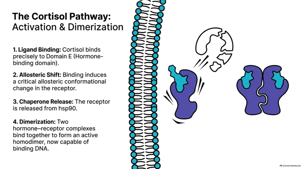
Hormone binding transforms the resting receptor and drives the Lipophilic Hormones Mechanism of Action forward. When cortisol enters the cytoplasm, it binds with high specificity to Domain E of the monomeric receptor. This binding triggers an allosteric conformational shift across the receptor, reorganizing its tertiary structure and altering its surface interactions with surrounding cytosolic proteins.
As a result of this conformational shift in the Lipophilic Hormones Mechanism of Action, the chaperone protein hsp90 dissociates from the receptor. Uncoupling from hsp90 exposes the previously hidden DNA-binding domain and nuclear localization sequence. Once freed from chaperone restraint, two activated hormone-receptor complexes pair together to form a stable homodimer, creating the functional unit required for DNA recognition.
This activation phase highlights the dynamic nature of the Lipophilic Hormones Mechanism of Action. Dimerization alters the spatial orientation of the recognition helices, enabling the complex to interact effectively with genetic regulatory elements. The newly assembled homodimer can cross the nuclear envelope through nuclear pore complexes, moving from the cytoplasm into the nucleus to target specific promoter regions.
Slide 8: Lipophilic Hormones Mechanism of Action — Target Acquisition: Hormone Response Elements (HREs)
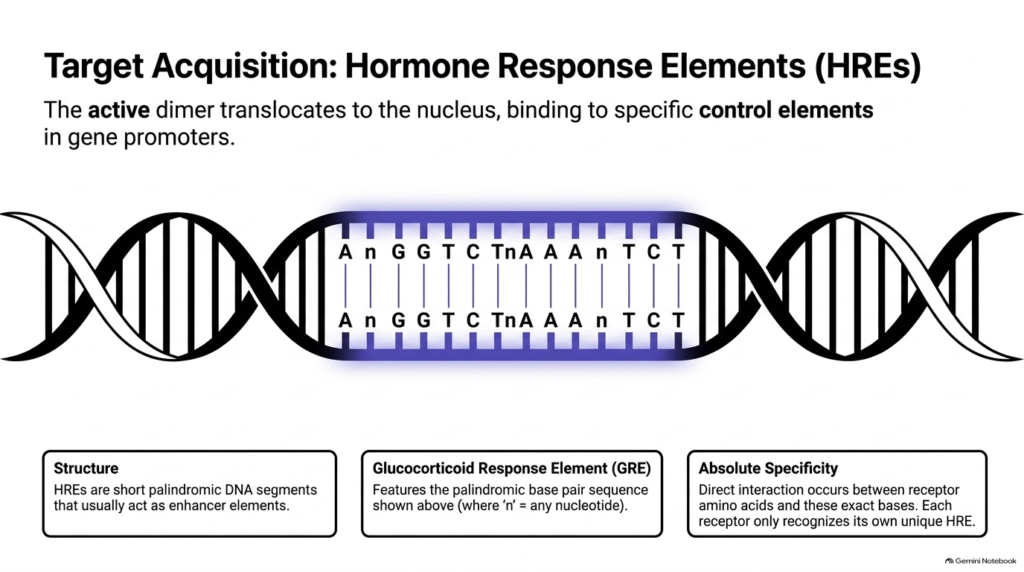
Once inside the nucleus, the activated homodimer acquires its target within the Lipophilic Hormones Mechanism of Action. The complex searches the genome for specific regulatory sequences called Hormone Response Elements (HREs). These HREs are short, palindromic DNA segments within gene promoters that primarily function as transcription enhancer elements, directing hormone activity to specific target genes.
For glucocorticoids, the Lipophilic Hormones Mechanism of Action targets a specific consensus sequence known as the Glucocorticoid Response Element (GRE). As displayed on the slide, this palindromic sequence consists of two conserved hexameric nucleotide repeats separated by a three-base spacer (AnGGTCTnAAAnTCT). Each receptor monomer in the homodimer interacts with one palindromic repeat, establishing symmetric contacts along the DNA backbone.
Target specificity in the Lipophilic Hormones Mechanism of Action relies on direct chemical interactions between amino acid residues in the receptor’s recognition helices and specific base pairs within the HRE. Because each nuclear receptor recognizes its own unique consensus sequence, hormones only stimulate genes containing matching response elements, ensuring precise genetic control without off-target gene activation.
Slide 9: Lipophilic Hormones Mechanism of Action — The Cortisol Pathway: Gene Transcription Control
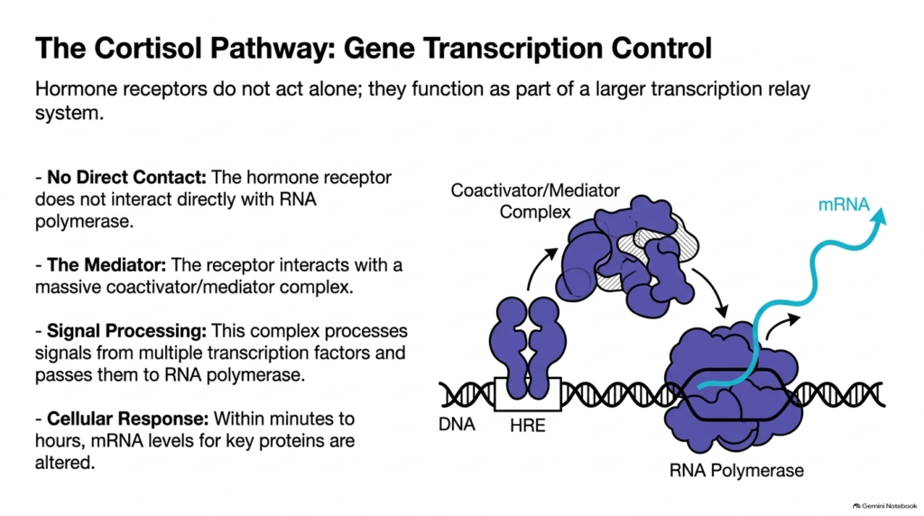
Regulating gene expression through the Lipophilic Hormones Mechanism of Action involves an indirect transcriptional relay system. After binding to the Hormone Response Element, the hormone-receptor homodimer does not directly contact RNA polymerase II. Instead, the receptor functions as an assembly platform that recruits large accessory protein complexes to the promoter region.
In this phase of the Lipophilic Hormones Mechanism of Action, the DNA-bound receptor dimer engages a massive coactivator and mediator complex. This multi-protein assembly processes biochemical signals from multiple transcription factors simultaneously, bridging the steroid receptor and RNA polymerase II to stimulate assembly of the basal transcription machinery at the target gene.
This coactivator recruitment completes the nuclear phase of the Lipophilic Hormones Mechanism of Action. By enhancing the rate of transcription initiation, the complex increases mRNA synthesis for specific functional proteins. Within minutes to hours following initial hormone exposure, altered mRNA levels translate into changed cellular protein concentrations, producing the characteristic physiological response of the target cell.
Slide 10: Lipophilic Hormones Mechanism of Action — Beyond Cortisol: Receptor Superfamily Diversity
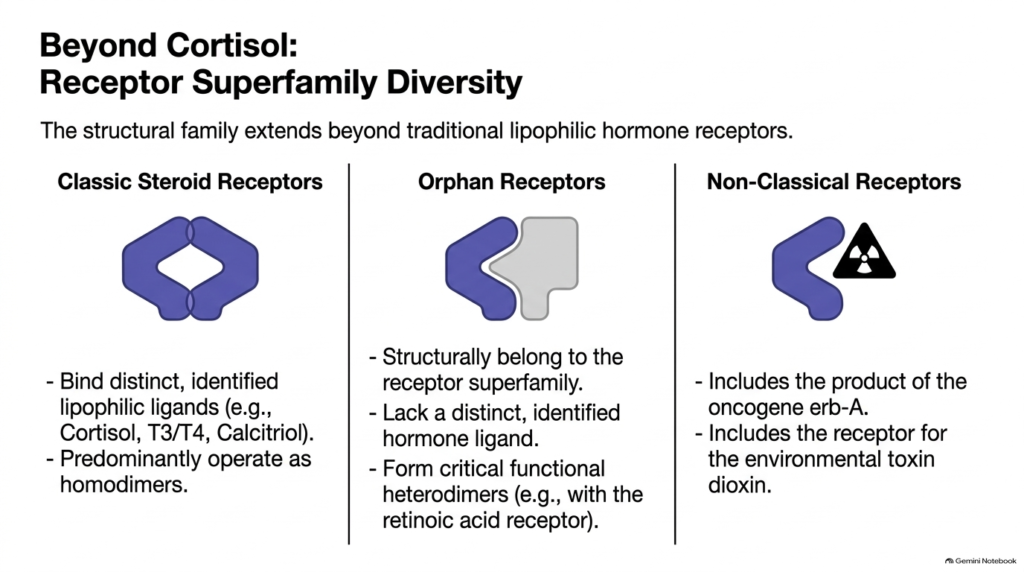
The structural design governing the Mechanism of Action of Lipophilic Hormones extends across a broad superfamily of nuclear receptors. Beyond classic steroid receptors that bind ligands such as cortisol, thyroid hormones, calcitriol, and retinoic acid, this protein family includes specialized receptor categories that expand cellular signaling across diverse biological processes.
A prominent group within the Lipophilic Hormones Mechanism of Action superfamily comprises orphan receptors. These proteins share the characteristic modular domain architecture of nuclear receptors but lack known endogenous hormone ligands. Rather than functioning solely as homodimers, many orphan receptors form functional heterodimers with other receptors, such as the retinoic acid receptor, to regulate distinct genetic pathways.
Additionally, non-classical receptors demonstrate the evolutionary versatility of the Lipophilic Hormones Mechanism of Action. This category includes the protein product of the erb-A oncogene and the receptor for the environmental pollutant dioxin. These examples illustrate how the modular nuclear receptor framework has adapted to accommodate oncogenic signaling and xenobiotic responses, reinforcing its central importance in modern cell biology.
Slide 11: Lipophilic Hormones Mechanism of Action — Synthesis: The Lipophilic Hormone Cascade
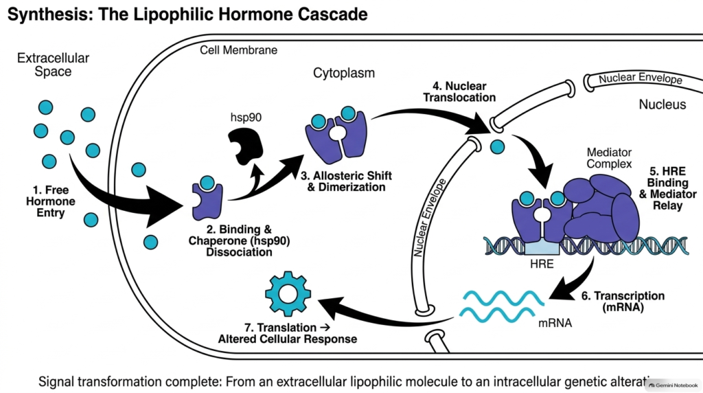
Integrating every stage of the Lipophilic Hormones Mechanism of Action reveals a seamless seven-step signal transduction cascade. First, free hormone diffuses across the plasma membrane into the cytoplasm. Second, the hormone binds its receptor, causing chaperone hsp90 dissociation. Third, an allosteric shift promotes receptor homodimerization, creating an active DNA-binding complex that can enter the nucleus.
The middle stage of the Lipophilic Hormones Mechanism of Action involves nuclear events. Fourth, the active homodimer translocates through the nuclear envelope. Fifth, the complex binds specific Hormone Response Elements on DNA and recruits the mediator/coactivator complex. Sixth, RNA polymerase transcribes target DNA into mRNA, completing the nuclear transmission of the original hormonal signal.
Finally, the seventh step of the Lipophilic Hormones Mechanism of Action occurs as newly synthesized mRNA exits the nucleus into the cytoplasm for ribosomal translation. The resulting operational proteins modify metabolic activities, cellular growth, or physiological function. This integrated cascade illustrates how extracellular lipid-soluble signals successfully convert into precise intracellular genetic responses to maintain homeostasis.
Please read our Content Disclaimer Statement.
Check out our social media channels: