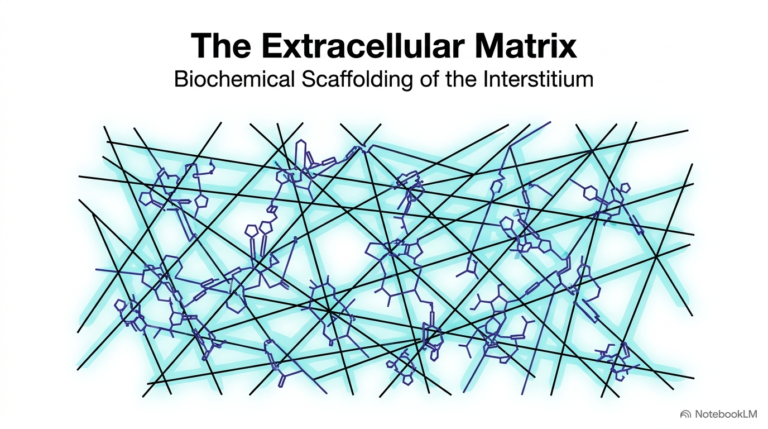
135. CNS Signal Transmission: The Biochemical Basis of Neural Communication
How does the human brain convert a fleeting thought into instant physical movement? Every sensory perception, memory, and reflex relies on precise communication between billions of specialized neural cells. This educational slide deck provides a comprehensive breakdown of CNS signal transmission, detailing the structural architecture, chemical messengers, and receptor mechanisms that enable neural signaling. Designed for college and medical school students, this resource clarifies the fundamental biochemistry driving central nervous system function and its profound impact on human physiology.
Slide 1: Overview of CNS Signal Transmission
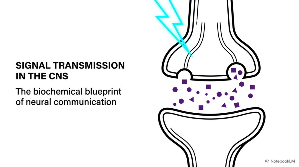
The brain operates through an intricate web of cellular pathways, where rapid communication maintains every thought, sensation, and physical movement. Understanding CNS signal transmission provides students with the foundational key to unlocking complex neurobiology and pharmacology. This initial slide introduces the core architecture of neuronal communication, highlighting how specialized cells pass critical biological information across microscopic gaps. Through precise molecular actions, chemical messengers transform electrical impulses into cellular responses that drive physiological function across neural networks.
Within the human nervous system, billions of neurons coordinate to form functional circuits. CNS signal transmission depends on a sophisticated interplay between electrical depolarization and chemical diffusion. As an action potential travels down a presynaptic neuron, it triggers neurotransmitter release into the narrow synaptic cleft. These chemical signals cross the fluid-filled gap to bind specific postsynaptic receptors, completing a crucial circuit link that propagates information smoothly between connected cells.
Mastering the biochemical mechanics of CNS signal transmission allows future scientists and medical clinicians to understand how therapeutic drugs alter neural function and how disease states disrupt cellular communication. From simple reflex arcs to high-level cognitive processing, information transfer across synapses remains the primary driver of central nervous system function. Studying these foundational principles bridges basic molecular cell biology with advanced clinical neurology and neuropharmacology.
Slide 2: Neuronal Morphology and CNS Signal Transmission
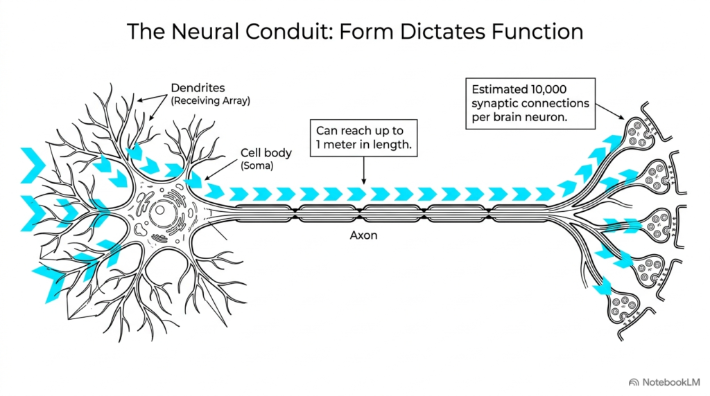
Form directly dictates function throughout the human nervous system, where unique cellular structures enable long-distance communication. A neuron’s physical shape is tailored to facilitate efficient CNS signal transmission across expansive cellular networks. Highly branched dendrites act as a massive receiving array, gathering incoming biochemical inputs from thousands of neighboring cells. These incoming signals travel inward toward the soma, or cell body, where the neuron integrates information before deciding whether to propagate an electrical impulse.
Once the cell body processes these incoming signals, an electrical surge travels along the axon toward downstream targets. In human anatomy, axons can reach up to one meter in length, demonstrating the incredible physical reach required for effective CNS signal transmission. Along this extended cellular pathway, specialized structural features preserve the electrical charge, ensuring that vital instructions move seamlessly from the brain and spinal cord to distant peripheral tissues throughout the organism.
At the far end of the axon, terminal branches split to form an estimated ten thousand synaptic connections per brain neuron. This vast anatomical arrangement highlights the enormous structural complexity of CNS signal transmission in the human brain. By distributing a single incoming signal across thousands of postsynaptic targets, individual neurons create highly flexible processing networks capable of managing complex physiological actions and cognitive functions.
Slide 3: Myelin Sheath Structure in CNS Signal Transmission
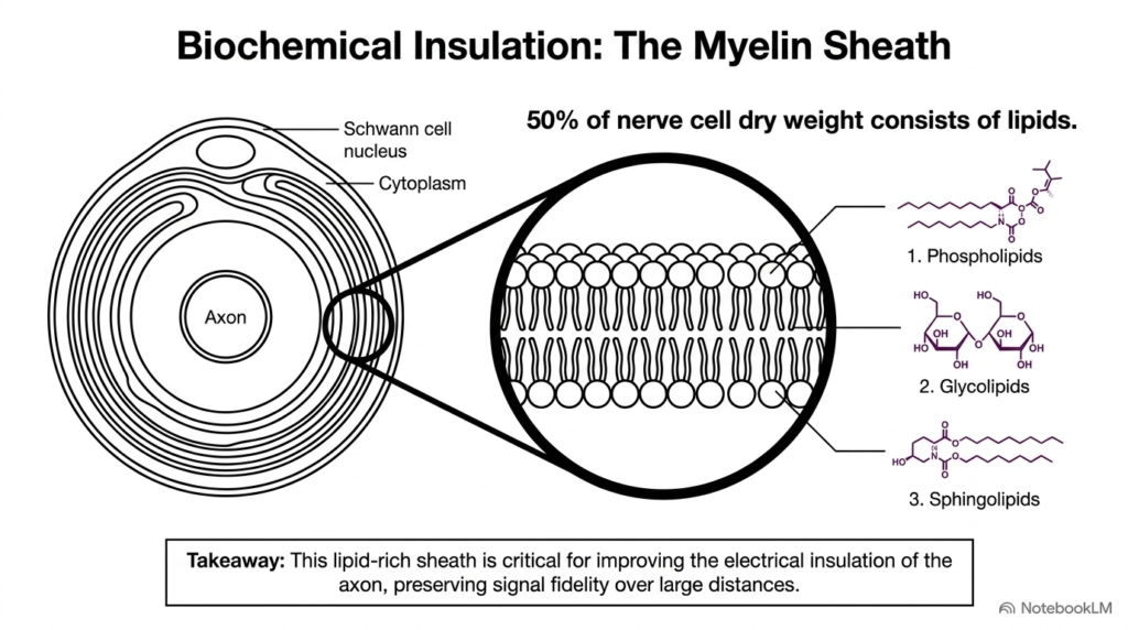
Rapid neural communication requires robust cellular insulation to prevent electrical charge leakage across long cellular distances. The myelin sheath serves as a specialized biochemical barrier that drastically increases the speed and efficiency of CNS signal transmission. Because it is composed predominantly of specialized cell membranes, approximately fifty percent of a nerve cell’s dry weight consists of complex lipids. These hydrophobic molecules wrap tightly around the axon, creating high electrical resistance that forces electrical impulses to jump rapidly between unmyelinated nodes.
Myelin’s molecular structure includes three primary classes of functional lipids: phospholipids, glycolipids, and sphingolipids. Together, these lipid components form a dense, hydrophobic layer that stabilizes the membrane structure during active CNS signal transmission. Glial cells, such as Schwann cells in the peripheral nervous system and oligodendrocytes in the central nervous system, repeatedly wrap their cytoplasm and plasma membranes around the axon to build this essential insulating sheath structure.
By preserving electrical fidelity over extended physical distances, myelin insulation prevents signal decay during high-speed neural processing. The loss or degradation of this lipid sheath severely impairs CNS signal transmission, leading to profound neurological deficits seen in demyelinating conditions like multiple sclerosis. Understanding the biochemical composition of myelin helps students appreciate how specialized plasma membranes protect electrical signals throughout the entire human nervous system.
Slide 4: Categorizing Neurosecretions in CNS Signal Transmission
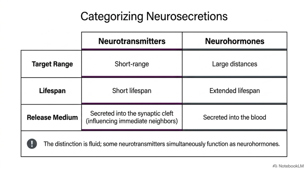
Chemical communication within the central nervous system relies on specialized signaling molecules that coordinate diverse cellular activities across tissues. Researchers categorize these secreted substances into neurotransmitters and neurohormones based on their operational range, functional lifespan, and release medium during CNS signal transmission. Neurotransmitters act locally over short distances, moving rapidly across the narrow synaptic cleft to influence nearby cells before undergoing swift enzymatic degradation or cellular reuptake by supporting astrocytes.
In contrast, neurohormones travel much larger physiological distances by entering the systemic bloodstream to regulate distant target organs over an extended functional lifespan. Despite these distinct operational differences, the boundary between these chemical categories remains remarkably fluid during actual CNS signal transmission. Certain signaling molecules, such as norepinephrine and vasopressin, serve dual roles as localized synaptic transmitters in one context and systemic neurohormones in another.
Distinguishing between local synaptic events and systemic hormonal actions clarifies how the brain integrates immediate reflex responses with long-term metabolic homeostasis. The versatility of chemical messengers expands the functional capacity of CNS signal transmission, enabling precise point-to-point electrical conversion alongside broad physiological regulation across the human body. Students analyzing these neurosecretory classes develop a comprehensive framework for understanding multi-tier chemical communication in medical science.
Slide 5: Synaptic Anatomy and CNS Signal Transmission
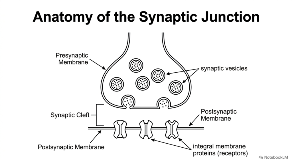
The synaptic junction represents the primary anatomical site where chemical communication occurs between adjacent neural cells in functional pathways. A precise physical arrangement of specialized cellular structures ensures that CNS signal transmission proceeds smoothly across the fluid-filled gap separating distinct neurons. On the presynaptic side, membrane-bound synaptic vesicles store concentrated amounts of chemical messengers, waiting for an arriving electrical signal to trigger their fusion and exocytotic release into the extracellular space.
The synaptic cleft separates the sending and receiving cells; it is a narrow extracellular space measuring only twenty to forty nanometers wide. This minimal physical distance is critical for efficient CNS signal transmission, as it allows released chemical messengers to diffuse rapidly across the gap without significant dilution or spatial dispersion. On the opposing side, the postsynaptic membrane contains specialized integral membrane proteins that function as highly selective receptors for specific neurotransmitter molecules.
This structured anatomical organization establishes a strict unidirectional pathway for neural information flow throughout the central nervous system. By positioning presynaptic storage vesicles directly opposite postsynaptic receptor proteins, the synapse optimizes the speed, accuracy, and overall precision of CNS signal transmission. Students studying this micro-architecture gain a clear structural framework for understanding subsequent biochemical and electrical events that drive neuronal computation in medical science.
Slide 6: Presynaptic Calcium Activation in CNS Signal Transmission
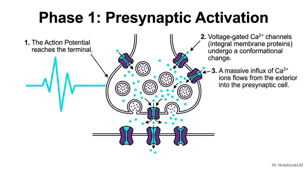
Chemical signaling begins when an electrical wave reaches the terminal end of a presynaptic neuron along the axon pathway. This arrival phase initiates presynaptic activation, a critical step that translates electrical excitation into chemical events during CNS signal transmission. As the incoming action potential depolarizes the presynaptic membrane, specialized voltage-gated calcium channels undergo a rapid conformational change, opening their selective pore structures to the surrounding extracellular environment.
Because calcium ion concentration is significantly higher outside the cell than inside, opening these channels causes a massive influx of calcium ions into the presynaptic terminal. This sudden rise in intracellular calcium concentration acts as an indispensable biochemical switch in CNS signal transmission. The incoming calcium ions bind directly to specialized sensor proteins like synaptotagmin, initiating a rapid cascade of intracellular vesicle movements toward the active presynaptic membrane release zone.
Without calcium influx, the presynaptic terminal cannot convert electrical depolarization into chemical messenger release across the junctional space. This total dependence on voltage-gated calcium channels highlights how ion flux couples electrical activity directly to chemical signaling during CNS signal transmission. Understanding presynaptic calcium dynamics provides medical students with essential physiological context for how channel-blocking clinical medications alter neuronal activity across central neural circuits in human patients. Analyzing these calcium-dependent mechanisms reinforces how synaptic transmission coordinates complex neural signaling.
Slide 7: Exocytosis Mechanics in CNS Signal Transmission
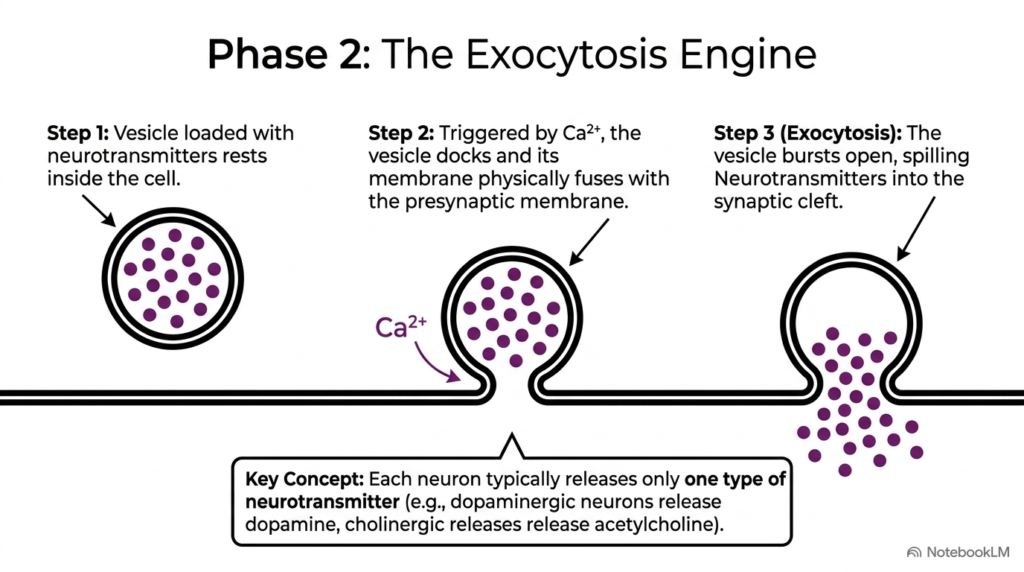
Once calcium enters the presynaptic terminal, a specialized mechanical engine drives the fusion of neurotransmitter vesicles with the plasma membrane. This process, known as exocytosis, releases stored chemical messengers to facilitate CNS signal transmission across the synaptic cleft. Initially, vesicles loaded with chemical compounds rest near the inner surface of the presynaptic membrane, tethered securely by specialized SNARE proteins that await calcium activation to initiate fusion.
Triggered by localized calcium elevation, the vesicle membrane docks tightly and physically fuses with the outer presynaptic lipid bilayer. The vesicle then bursts open, spilling its chemical payload directly into the synaptic gap to advance CNS signal transmission. Under standard physiological conditions, each neuron releases primarily one specific neurotransmitter type, such as dopamine in dopaminergic cells or acetylcholine in cholinergic neurons.
This selective chemical release ensures neural pathways maintain distinct functional identities and target specific downstream receptor classes. The rapid execution of membrane fusion and chemical release renders exocytosis a cornerstone mechanism of CNS signal transmission. Students analyzing exocytosis gain valuable insight into how synaptic vesicle protein mutations can cause severe neurological disorders, impaired synaptic signaling, and movement abnormalities in clinical human patients. Studying exocytotic pathways highlights the essential role of precise vesicle fusion in maintaining healthy neural communication.
Slide 8: Diffusion and Target Binding in CNS Signal Transmission
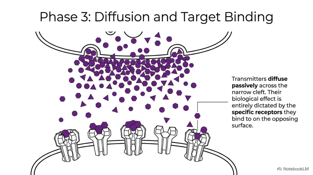
Following exocytosis, thousands of neurotransmitter molecules flood into the narrow extracellular space separating the communicating cells. These chemical messengers diffuse passively across the synaptic cleft, moving down their concentration gradient to sustain CNS signal transmission across the junction. Because the synaptic cleft spans a minimal physical distance of mere nanometers, passive diffusion occurs in a fraction of a millisecond, ensuring rapid delivery of chemical information to target postsynaptic structures.
Once chemical messengers cross the cleft, their ultimate biological effect depends entirely on the specific receptor proteins they engage on the postsynaptic membrane. The neurotransmitter acts as a chemical key, while the receptor determines whether the receiving cell responds with excitation, inhibition, or metabolic modulation during CNS signal transmission. Matching specific chemical ligands to complementary binding sites ensures precise target cell recognition.
This separation of ligand identity from cellular outcome allows a single chemical messenger to produce diverse physiological effects across different brain regions. By varying postsynaptic receptor distribution, the central nervous system fine-tunes signal transmission to meet specific neural circuit requirements. Studying receptor-ligand interactions helps future pharmacologists design targeted medications with minimal unwanted side effects in clinical medicine. Understanding passive diffusion dynamics clarifies how synapses achieve rapid signal processing across complex central networks.
Slide 9: Postsynaptic Receptor Diversity in CNS Signal Transmission
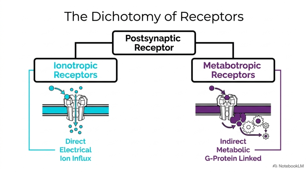
Target cells interpret chemical signals through two major classes of postsynaptic receptors, each operating through distinct molecular mechanisms. Recognizing this functional dichotomy is essential to understanding how neurons process information during CNS signal transmission. Ionotropic receptors function as ligand-gated ion channels, providing a direct physical pathway for rapid ion entry immediately upon chemical ligand binding to extracellular protein domains.
In contrast, metabotropic receptors lack an internal pore and instead couple to intracellular G-protein complexes to trigger indirect metabolic cascades within the cell. While ionotropic pathways mediate fast electrical changes lasting only milliseconds, metabotropic pathways drive slower, long-lasting cellular modifications during CNS signal transmission. This division of labor enables neural circuits to combine high-speed reflex actions with sustained physiological modulation across target networks.
By deploying both receptor types at various synaptic junctions, the brain achieves remarkable operational flexibility across diverse functional pathways. The balance between direct ion gating and indirect metabolic signaling shapes overall CNS signal transmission throughout central brain networks. Medical students must master this receptor division to comprehend how neuroactive medications produce fast versus prolonged biological responses in clinical patient care. Mastering these distinct receptor mechanisms provides a solid biochemical foundation for advanced neurophysiological studies.
Slide 10: Ionotropic Channel Mechanics in CNS Signal Transmission
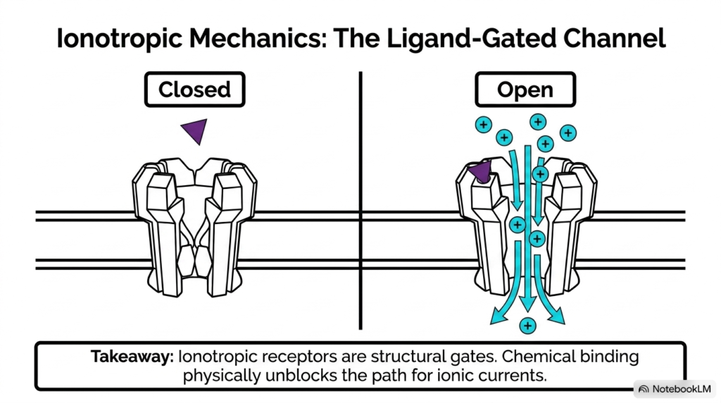
Ionotropic receptors act as molecular gates, converting ligand binding into immediate ionic currents across the postsynaptic membrane. These ligand-gated channels play a vital role in fast-acting CNS signal transmission by physically opening in direct response to specific neurotransmitters. In the resting or closed state, structural protein domains block the central pore, preventing charged ions from passing across the lipid bilayer.
When a complementary neurotransmitter binds to the extracellular binding site, the channel protein undergoes an instantaneous conformational change. This movement unblocks the central pore, allowing specific ions to rush down their electrochemical gradients and drive CNS signal transmission. Because the receptor protein and ion channel form a single integrated protein structure, cellular responses occur almost instantaneously, within fractions of a millisecond.
This direct mechanical opening makes ionotropic receptors ideal for rapidly propagating electrical information across active neural circuits. Chemical binding physically unblocks the path for ionic currents, demonstrating a direct conversion of chemical energy into electrical work during CNS signal transmission. Understanding ionotropic gating mechanics helps students grasp how rapid reflex pathways achieve remarkable speed across complex physiological circuits in the body. Exploring these channel mechanics helps students appreciate the physical basis of rapid electrical signaling in central nervous pathways.
Slide 11: Ionotropic Depolarization and Hyperpolarization in CNS Signal Transmission
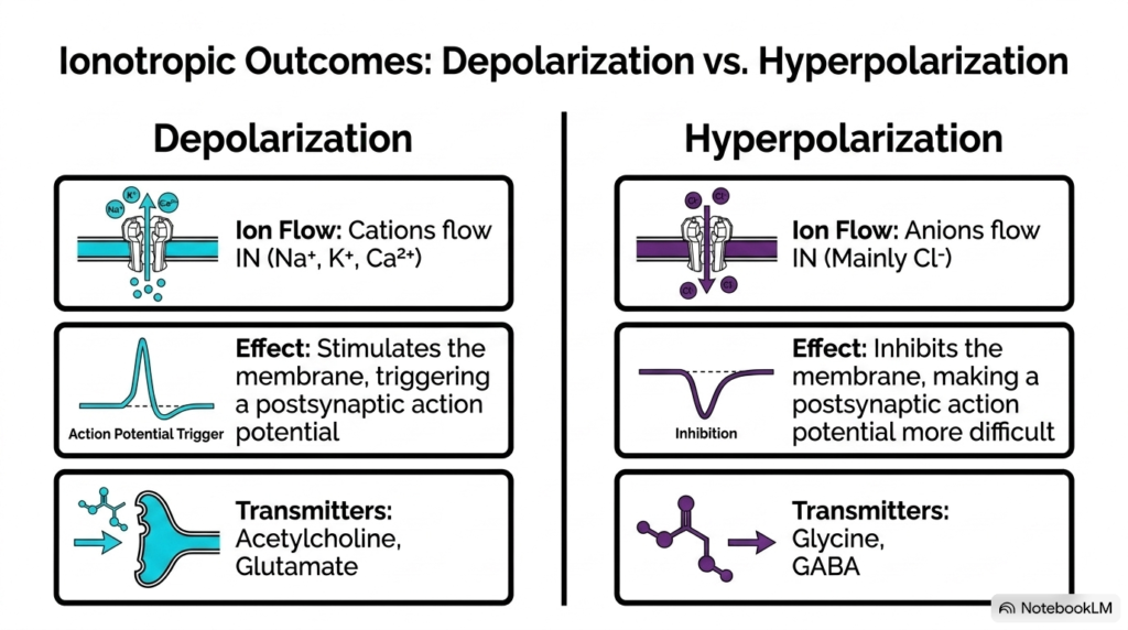
Ion influx through open ligand-gated channels produces two opposing electrical outcomes in the receiving postsynaptic neuron. The specific charge of the flowing ions determines whether CNS signal transmission stimulates or suppresses subsequent electrical firing in target cells. Depolarization occurs when positive cations, such as sodium, potassium, or calcium, enter the cell, shifting the interior membrane potential closer to the action potential firing threshold.
Excitatory neurotransmitters like glutamate and acetylcholine trigger cation influx, stimulating the membrane and increasing the probability of generating a postsynaptic action potential during CNS signal transmission. Conversely, hyperpolarization occurs when negative anions, primarily chloride, flow into the cell through channels opened by inhibitory neurotransmitters like GABA or glycine. This influx makes the interior membrane potential more negative, effectively inhibiting electrical firing across the postsynaptic membrane.
The dynamic balance between excitatory depolarization and inhibitory hyperpolarization governs signal integration across central neural networks. By summing these opposing electrical inputs, the postsynaptic cell decides whether to propagate CNS signal transmission downstream. Studying these dual electrical outcomes provides students with a foundational model for how complex neural circuits filter and compute physiological information across central pathways. Comprehending these electrical mechanisms equips future researchers to analyze complex neuronal integration and synaptic summation.
Slide 12: Metabotropic G-Protein Cascades in CNS Signal Transmission
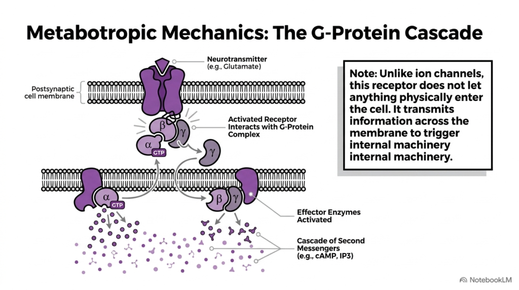
Metabotropic receptors utilize intricate intracellular protein cascades rather than direct pore opening to alter postsynaptic cell behavior. This indirect mechanism broadens the temporal scope of CNS signal transmission, generating metabolic changes that outlast initial ligand-binding events. Unlike ion channels, metabotropic receptors do not allow physical ion entry; instead, they transmit information across the cell membrane to activate attached G-protein complexes.
When a ligand like glutamate binds the receptor, the attached G-protein complex dissociates into active alpha and beta-gamma protein subunits. These activated subunits move along the inner membrane surface to stimulate specialized effector enzymes, driving key biochemical pathways in CNS signal transmission. These effector enzymes then catalyze the mass production of intracellular second messenger molecules, such as cyclic AMP or inositol trisphosphate, inside the cytoplasm.
These internal second messengers diffuse throughout the cytoplasm, activate downstream kinases, and initiate broad regulatory routines within the target cell. This multi-step molecular relay demonstrates how metabotropic CNS signal transmission achieves long-range metabolic modulation. Studying G-protein signaling helps medical students understand the molecular targets of numerous psychiatric and neurological therapeutic drugs used in clinical medicine today. Mastering these metabolic cascade steps offers essential insights into how long-term cellular modulation shapes brain function and neural plasticity.
Slide 13: Signal Amplification and Cellular Modulation in CNS Signal Transmission
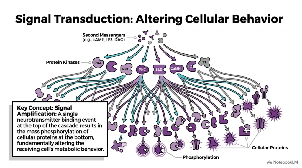
A defining feature of metabotropic receptor signaling is its ability to produce massive signal amplification within target receiving cells. Through multi-tier enzyme activation, a single ligand binding event triggers widespread physiological changes during CNS signal transmission across neural circuits. Effector enzymes generate second messengers that activate various protein kinases, including protein kinase A, protein kinase C, and calcium/calmodulin-dependent protein kinase II.
Once activated, these kinases catalyze the widespread phosphorylation of numerous functional proteins throughout the receiving cell’s cytoplasm and membrane structures. Phosphorylation fundamentally alters protein shape, enzymatic activity, and ion channel sensitivity, reshaping cellular behavior during CNS signal transmission. Consequently, a single chemical messenger binding at the membrane surface triggers widespread downstream modification of thousands of functional target proteins within the cell.
This profound biochemical amplification allows neurons to modify gene expression, adjust synaptic strength, and regulate long-term metabolic states. Signal amplification turns localized synaptic events into lasting structural and functional adaptations during CNS signal transmission. Understanding these cascade dynamics helps students appreciate the molecular foundations of learning, memory, and neuronal plasticity in the human central nervous system. Investigating signal amplification cascades provides critical context for understanding how persistent synaptic changes support cognitive processes in neurobiology.
Slide 14: Modality Transduction Cycles in CNS Signal Transmission
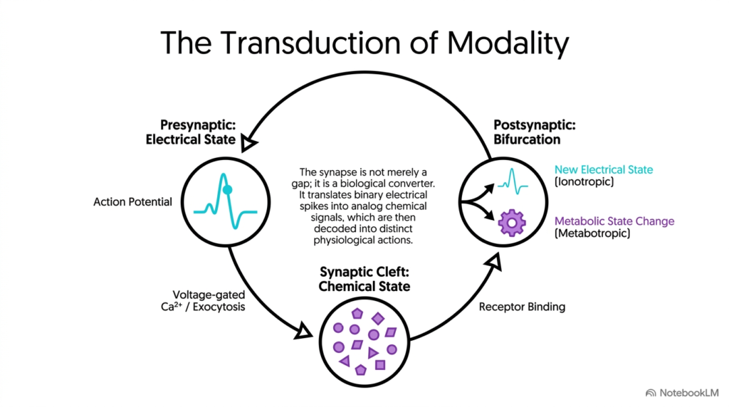
Synapses are not merely simple gaps between cells; they function as sophisticated biological converters that transform physical signal states. The complete cycle of CNS signal transmission illustrates how electrical energy shifts into chemical communication before restoring electrical or metabolic actions in target cells. The presynaptic cell initiates this sequence in an electrical state, converting digital action potentials into calcium-driven exocytosis at the axon terminal.
Within the synaptic cleft, the signal exists briefly in a chemical state as neurotransmitter molecules diffuse passively across the narrow gap. At the postsynaptic membrane, receptor binding converts the chemical message into either a new electrical state via ionotropic channels or a metabolic state change via metabotropic receptors. This cyclic state transition defines the overall process of CNS signal transmission throughout neural circuits.
By translating binary electrical spikes into analog chemical signals, the central nervous system achieves remarkable computational nuance and operational flexibility. Studying this complete circuit of CNS signal transmission allows students to synthesize structural, biochemical, and physiological concepts into a unified model of neural communication. Mastering these foundational principles prepares future clinicians and researchers to solve complex neurological challenges in medical science. Understanding this modality cycle provides an integrated framework for exploring complex central nervous system functions.
💡 Next Step: Would you like to create study flashcards or a practice quiz based on these 14 slides to test key concepts like exocytosis, ionotropic channels, and G-protein cascades?
Please read our Content Disclaimer Statement.
Check out our social media channels: