150. Hydrophilic Hormones: Biochemistry, Receptors, and Signaling
Imagine sending an urgent message to a fortress without ever stepping through its heavy stone gates. Cells perform this exact feat every second using chemical messengers that travel through the bloodstream. This comprehensive slide deck explores the fundamental biochemistry governing water-soluble signaling molecules. By examining their structural classifications, membrane receptor interactions, and intracellular cascade pathways, college and medical students can master how these signals regulate human metabolism and maintain vital body stability.
Slide 1: Biochemistry of Hydrophilic Hormones – Clinical Blueprint, Signaling, and Pathways
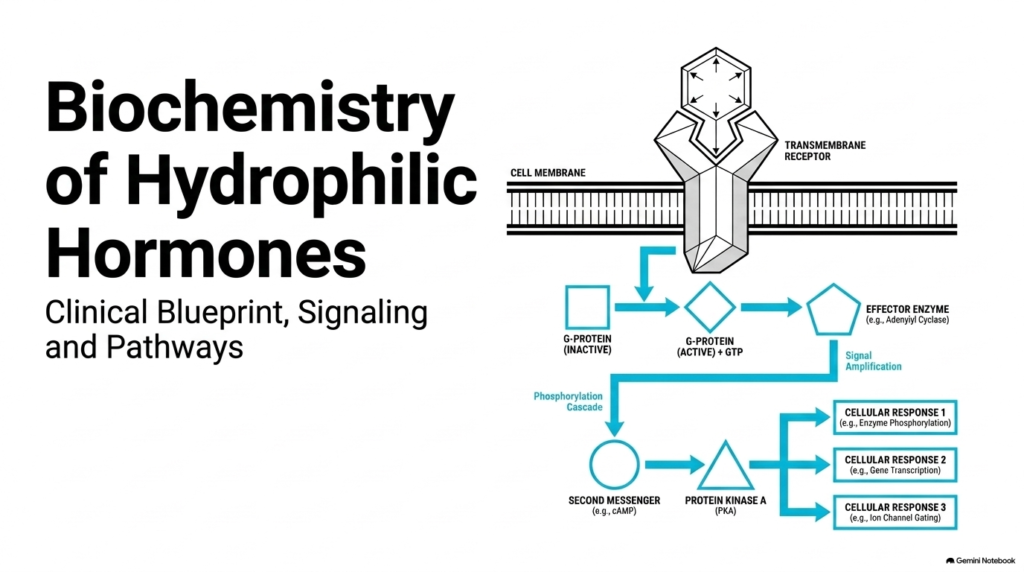
Cells require precise molecular communication networks to coordinate complex physiological processes across distant organ systems. Hydrophilic Hormones serve as primary chemical messengers that circulate freely within the water-based environment of the bloodstream. Because lipid membranes act as physical barriers to polar substances, Hydrophilic Hormones cannot diffuse into target cells and must instead bind to specialized transmembrane receptors embedded in the cell membrane. This extracellular binding alters receptor shape, triggering activation of intracellular G-proteins by promoting the exchange of bound GDP for GTP.
Once stimulated, active G-proteins engage downstream effector enzymes, such as adenylyl cyclase, to produce secondary chemical messengers like cyclic AMP. These second messengers quickly activate intracellular enzymes like Protein Kinase A, initiating a continuous phosphorylation cascade throughout the cytoplasm. This sequential cascade achieves massive signal amplification, allowing minute concentrations of Hydrophilic Hormones to generate profound biological responses. Ultimately, these signal transduction pathways modify metabolic enzyme activity, regulate nuclear gene transcription, and adjust ion channel gating to preserve physiological balance.
Medical students must recognize that this rapid signaling mechanism relies on pre-existing intracellular machinery. By utilizing membrane receptors and enzymatic cascades, Hydrophilic Hormones rapidly adjust cellular functions within seconds or minutes.
Slide 2: The Extracellular Barrier Concept for Hydrophilic Hormones
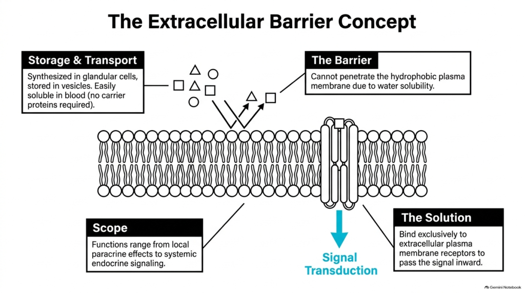
Endocrine organs synthesize chemical messengers in specialized glandular cells and store them within membrane-bound secretory vesicles until physiological triggers stimulate release. Because Hydrophilic Hormones dissolve readily in aqueous environments, they travel through blood plasma without requiring specialized transport proteins. This water solubility allows Hydrophilic Hormones to diffuse rapidly through systemic blood circulation to reach target tissues. However, this same solubility creates a fundamental physical limitation when these signaling molecules reach target cell boundaries.
The hydrophobic lipid bilayer of the cell plasma membrane creates an impassable physical barrier to water-soluble compounds. Consequently, Hydrophilic Hormones cannot cross the plasma membrane to enter the cytoplasm directly. To overcome this obstacle, target tissues express specialized cell-surface membrane receptors that extend into the extracellular space. Hydrophilic Hormones bind exclusively to these external receptor sites, transmitting their physiological instructions into the cell interior strictly through signal transduction mechanisms.
This receptor-mediated signaling mechanism allows water-soluble messengers to exert diverse physiological effects across varying distances. The functional scope of Hydrophilic Hormones ranges from localized paracrine signaling, where molecules act on adjacent neighboring cells, to broad endocrine signaling that coordinates whole-body homeostasis. Understanding this barrier concept is essential for comprehending how target cells remain selective while processing extracellular chemical signals.
Slide 3: Bifurcation of Hydrophilic Hormones into Structural Classes
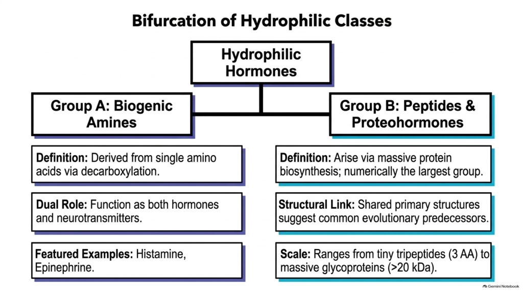
Water-soluble endocrine messengers display significant structural diversity, yet biochemists categorize them into two principal groups based on their synthetic origins. The overarching category of Hydrophilic Hormones splits into Group A, known as biogenic amines, and Group B, comprising peptides and proteohormones. Biogenic amines represent small molecules derived from single amino acids through specific enzymatic decarboxylation reactions. Because these amine molecules retain charged functional groups, Hydrophilic Hormones in this class remain fully soluble in water while acting as both endocrine signals and neural transmitters.
In contrast, Group B includes peptide hormones and larger proteohormones synthesized through classical gene transcription and ribosome translation pathways. This group represents the numerically largest class of hydrophilic hormones in human physiology. Structural analysis reveals striking primary-sequence similarities among many peptide signals, suggesting these molecules evolved from common precursor proteins over time. The structural scale within Group B varies widely, from tiny tripeptides with just three amino acids to complex dimeric glycoproteins exceeding twenty kilodaltons in molecular mass.
Despite their vast difference in molecular size, both biogenic amines and complex proteohormones share core functional characteristics. All Hydrophilic Hormones depend on extracellular receptor interactions because their water-soluble structures prevent passive membrane diffusion. Recognizing this structural classification helps medical students organize dozens of individual chemical signals into logical functional families.
Slide 4: Histamine Profile as Amine-Based Hydrophilic Hormones
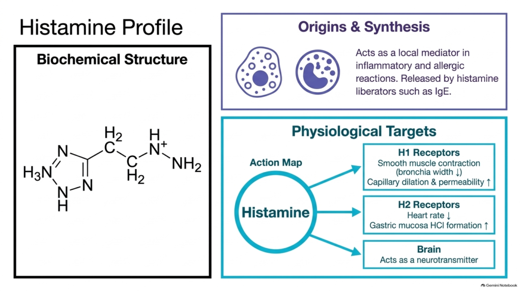
Histamine is a prominent example of an amine-derived Hydrophilic Hormone that regulates vital local physiological and immune processes. Synthesized from the amino acid histidine through enzymatic decarboxylation, histamine is concentrated within tissue mast cells and circulating basophilic granulocytes. Specific triggering stimuli, including tissue injury, immunoglobulin E antibodies, and chemical histamine liberators, prompt these storage vesicles to release histamine into surrounding tissues. As water-soluble Hydrophilic Hormones, these amine molecules dissolve instantly in interstitial fluids to engage nearby cell membrane receptors.
Histamine’s biological actions depend on which receptor subtype is expressed on the target cell membrane. Binding to H1 receptors stimulates smooth muscle contraction in bronchial airways while dilating blood capillaries and increasing vascular permeability. These responses drive classical acute inflammatory and allergic reactions. Conversely, histamine binding to H2 receptors reduces cardiac rate and stimulates parietal cells within the gastric mucosa to secrete hydrochloric acid for digestion.
Beyond its localized tissue roles, histamine acts directly within the central nervous system as an important neurotransmitter. This dual functionality highlights how amino acid-derived hydrophilic hormones can serve simultaneous roles as endocrine, paracrine, and neural signaling agents. Understanding histamine receptor distribution helps clinicians select targeted antihistamines that selectively block specific receptor pathways without disrupting other physiological systems.
In summary, Hydrophilic Hormones like histamine show how simple chemical modifications create versatile signaling molecules that coordinate complex immune, digestive, and neurological functions.
Slide 5: Epinephrine Profile as Stress-Responsive Hydrophilic Hormones
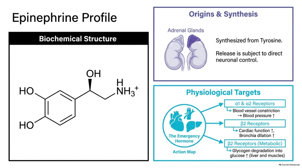
Epinephrine, commonly known as adrenaline, is a central catecholamine belonging to the family of Hydrophilic Hormones that coordinates rapid physiological adaptations during acute stress. Synthesized from the amino acid tyrosine within the adrenal medulla, its secretion is governed by direct sympathetic neural pathways. During acute stress or physical danger, neural impulses trigger the rapid release of epinephrine into systemic circulation. As water-soluble, Hydrophilic Hormones, catecholamines travel rapidly through blood plasma without transport proteins to initiate immediate systemic responses across multiple target organs.
Epinephrine exerts its physiological actions by binding to distinct alpha and beta adrenergic membrane receptors distributed throughout the body. Binding to alpha-1 and alpha-2 receptors causes vascular smooth muscle constriction, which increases systemic blood pressure. Simultaneously, stimulation of beta-2 adrenergic receptors increases heart rate and cardiac output while dilating bronchial airways to maximize tissue oxygenation. These combined cardiovascular and respiratory adjustments ensure optimal delivery of oxygenated blood to essential skeletal muscles.
In addition to cardiovascular changes, Hydrophilic Hormones like epinephrine drive profound metabolic reprogramming during emergencies. Epinephrine binds to hepatic and muscle beta-2 receptors to accelerate glycogen breakdown into free glucose while stimulating lipolysis in adipose tissue. This metabolic shift floods the bloodstream with energy substrates to fuel physical exertion. Because hydrophilic hormones act through cell-surface receptors and rapid enzymatic cascades, these metabolic and cardiovascular changes occur within seconds, providing a survival advantage during acute stress.
Slide 6: The Scale of Proteohormones among Hydrophilic Hormones
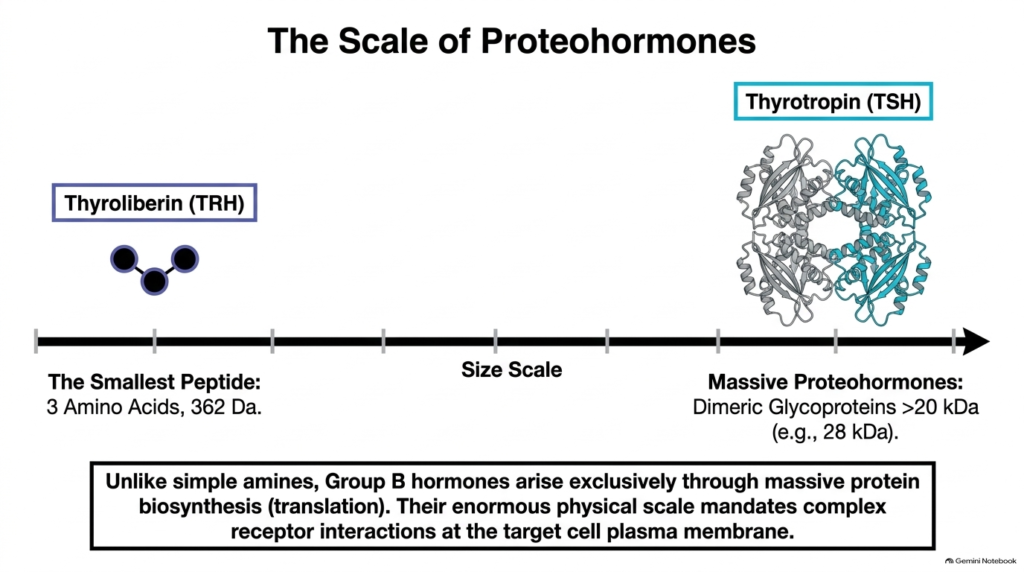
Peptide and protein signaling molecules vary widely in size, illustrating the broad structural spectrum of Hydrophilic Hormones. Unlike biogenic amines derived from single amino acids, these larger messengers are generated exclusively through gene transcription and ribosomal protein translation. The smallest representative in this category is thyroliberin, a tiny tripeptide composed of just three modified amino acids with a molecular mass of 362 Daltons. Because Hydrophilic Hormones of this size are so compact, they interact efficiently with specialized pituitary membrane receptors.
At the opposite extreme of this size continuum are massive proteohormones like thyrotropin, a dimeric glycoprotein with a molecular mass exceeding twenty-eight kilodaltons. Thyrotropin consists of two distinct peptide chains: an alpha chain composed of ninety-two amino acids and a beta chain containing one hundred twelve amino acids. Despite this massive physical structure, thyrotropin remains fully soluble in aqueous blood plasma because its outer protein surface displays abundant hydrophilic amino acid side chains and carbohydrate moieties.
This immense physical scale forces large Hydrophilic Hormones to utilize highly specific, multi-point binding interactions at target cell plasma membranes. Their size prevents membrane penetration, reinforcing total reliance on extracellular signal transduction. Examining this structural spectrum allows students to appreciate how ribosomal synthesis generates functional diversity among Hydrophilic Hormones while maintaining strict solubility and receptor specificity across all molecular sizes.
Slide 7: The Endocrine Cascade Part I – Hypothalamic Hydrophilic Hormones
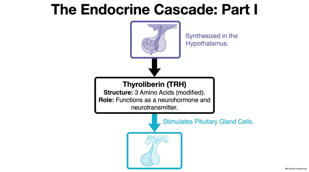
The endocrine system relies on multi-tiered signaling cascades where neurohormones from the brain trigger downstream hormone release in peripheral glands. At the top of this regulatory hierarchy is the hypothalamus, which synthesizes small peptide neurohormones like thyroliberin. Thyroliberin consists of three modified amino acids and functions as both a hypothalamic neurohormone and a central nervous system neurotransmitter. As water-soluble Hydrophilic Hormones, hypothalamic releasing factors diffuse through local blood vessels without carrier proteins to reach nearby target cells rapidly.
After synthesis, thyroliberin travels through the hypophyseal portal system directly to the anterior pituitary gland, also known as the adenohypophysis. Here, thyroliberin binds to specific cell-surface receptors on pituitary cells, activating intracellular signal transduction pathways. This binding event triggers the synthesis and exocytosis of secondary trophic hormones into the systemic circulation. Because hydrophilic hormones act through receptor-mediated mechanisms, minute amounts of hypothalamic releasing factors can induce significant glandular responses.
This initial step demonstrates how Hydrophilic Hormones orchestrate complex neuroendocrine feedback loops. By converting neural signals into endocrine secretions, hypothalamic peptide messengers precisely control downstream endocrine organs. Understanding this primary cascade stage helps medical students trace how brain inputs regulate metabolic rate, growth, and physiological adaptation throughout the entire human body.
Furthermore, the high solubility of Hydrophilic Hormones in portal blood ensures rapid transport from hypothalamic nerve terminals to anterior pituitary target cells.
Slide 8: The Endocrine Cascade Part II – Pituitary Hydrophilic Hormones
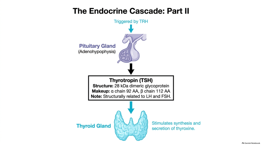
Building upon the initial hypothalamic trigger, the second tier of the neuroendocrine cascade involves the secretion of complex pituitary proteohormones. In response to thyroliberin stimulation, adenohypophyseal cells release thyrotropin, also known as thyroid-stimulating hormone. Thyrotropin is a 28-kilodalton dimeric glycoprotein composed of an alpha chain with 92 amino acids and a beta chain with 112 amino acids. These large Hydrophilic Hormones share structural homology with other pituitary glycoprotein signals, including luteinizing hormone and follicle-stimulating hormone.
Once secreted into systemic blood vessels, thyrotropin circulates freely to reach its primary target, the thyroid gland. Upon arrival, thyrotropin binds specifically to thyroid cell membrane receptors, activating G-protein-coupled signaling pathways that promote the synthesis and release of thyroid hormones like thyroxine. Because Hydrophilic Hormones like thyrotropin cannot cross cell membranes, their extracellular interaction with thyroid receptors is mandatory to stimulate key enzymatic processes involved in thyroid hormone production.
This secondary tier highlights the amplification capability inherent in hierarchical endocrine systems. A small signal from hypothalamic neurohormones induces larger secretions of pituitary proteohormones, which in turn drive broad metabolic modulation via thyroid hormones. Recognizing how Hydrophilic Hormones function within multi-step cascades helps students comprehend clinical endocrine disorders caused by receptor mutations or pituitary dysfunction.
In conclusion, Hydrophilic Hormones operating at the pituitary level serve as indispensable regulatory bridges linking central hypothalamic command to target organ hormone secretion.
Slide 9: Insulin Profile and Metabolic Regulation by Hydrophilic Hormones
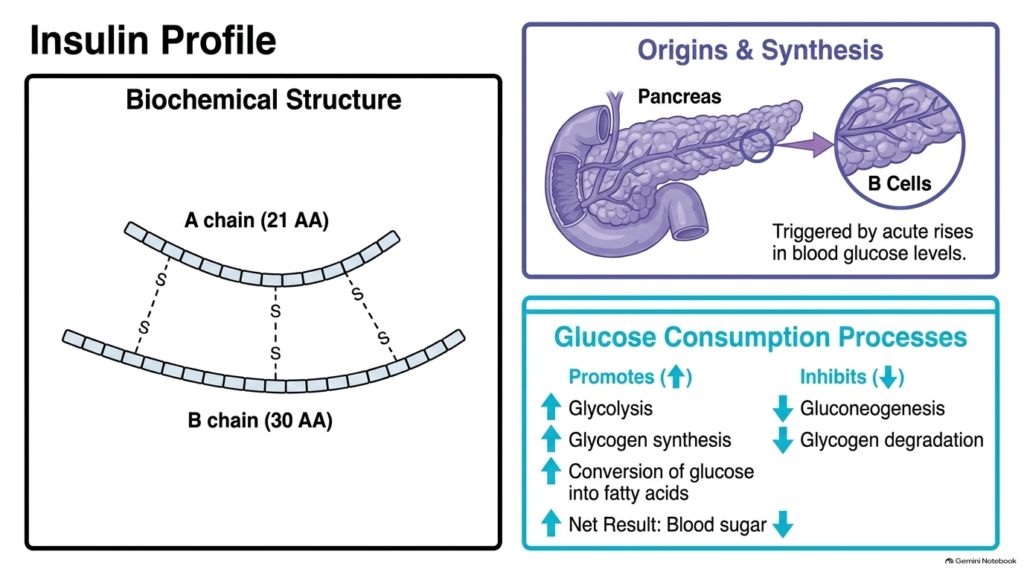
Insulin is a vital pancreatic peptide hormone that drives glucose storage and energy conservation in human physiology. Synthesized within the beta cells of the pancreatic islets, insulin consists of two distinct peptide chains: an A chain of twenty-one amino acids and a B chain of thirty amino acids linked by disulfide bonds. Acute elevations in blood glucose levels trigger pancreatic beta cells to release stored insulin into the bloodstream. As key Hydrophilic Hormones, insulin molecules travel rapidly through blood plasma without transport proteins to act on liver, muscle, and adipose tissues.
Insulin lowers blood glucose by binding to tyrosine kinase receptors on target cell plasma membranes, stimulating intracellular glucose uptake and metabolic utilization. This signaling cascade upregulates glucose consumption pathways, promoting glycolysis, glycogen synthesis, and the conversion of surplus glucose into fatty acids. Concurrently, insulin signaling suppresses energy-mobilizing pathways by inhibiting gluconeogenesis and glycogen breakdown. Because Hydrophilic Hormones like insulin bind externally, they alter metabolic enzyme activities through rapid phosphorylation cascades without requiring initial gene transcription.
Insulin’s physiological impact shows how Hydrophilic Hormones coordinate systemic nutrient storage after meals. By directing cells to store excess nutrients as glycogen and triglycerides, insulin maintains normoglycemia and prevents glucose toxicity. Understanding insulin biochemistry provides medical students with a foundation for diagnosing and treating metabolic diseases such as type 1 and type 2 diabetes mellitus.
Proper regulation by Hydrophilic Hormones ensures that cellular energy intake matches whole-body metabolic demand seamlessly.
Slide 10: Glucagon Profile and Glucose Mobilization by Hydrophilic Hormones
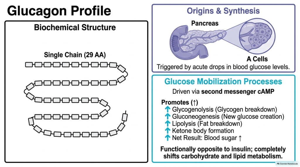
Glucagon is a single-chain peptide hormone composed of twenty-nine amino acids that acts as the primary physiological antagonist to insulin. Produced and secreted by alpha cells within the pancreatic islets, glucagon release is triggered by acute drops in blood glucose levels. When blood sugar falls below normal physiological thresholds, pancreatic alpha cells discharge stored glucagon into circulation. As water-soluble, Hydrophilic Hormones, glucagon peptides dissolve easily in blood plasma and travel swiftly to primary target-cell receptors in hepatic tissue.
Upon reaching liver cells, glucagon binds to specific G-protein-coupled membrane receptors, activating adenylyl cyclase to generate the intracellular second messenger cyclic AMP. Increased cyclic AMP levels stimulate protein kinase A, triggering enzymatic cascades that promote glucose mobilization. Glucagon signaling accelerates glycogen breakdown and stimulates new glucose synthesis via gluconeogenesis. Additionally, it enhances fat breakdown and drives ketone body formation in the liver. Because Hydrophilic Hormones like glucagon use second-messenger cascades, hepatic glucose release increases within minutes of hormone binding.
This metabolic mobilization restores circulating blood glucose to safe physiological levels during periods of fasting or intense exercise. Hydrophilic Hormones such as glucagon prevent dangerous hypoglycemia by converting stored hepatic glycogen into free glucose for brain and peripheral tissue energy. Mastering glucagon signaling pathways is crucial for medical students analyzing endocrine responses to starvation and diabetic ketoacidosis.
In summary, Hydrophilic Hormones responsible for glucose mobilization ensure continuous energy availability despite fluctuating dietary nutrient intake.
Slide 11: The Pancreatic Antagonism Matrix of Hydrophilic Hormones
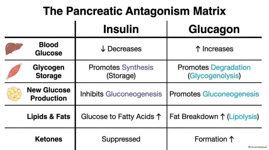
Metabolic homeostasis relies on dynamic antagonism between counter-regulatory pancreatic peptides that oppose each other in controlling nutrient flux. Insulin and glucagon represent classic paired Hydrophilic Hormones that balance blood glucose concentration through reciprocal metabolic pathways. Insulin lowers blood glucose by promoting cellular glucose uptake and nutrient storage, while glucagon raises circulating blood sugar by stimulating hepatic glucose release. These opposing actions keep blood glucose within a narrow physiological range regardless of feeding or fasting.
This functional antagonism extends across carbohydrate, lipid, and ketone metabolic pathways. In glycogen metabolism, insulin promotes glycogen synthesis while glucagon accelerates glycogen degradation into glucose. Regarding new glucose production, insulin inhibits gluconeogenesis, whereas glucagon strongly stimulates gluconeogenic enzyme activity through distinct hydrophilic hormone receptor pathways. For lipid metabolism, insulin directs excess glucose into fatty acid synthesis, while glucagon promotes adipose fat breakdown via lipolysis. Furthermore, insulin suppresses ketone production, whereas glucagon stimulates hepatic ketone body formation during prolonged energy deficits.
Because both pancreatic messengers are water-soluble, Hydrophilic Hormones, their circulating ratio determines the net metabolic state of target organs. Target liver cells continuously integrate signals from both hormone receptors, adjusting intracellular metabolic flux accordingly. Understanding this antagonism matrix provides medical students with an intuitive framework for evaluating metabolic diseases, therapeutic insulin administration, and physiological adaptations to nutrient fasting.
Ultimately, balance between these Hydrophilic Hormones prevents extreme metabolic swings between hyperglycemia and hypoglycemia.
Slide 12: Master Classification Matrix of Hydrophilic Hormones
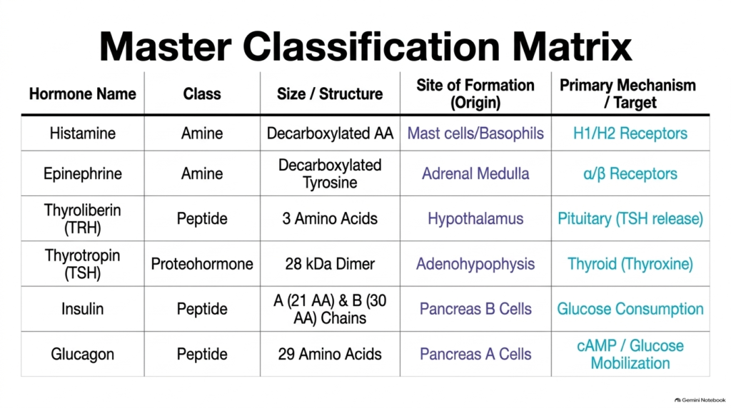
To synthesize these diverse endocrine concepts, biochemists utilize a master classification matrix that organizes water-soluble signaling molecules by structure, origin, and mechanism. This integrated summary shows how Hydrophilic Hormones span distinct chemical classes, from simple amine derivatives to complex proteohormones. Biogenic amines like histamine and epinephrine come from decarboxylated amino acids and act on cell-surface adrenergic or histamine receptors. Small peptides like thyroliberin and glucagon consist of short amino acid chains and operate through hypothalamic or pancreatic pathways to regulate trophic secretions and blood sugar balance.
Larger proteohormones, including thyrotropin and insulin, have complex multi-chain or dimeric structures synthesized through ribosomal translation. Thyrotropin exists as a twenty-eight-kilodalton dimer produced by the adenohypophysis, whereas insulin features two disulfide-linked peptide chains produced by pancreatic beta cells. Despite structural differences, all hydrophilic hormones must bind to extracellular plasma membrane receptors to initiate intracellular signal transduction cascades.
Comparing these signaling molecules in a unified matrix reveals the logical design governing human endocrine control. By mapping structural dimensions to specific glandular origins and receptor mechanisms, students can quickly predict how different Hydrophilic Hormones alter cellular metabolism. This master matrix serves as an essential reference tool for medical students preparing for clinical examinations and endocrinology rotations.
In conclusion, mastering this classification enables clinicians to diagnose receptor defects, hormone deficiencies, and metabolic imbalances across all Hydrophilic Hormones.
Please read our Content Disclaimer Statement.
Check out our social media channels: