152. Peptide Hormone Metabolism: Biosynthesis, Cleavage, and Degradation
Endocrine signaling keeps the human body balanced, but how do cells manufacture and clear precise chemical messages? Understanding Peptide Hormone Metabolism explains how tissue signals originate, function, and dissipate. This slide deck breaks down the full life cycle of water-soluble signals, moving step by step from nuclear gene expression to final lysosomal destruction. Students will gain a clear biochemical framework for precursor polyprotein processing, regulated exocytosis, and rapid systemic clearance. Let us explore these essential pathways that preserve metabolic harmony across organs.
Slide 1: An Overview of Peptide Hormone Metabolism
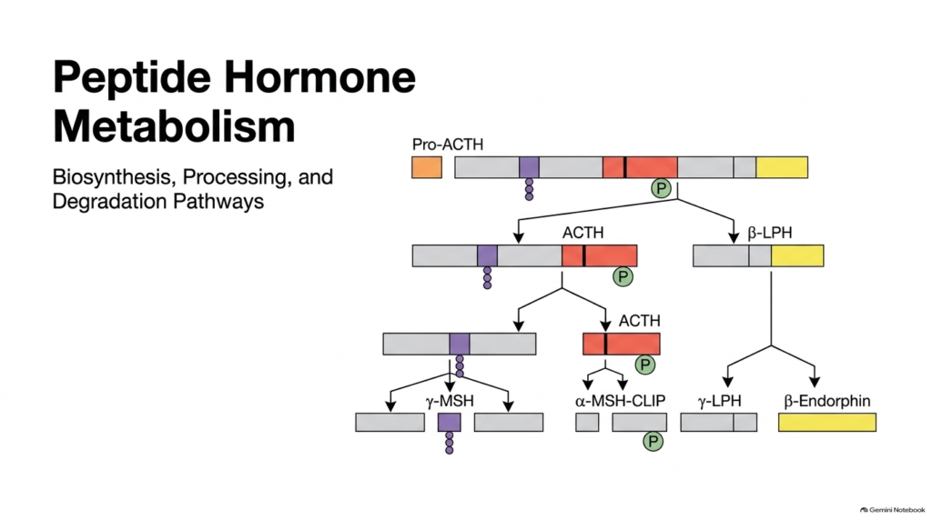
The complete framework of Peptide Hormone Metabolism spans every vital stage of a signaling molecule’s life cycle, from initial nuclear gene transcription to final enzymatic breakdown. Cells manufacture large precursor proteins, process them into biologically active forms, store them in secretory vesicles, and eventually dismantle them to terminate signal transmission. This comprehensive overview highlights how a single polyprotein precursor gives rise to distinct regulatory peptides through targeted cleavage events. Understanding Peptide Hormone Metabolism requires analyzing both intracellular synthesis and extracellular clearance.
Inside the cell’s secretory pathway, specific enzymes break complex precursor chains into functional fragments such as adrenocorticotropic hormone, melanocyte-stimulating hormones, and various endorphins. Once these signaling peptides complete their physiological work in systemic circulation, target tissues and excretory organs rapidly inactivate them. Overall, Peptide Hormone Metabolism coordinates precise cellular timing so that endocrine signals act only as long as necessary. Plasma peptidases and cell membrane receptors prevent overstimulation by systematically removing circulating signaling molecules.
By studying these linked pathways, students discover how cellular production and systemic breakdown work together to preserve physiological balance across human organ systems. The entire lifecycle relies on tight enzymatic control, ensuring that hormone concentrations match real-time metabolic demands without causing prolonged cellular activation across the body.
Slide 2: Taxonomy of Hydrophilic Molecules in Peptide Hormone Metabolism
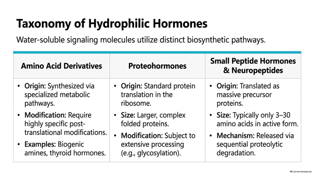
Water-soluble signaling molecules fall into three distinct structural classes, each following specific production pathways in peptide hormone metabolism. Amino acid derivatives arise through specialized metabolic steps or post-translational modifications of single amino acid residues. Examples include catecholamines and thyroid hormones, which regulate metabolic rate and physiological stress responses. In contrast, larger proteohormones undergo standard ribosomal translation like typical cellular proteins before undergoing folding and extensive post-translational modifications such as glycosylation.
Small peptide hormones and neuropeptides represent a unique category within Peptide Hormone Metabolism. These active molecules typically consist of only three to thirty amino acids in their functional state. However, cells do not synthesize these short sequences directly as individual small units. Instead, ribosomes translate large precursor proteins that embed the active sequences within longer polypeptide chains. Sequential proteolytic degradation then releases the functional peptides when the cell requires them.
Understanding this taxonomy clarifies why different signaling molecules exhibit distinct lifespans and regulatory mechanisms. While simple amino acid derivatives rely on dedicated synthetic enzymes, short neuropeptides require elaborate precursor-processing cascades within specialized organelles. Broadly speaking, Peptide Hormone Metabolism uses these varied structural frameworks to match the specific physiological demands of diverse endocrine and neural communication pathways throughout the human body.
Slide 3: Proopiomelanocortin as a Model in Peptide Hormone Metabolism
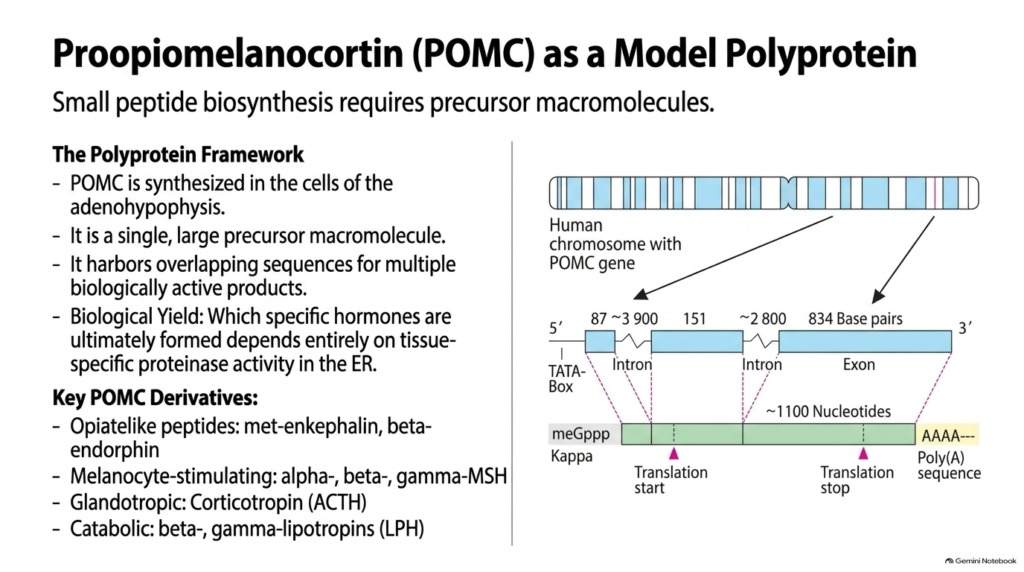
Proopiomelanocortin, commonly abbreviated as POMC, serves as a classic model for studying precursor polyproteins in Peptide Hormone Metabolism. Cells in the adenohypophysis, or anterior pituitary gland, synthesize this single large precursor macromolecule. The POMC sequence harbors overlapping regions that encode multiple biologically active signaling units. These derivative products include opiate-like peptides such as met-enkephalin and beta-endorphin, melanocyte-stimulating hormones, adrenocorticotropic hormone, and catabolic lipotropins that act on adipose tissue.
The genomic architecture of POMC illustrates how cells store multiple hormonal messages within one gene during Peptide Hormone Metabolism. On human chromosomes, the POMC gene is transcribed to produce a pre-mRNA transcript. Splicing removes non-coding introns spanning thousands of base pairs, generating a mature messenger RNA transcript of roughly eleven hundred nucleotides. This modified transcript features a 5-prime cap and a 3-prime poly-A tail to ensure mRNA stability during cytosolic transport to ribosomes.
Which specific hormones are generated from POMC depends entirely on the tissue-specific proteinases active within the endoplasmic reticulum and secretory granules. Different cell types express distinct cleavage enzymes, allowing pituitary cells to produce stress hormones while hypothalamic neurons yield appetite-regulating peptides. Thus, Peptide Hormone Metabolism relies on selective cleavage to extract customized chemical messages from a single universal polyprotein template.
Slide 4: Genomic Transcription and Splicing in Peptide Hormone Metabolism
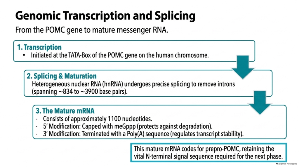
The initial phase of Peptide Hormone Metabolism begins inside the nucleus with gene transcription and RNA processing. Transcription initiates at the TATA-box promoter region of the POMC gene on the chromosome. RNA polymerase reads the DNA template to construct a primary heterogeneous nuclear RNA transcript. This initial transcript contains both coding exon regions and non-coding intron sequences that span between eight hundred and nearly four thousand base pairs.
To prepare the transcript for protein translation during Peptide Hormone Metabolism, nuclear spliceosomes precisely excise the intron sequences. Splicing joins the exon fragments, reducing the transcript to a mature messenger RNA molecule of about 1,100 nucleotides. Enzymatic modifications at both ends protect the mRNA from premature degradation in the cytoplasm. A modified guanosine cap attaches to the 5-prime end, while a poly-A tail attaches to the 3-prime end to regulate transcript longevity and translation efficiency.
This mature messenger RNA carries the exact genetic code required to synthesize prepro-POMC. Crucially, the transcript retains the sequence for an N-terminal signal peptide that directs the nascent protein into the cellular secretory pathway. Without these initial nuclear processing steps, Peptide Hormone Metabolism could not successfully direct newly translated polypeptide chains toward specialized ER compartments for downstream processing and maturation.
Slide 5: Translating Prepro-POMC in Peptide Hormone Metabolism
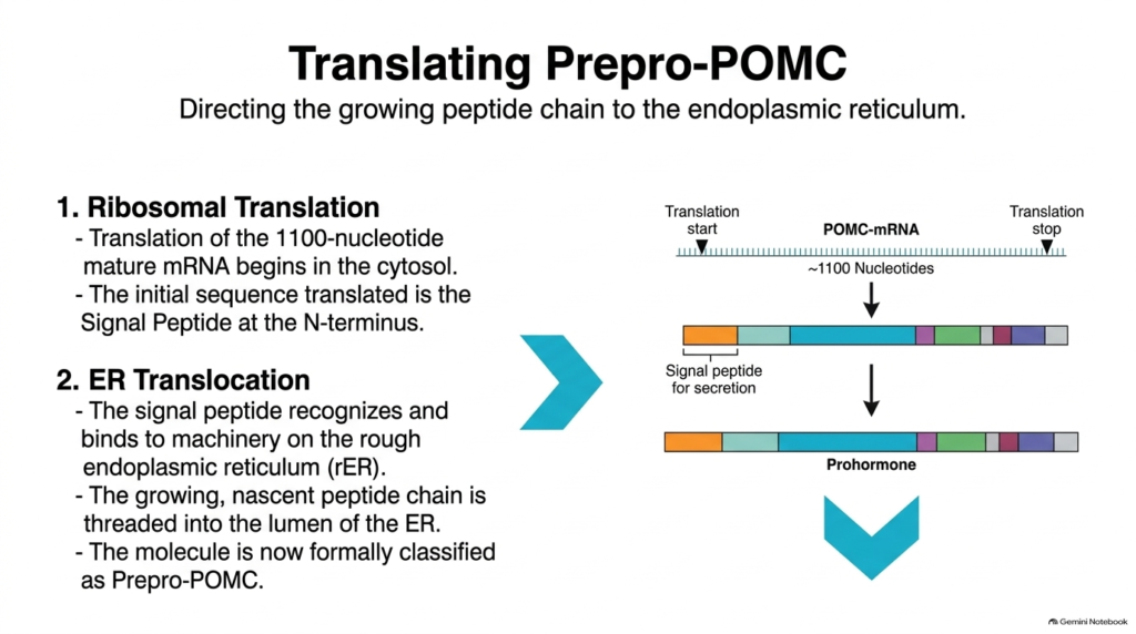
Protein synthesis marks the transition from nuclear genetic encoding to active polypeptide assembly in Peptide Hormone Metabolism. Translation begins when a cytosolic ribosome binds the mature eleven-hundred-nucleotide mRNA near its 5-prime cap. As the ribosome reads the transcript starting at the initiation codon, it first translates a specialized hydrophobic amino acid sequence located at the N-terminus. This leading sequence is known as the signal peptide for secretion.
The N-terminal signal peptide acts as a molecular passport during Peptide Hormone Metabolism. Signal recognition particles recognize this hydrophobic sequence as it emerges from the ribosome and temporarily pause translation. The ribosome-protein complex then docks onto receptor machinery located on the membrane of the rough endoplasmic reticulum. Once docked, translation resumes, and the growing polypeptide chain is threaded directly through a protein translocon channel into the endoplasmic reticulum lumen.
At this stage, the growing protein is classified as prepro-POMC. Co-translational insertion into the ER lumen segregates the hormone precursor away from cytosolic enzymes and directs it into the secretory pathway. By isolating the polypeptide within membrane-bound organelles, Peptide Hormone Metabolism ensures that early precursor processing occurs in a controlled environment tailored for protein folding and modification.
Slide 6: ER Modifications and Prohormone Formation in Peptide Hormone Metabolism
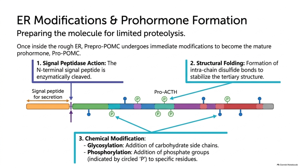
Once prepro-POMC enters the rough endoplasmic reticulum, crucial structural changes transform the molecule during Peptide Hormone Metabolism. First, a membrane-bound signal peptidase enzyme clips off the N-terminal signal peptide. Removing this signal sequence converts the precursor into the mature prohormone, known as pro-POMC or pro-ACTH. Freed from its hydrophobic targeting sequence, the prohormone folds into its native three-dimensional conformation within the endoplasmic reticulum lumen.
Structural stabilization requires specific covalent modifications essential to Peptide Hormone Metabolism. Resident ER enzymes form intra-chain disulfide bonds between cysteine residues, locking the prohormone into a stable tertiary structure. In addition, glycosylation enzymes attach carbohydrate side chains to specific amino acid residues along the polypeptide backbone. Phosphorylation also occurs, adding phosphate groups to target sites to influence protein stability and target recognition.
These chemical modifications protect the prohormone from nonspecific cytosolic degradation and prepare it for selective enzymatic cleavage in secretory vesicles. Phosphorylation and glycosylation mark specific regions of the precursor polypeptide, guiding downstream proteases toward correct cleavage sites. Through these coordinated ER modifications, Peptide Hormone Metabolism refines the precursor molecule so that subsequent limited proteolysis yields precise, biologically active hormone products ready for cellular secretion.
Slide 7: Limited Proteolysis and POMC Cleavage in Peptide Hormone Metabolism
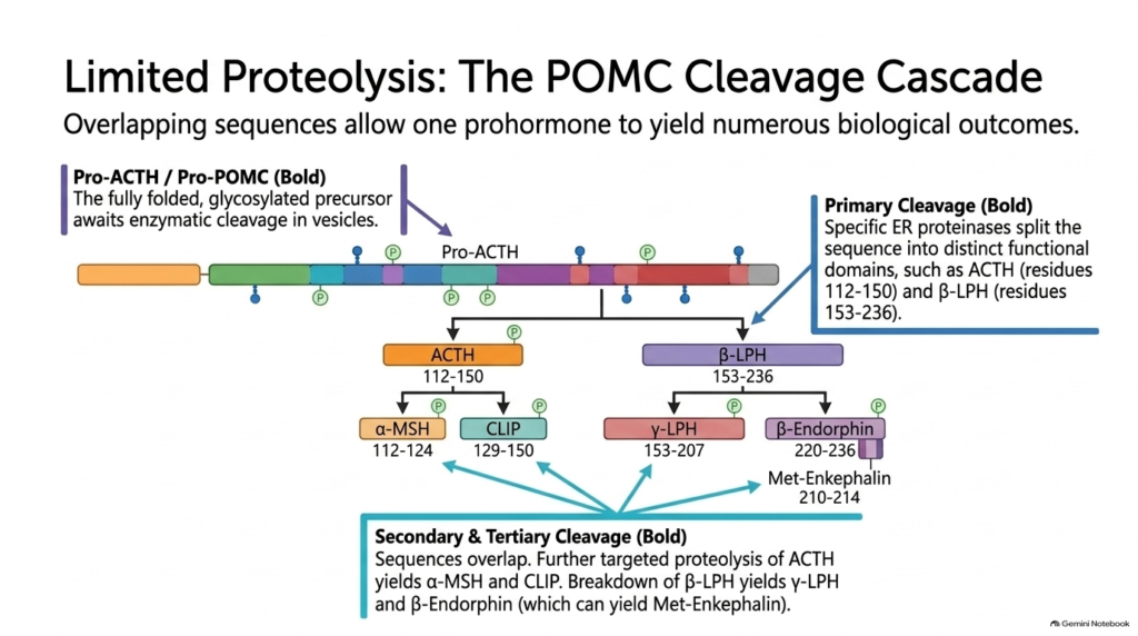
The central mechanism for producing functional signaling units in Peptide Hormone Metabolism is limited proteolysis. Unlike non-specific protein digestion, limited proteolysis breaks specific peptide bonds at designated cleavage sites while leaving the rest of the protein intact. Pro-POMC moves into specialized Golgi compartments and dense-core secretory granules, where tissue-specific endopeptidases perform primary cleavage. This primary cleavage splits pro-POMC into adrenocorticotropic hormone (residues 112 to 150) and beta-lipotropin (residues 153 to 236).
Depending on the cell type and its enzyme profile, secondary and tertiary cleavage steps occur during peptide hormone metabolism. In the intermediate pituitary or hypothalamus, further targeted proteolysis of adrenocorticotropic hormone yields alpha-melanocyte-stimulating hormone and corticotropin-like intermediary peptide. Simultaneously, enzymatic breakdown of beta-lipotropin produces gamma-lipotropin and beta-endorphin. Beta-endorphin can undergo additional processing to generate met-enkephalin, a potent opioid peptide that modulates pain perception.
Because the precursor polypeptide harbors overlapping functional sequences, one prohormone template yields diverse regulatory products. The specific mixture of hormones secreted depends entirely on which cleavage enzymes a particular tissue expresses. Thus, Peptide Hormone Metabolism uses limited proteolysis to generate distinct physiological responses across organ systems, ensuring cells produce the signaling peptides needed for homeostatic control.
Slide 8: Regulated Hormone Exocytosis in Peptide Hormone Metabolism
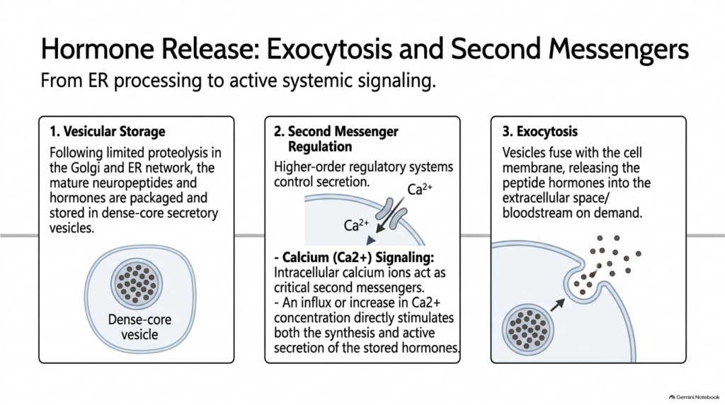
Following limited proteolysis in the Golgi apparatus and secretory network, newly generated signaling peptides enter the storage phase of Peptide Hormone Metabolism. Cells package mature neuropeptides and hormones into specialized dense-core secretory vesicles. These vesicles accumulate in the cytoplasm, holding concentrated hormone supplies until the cell receives an explicit extracellular signal to release them. Storage in secretory vesicles prevents continuous, unregulated hormone leakage into circulation.
Releasing stored hormones requires tight second-messenger regulation, a fundamental feature of Peptide Hormone Metabolism. Higher-order regulatory systems, such as neural inputs or hypothalamic releasing factors, control secretion by altering intracellular ion concentrations. Calcium ions act as critical second messengers in this process. When an activating signal arrives, calcium channels open, allowing calcium ions to enter the cytosol. Elevated calcium concentrations stimulate vesicle trafficking and trigger both hormone synthesis and active secretion.
In response to rising intracellular calcium levels, dense-core vesicles undergo exocytosis. The vesicle membranes fuse with the plasma membrane, releasing their soluble hormone contents directly into the extracellular space and surrounding blood capillaries. Through this regulated mechanism, Peptide Hormone Metabolism ensures rapid, demand-based hormone delivery into systemic circulation while keeping baseline hormone levels low between physiological stimulation events.
Slide 9: Systemic Degradation and Inactivation in Peptide Hormone Metabolism
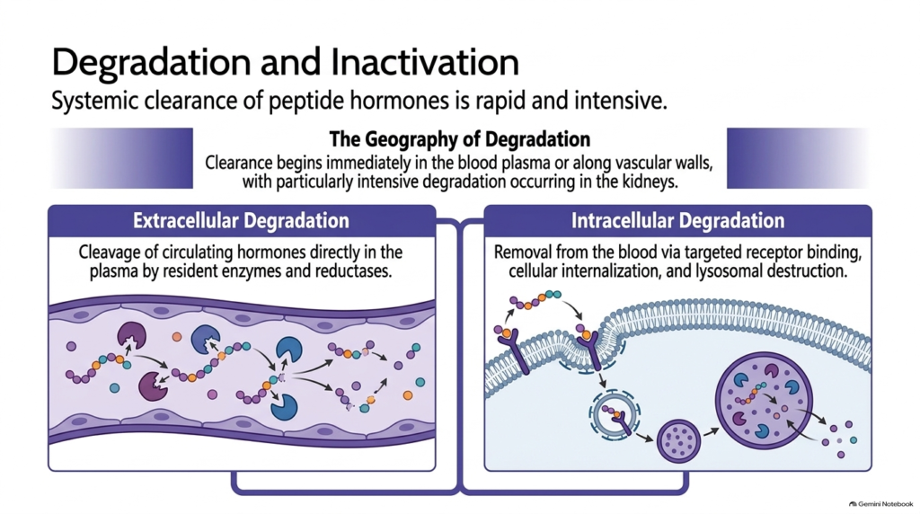
Secretion is only half the story; efficient signal termination is equally vital in Peptide Hormone Metabolism. Once peptide hormones circulate and deliver their chemical messages to target tissues, they must be rapidly cleared to prevent continuous cellular stimulation. Systemic clearance begins immediately in the blood plasma and along vascular endothelial walls. Excretory and metabolic organs, particularly the kidneys and liver, exhibit intense inactivating activity, rapidly removing active peptides from circulation.
The clearance pathways of peptide hormone metabolism fall into two primary modes: extracellular degradation and intracellular degradation. Extracellular degradation occurs directly within circulating blood plasma or on tissue surfaces. Resident plasma proteinases, peptidases, and reductases cleave peptide bonds or disulfide linkages, converting active hormones into inactive peptide fragments. This extracellular breakdown ensures that circulating hormone concentrations decline rapidly once glandular secretion stops.
In contrast, intracellular degradation relies on active involvement of target cells. Target tissues clear circulating hormones by binding them to specific cell membrane receptors, internalizing the hormone-receptor complexes, and routing them to lysosomes for complete enzymatic destruction. Together, these complementary clearance mechanisms in Peptide Hormone Metabolism maintain tight systemic control over hormone half-lives, allowing endocrine signals to switch off promptly when physiological conditions normalize.
Slide 10: Extracellular Proteolysis and Reduction in Peptide Hormone Metabolism
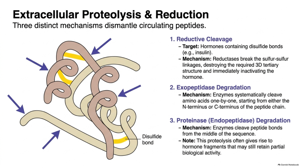
Extracellular inactivation employs three distinct enzymatic mechanisms to dismantle circulating signaling peptides during Peptide Hormone Metabolism. First, hormones that rely on intra-chain or inter-chain disulfide bonds for their active tertiary structure undergo reductive cleavage. Specific reductase enzymes break these sulfur-sulfur linkages. For example, insulin loses its functional three-dimensional shape and biological activity immediately upon disulfide-bond reduction, rendering it incapable of binding target cell receptors.
Second, exopeptidase degradation systematically dismantles peptide chains during Peptide Hormone Metabolism. Exopeptidases attack the ends of circulating peptides, trimming amino acids one by one from either the N-terminus or C-terminus. Third, endopeptidases, also called proteinases, cleave peptide bonds in the middle of the amino acid sequence. Proteinase activity splits long hormones into smaller peptide fragments, drastically reducing their affinity for target cell receptors.
An interesting feature of endopeptidase degradation is that initial cleavage fragments sometimes retain partial or modified biological activity before complete breakdown occurs. However, ongoing cleavage by surrounding exopeptidases rapidly reduces these intermediary fragments to inactive short oligopeptides. Through these coordinated enzymatic steps, Peptide Hormone Metabolism ensures that circulating hormones are broken down effectively within blood plasma, vascular walls, and renal filtering tissues.
Slide 11: Receptor-Mediated Intracellular Degradation in Peptide Hormone Metabolism
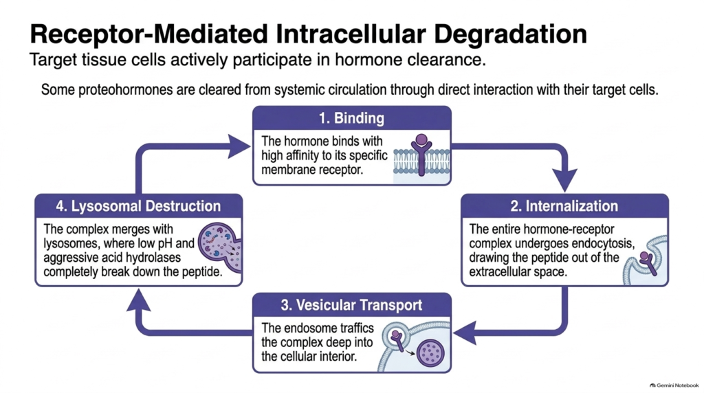
In addition to plasma proteolysis, target tissues actively clear hormones through receptor-mediated endocytosis, a key intracellular branch of Peptide Hormone Metabolism. The process begins when a circulating peptide hormone binds with high affinity to its specific transmembrane receptor on the outer plasma membrane of a target cell. Receptor binding triggers local cell membrane invagination, engulfing the entire hormone-receptor complex into a specialized endocytic vesicle.
Internalization removes the active hormone from the extracellular fluid, immediately terminating its signaling action at the cell surface during Peptide Hormone Metabolism. Once inside the cytoplasm, the endocytic vesicle fuses with early endosomes, which traffic the internalized hormone-receptor complex deeper into the cellular interior. As the endosome matures, active proton pumps lower the internal pH, causing many peptide hormones to dissociate cleanly from their membrane receptors.
Finally, the endosome fuses with a lysosome, exposing the internalized peptide to an acidic environment filled with aggressive acid hydrolases. These lysosomal enzymes rapidly degrade the peptide hormone into individual amino acid components. Meanwhile, freed receptors are often recycled back to the cell surface. Through this endocytic pathway, Peptide Hormone Metabolism couples target tissue response directly with hormone destruction, ensuring efficient receptor turnover and signal termination.
Slide 12: The Metabolic Loop in Peptide Hormone Metabolism
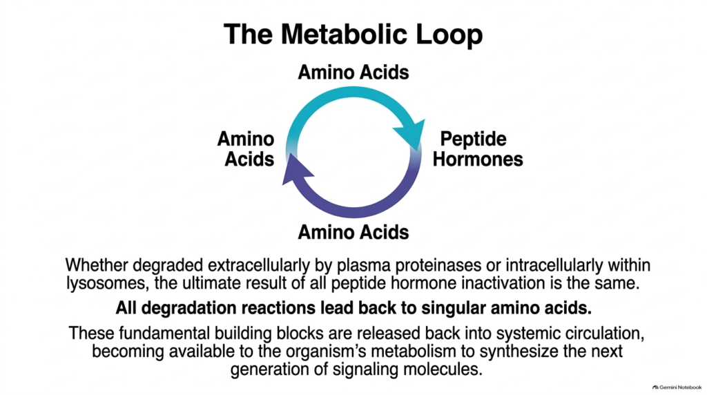
The complete lifecycle of water-soluble signaling molecules concludes in a continuous biochemical cycle central to Peptide Hormone Metabolism. Whether degraded extracellularly by plasma proteinases or intracellularly within lysosomal compartments, all peptide hormone inactivation ultimately yields the same biochemical result. Each cleavage pathway reduces complex signaling molecules to individual free amino acids. This highly efficient biological recycling system wastes nothing.
These liberated amino acids re-enter systemic circulation and join the body’s general metabolic nutrient pools during Peptide Hormone Metabolism. Cells throughout the organism import these basic building blocks through membrane transporters to support ongoing cellular processes, energy generation, and protein translation. Specifically, endocrine cells reuse these recycled amino acids to construct new preprohormone chains, completing the continuous metabolic loop that connects hormone synthesis, secretion, cellular action, and eventual degradation.
Understanding this closed loop reinforces why clearance is just as critical as synthesis in endocrine control. Systemic homeostasis depends on the rapid turnover of chemical messengers so that body tissues can respond dynamically to changing internal and external demands. Ultimately, Peptide Hormone Metabolism demonstrates how organisms efficiently conserve precious molecular resources while maintaining precise, real-time control over systemic endocrine communication across all organ systems.
Please read our Content Disclaimer Statement.
Check out our social media channels: