134. The Extracellular Matrix: Structure, Components, and Functions
Cells do not float in empty space inside human tissues. Instead, cells depend on a supportive physical network that surrounds them, provides structural stability, and directs cellular behavior. This slide deck explores the fundamental biochemistry of tissue scaffolding. By examining key structural proteins, adhesive linkers, and hydrated carbohydrate complexes, students will gain a thorough understanding of how extracellular molecular architecture supports cellular survival, tissue organization, and organ function.
Slide 01: Introduction to the Extracellular Matrix
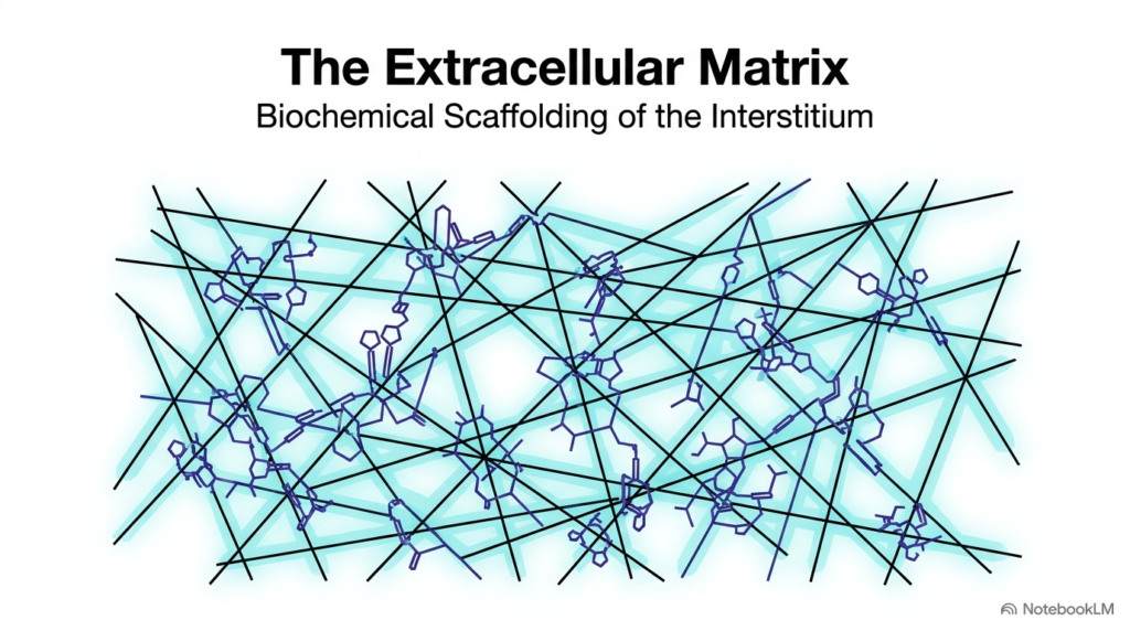
Biological tissues depend heavily on a sophisticated structural network that fills the spaces between individual cells. This complex acellular meshwork is known as the Extracellular Matrix. Far from being an inert backdrop, the Extracellular Matrix serves as a dynamic scaffold that holds cells together, organizes tissue architecture, and mediates vital physiological processes. It consists of an intricate web of fibrous proteins and complex carbohydrate chains that form a cohesive mesh throughout the interstitium. This arrangement enables tissues to resist severe mechanical stresses while maintaining the flexibility needed across various physiological conditions.
The specific molecular composition of the Extracellular Matrix varies significantly across different human organs to satisfy distinct physiological demands. In soft glandular tissues, it forms a delicate supportive meshwork, whereas in rigid bones and resilient cartilage, it forms a dense functional lattice. Cells anchor themselves directly to this specialized mesh, which provides physical support and transmits extracellular mechanical signals directly into the cytoplasm.
Understanding the fundamental structure of the Extracellular Matrix gives medical students a clear foundation for explaining how tissues establish anatomical boundaries, maintain structural integrity, and coordinate cell movement during growth and tissue repair. This foundational knowledge is essential for understanding how cellular function relies on continuous extracellular support throughout development, physiological adaptation, and tissue homeostasis in human health.
Slide 02: Physiological Roles of the Extracellular Matrix
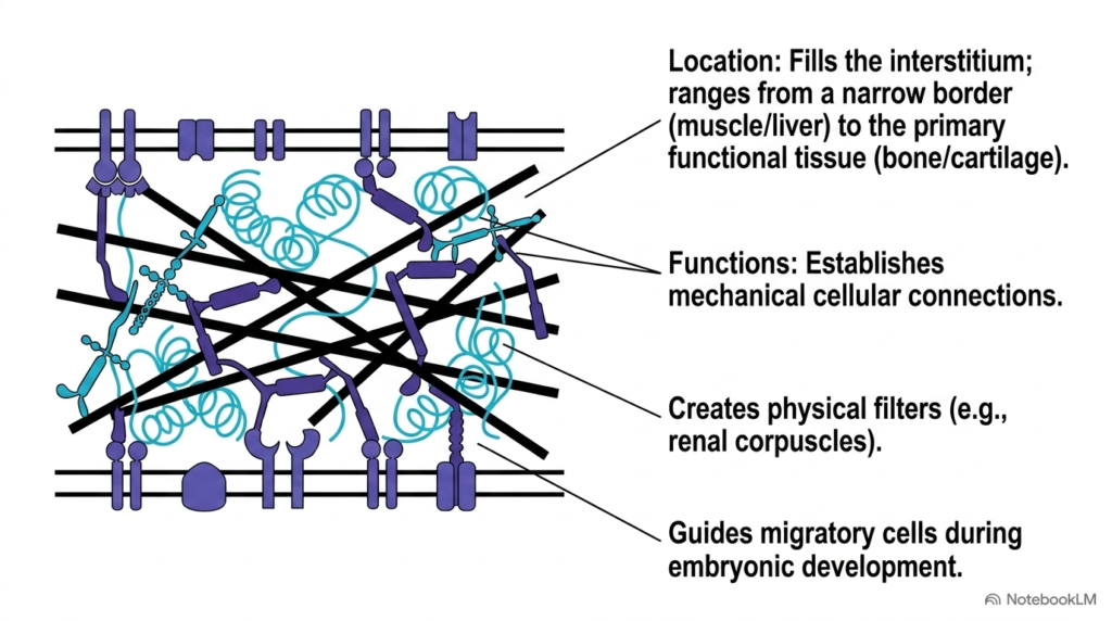
The Extracellular Matrix fulfills several essential physiological roles that maintain overall tissue function and cellular organization across the human body. Located within the interstitial space, the Extracellular Matrix exhibits remarkable anatomical diversity in human organs. Its presence ranges from a narrow structural border surrounding muscle fibers and liver parenchymal cells to the primary functional mass of hard connective tissues like bone and cartilage. One primary function of this molecular network is establishing robust mechanical connections between neighboring cells. These physical bridges stabilize overall tissue architecture and allow groups of cells to function as unified tissue units under continuous physical strain.
Beyond physical support, the Extracellular Matrix creates selective molecular filters that regulate the passage of fluids, nutrients, and solutes between biological compartments. A classic physiological example occurs within renal corpuscles, where a specialized basement membrane matrix sheet filters blood plasma based strictly on molecular size and electrical charge, preventing essential plasma proteins from entering urinary filtration streams.
Furthermore, the Extracellular Matrix acts as a guided pathway for cell migration during embryonic development. Migrating cells rely on specific chemical cues and structural tracks within this matrix to navigate to their designated anatomical destinations. This guided cellular movement ensures proper tissue patterning, organ formation, and structural development throughout human embryogenesis, fetal organogenesis, and postnatal tissue growth.
Slide 03: The Three Pillar Components of the Extracellular Matrix
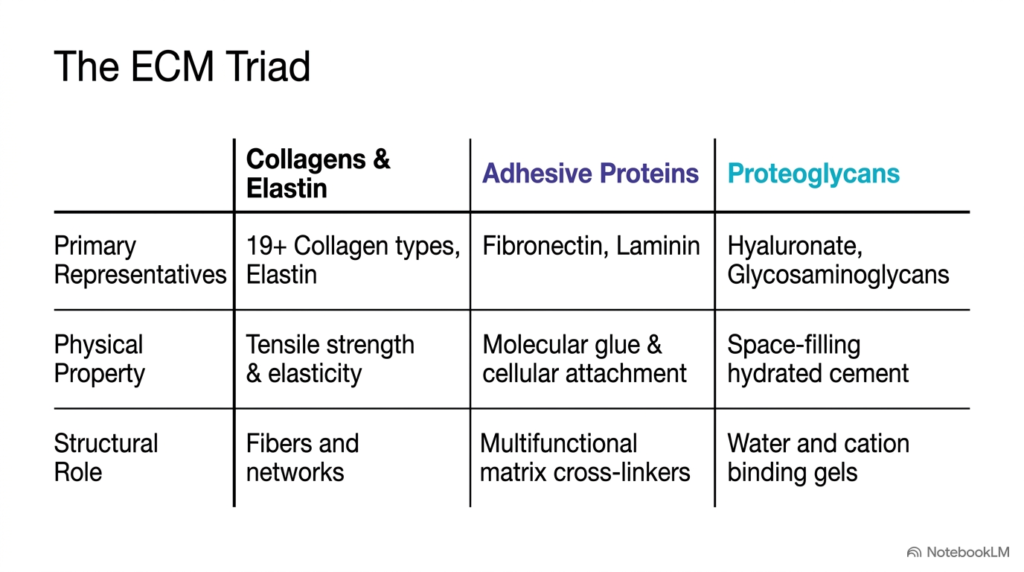
Biochemists categorize the primary structural components of the Extracellular Matrix into three distinct functional classes, collectively known as the matrix triad. The first category comprises collagens and elastin, which form extensive fibrous networks throughout human connective tissues. With over nineteen collagen types identified in human anatomy, these structural proteins provide tensile strength and elastic recoil to organs subjected to continuous mechanical stretching. The second category includes adhesive proteins like fibronectin and laminin. These multifunctional cross-linking molecules act as molecular glue, attaching cells directly to structural fibers within the Extracellular Matrix.
The third essential class consists of proteoglycans and hyaluronate, which form a space-filling hydrated cement. These specialized carbohydrate-rich complexes form water-binding gels that retain positively charged cations and resist high compressive forces during physical impact across various skeletal joints and load-bearing tissues. They maintain constant tissue turgor and lubricate articular surfaces during mechanical motion. This hydrated matrix cushions mechanical joints against heavy mechanical loads.
Together, these three component classes assemble into a highly coordinated structural framework within interstitial spaces. Each class contributes distinct mechanical and chemical properties, ensuring the Extracellular Matrix can withstand pulling forces, maintain optimal tissue hydration, and support robust cellular adhesion across diverse anatomical sites throughout the human body during normal physiological activity, physical exercise, and mechanical adaptation. Medical students should appreciate how this three-part biochemical triad maintains structural integrity in healthy connective tissues.
Slide 04: Domain Architecture of Fibronectin in the Extracellular Matrix
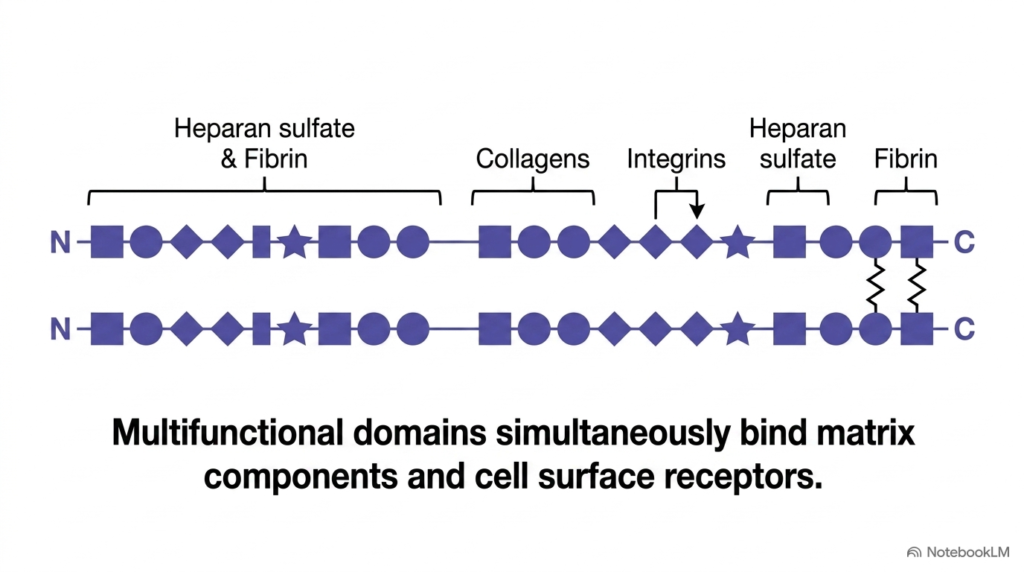
Fibronectin is a prominent adhesive glycoprotein that cross-links various structural elements within the Extracellular Matrix. Structurally, fibronectin exists as a large dimeric protein composed of two nearly identical peptide subunits joined near their carboxyl terminals by covalent disulfide bonds. Each polypeptide chain contains repeating modular sequences that fold into distinct functional domains. These modular domains allow fibronectin to bind simultaneously to multiple structural fibers and cell membrane receptors within the Extracellular Matrix, creating a integrated network throughout connective tissues.
Specific regions along the fibronectin polypeptide chain contain distinct binding sites for heparan sulfate, fibrin, collagen fibers, and cell-surface integrin receptors. With these specialized binding sites, fibronectin connects fibrous collagen directly to cell membranes and adjacent matrix components in a highly organized, spatial manner across extracellular spaces. This precise spatial arrangement promotes efficient cell alignment and structural continuity across tissue boundaries. This organized spatial alignment promotes proper cellular polarization and migration.
This multivalent binding capability makes fibronectin an indispensable structural bridge within the Extracellular Matrix. It links the internal cell cytoskeleton to the surrounding extracellular network, facilitating efficient mechanical force transmission, cellular communication, and structural stability across tissue layers during mechanical deformation and dynamic organ movement in health, physiological growth, development, and active tissue repair. Consequently, fibronectin serves as a key functional link between physical extracellular structures and internal cytoplasmic activities.
Slide 05: Genomic Splicing and Diversity in the Extracellular Matrix
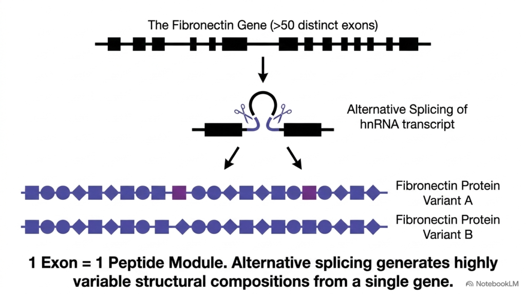
The remarkable structural diversity of fibronectin within the Extracellular Matrix stems from sophisticated genomic organization and pre-messenger RNA processing. A single fibronectin gene contains over fifty distinct exons, each encoding a specific functional peptide module. Primary heterogeneous nuclear RNA transcripts undergo alternative splicing, allowing human cells to generate multiple fibronectin protein variants from a single gene sequence. This flexible genetic mechanism provides extraordinary adaptability to the Extracellular Matrix across different tissue contexts and developmental stages.
By selectively including or excluding specific exon modules during alternative splicing, cells synthesize customized fibronectin isoforms tailored for distinct physiological environments. For instance, soluble fibronectin produced by liver hepatocytes circulates in blood plasma to help stabilize clots during vascular injury, whereas insoluble fibronectin variants assemble directly into solid tissue structures to support local cellular adhesion and matrix cross-linking. This functional versatility highlights the importance of post-transcriptional regulation in matrix assembly.
Alternative splicing thus enables precise cellular control over matrix properties across different anatomical sites. Through this mechanism, the Extracellular Matrix fine-tunes its adhesive properties to meet changing physiological demands during embryonic growth, tissue repair, and ongoing organ remodeling throughout human life under both normal physiological health and pathological disease states across diverse human tissues. Medical researchers study these splicing patterns to understand how matrix diversity influences tissue repair, tissue engineering, and tumor metastasis.
Slide 06: Cell Adhesion Modules and Integrins in the Extracellular Matrix
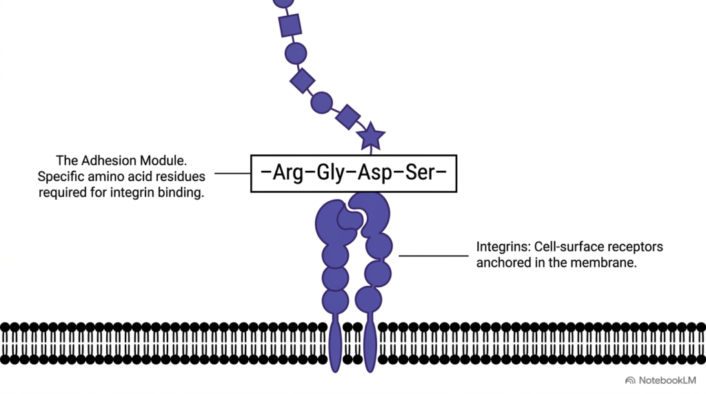
Cellular attachment to the Extracellular Matrix relies on precise amino acid recognition sequences embedded within adhesive glycoproteins. Fibronectin contains a key adhesion module with a specific tetrapeptide sequence of arginine, glycine, aspartate, and serine. This specific sequence, abbreviated as RGDS, is required for binding to cell-surface receptors called integrins. Through this conserved peptide motif, the Extracellular Matrix directly engages transmembrane integrin heterodimers anchored within the phospholipid bilayer.
Integrins function as bidirectional mechanical transducers that span the plasma membrane. When integrins bind the RGDS sequence in the Extracellular Matrix, they initiate intracellular signaling pathways and connect extracellular matrix fibers directly to internal actin microfilaments in the cell cytoplasm. This physical linkage establishes a direct route for mechanotransduction across the cell boundary, allowing external physical forces to alter internal enzymatic activity. This direct physical link enables cells to convert mechanical stresses into intracellular biochemical signals that alter gene transcription.
This physical connection enables cells to sense environmental mechanical forces and adjust their gene expression accordingly. Without this specific peptide-recognition module in the Extracellular Matrix, cells cannot attach properly to their surroundings, triggering detachment and programmed cell death in unattached tissue cells across various human organ systems during normal tissue growth, development, and cellular homeostasis. Understanding integrin-matrix interactions is essential for studying cancer cell invasion, tissue engineering, and cell survival mechanisms in clinical biology.
Slide 07: Proteoglycan Complexes in the Extracellular Matrix
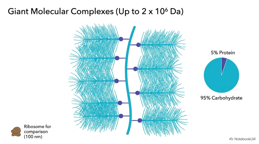
Proteoglycans form giant molecular aggregates that provide essential compressive resistance within the Extracellular Matrix. These massive molecular complexes reach molecular weights up to two million Daltons, dwarfing standard cellular structures such as ribosomes. Visually and structurally, a proteoglycan aggregate resembles a large bottle brush, where a central hyaluronan glycosaminoglycan chain acts as the main structural backbone. Multiple core proteins attach along this central backbone, with hundreds of glycosaminoglycan chains radiating outward into the Extracellular Matrix.
A defining biochemical feature of proteoglycan aggregates in the Extracellular Matrix is their overwhelming carbohydrate content. By total dry weight, these complexes consist of ninety-five percent carbohydrate and only five percent core protein. This dominant carbohydrate fraction renders the complex extremely hydrophilic, allowing it to occupy large aqueous volumes in tissue spaces and swell into a firm, water-rich gel. This high density of carbohydrate chains enables proteoglycans to absorb massive amounts of tissue fluid and maintain high turgor pressure.
Consequently, these giant complexes act as space-filling hydrated cushions within the Extracellular Matrix, protecting delicate tissue structures from heavy physical impacts, mechanical compression, and structural deformation during strenuous physiological activity across human tissue environments throughout life in healthy musculoskeletal organs and articular joint spaces during continuous weight-bearing movements. In weight-bearing cartilage, these proteoglycan aggregates help joints withstand repetitive impacts without structural damage.
Slide 08: Glycosaminoglycan Structure in the Extracellular Matrix
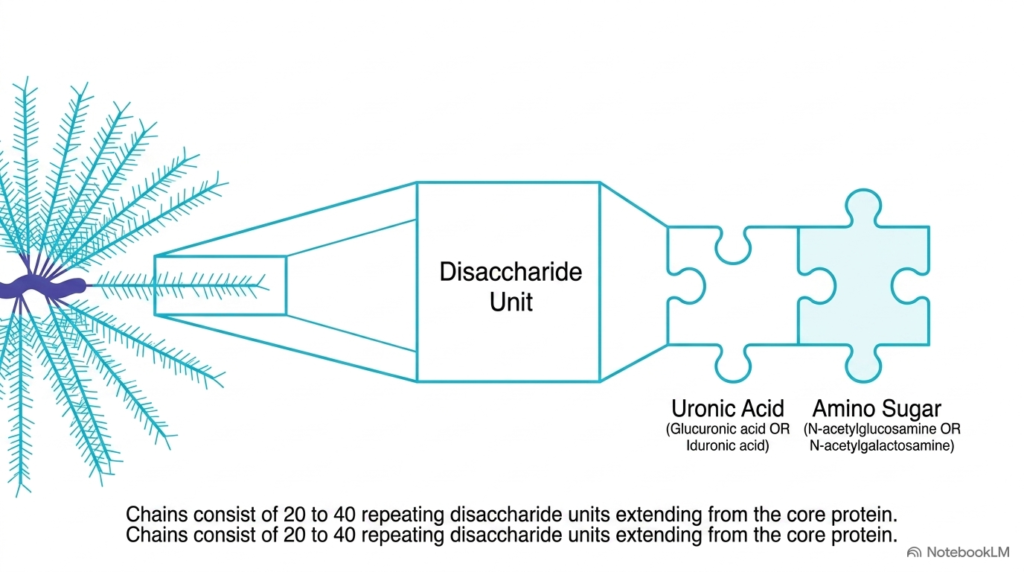
The extensive carbohydrate chains attached to proteoglycan core proteins in the Extracellular Matrix are called glycosaminoglycans. These long, unbranched polysaccharide chains consist of repeating disaccharide units extending outward from a central core protein backbone. Typically, a single glycosaminoglycan chain contains twenty to forty repeating disaccharide pairs linked covalently. Within the Extracellular Matrix, these long carbohydrate polymers create a dense molecular mesh that retains large amounts of water and fills intercellular spaces across biological tissues.
Each disaccharide unit within a glycosaminoglycan chain consists of two specific modified sugar residues. The first sugar is a uronic acid, such as glucuronic acid or iduronic acid. The second sugar is an amino sugar, such as N-acetylglucosamine or N-acetylgalactosamine. This specific disaccharide pairing repeats predictably along the carbohydrate chain, forming a rigid linear sugar polymer that resists mechanical compression. The structural uniformity of this sugar backbone provides the physical blueprint for specialized sulfation patterns.
The precise arrangement of these repeating sugar units forms the backbone on which enzymes add modifications, giving different glycosaminoglycan classes unique functions as they assemble into mature tissue scaffolds during development, growth, repair, and cellular differentiation across organ systems. By organizing these carbohydrate polymers into larger complexes, tissues establish specialized extracellular microenvironments that support diverse biological functions.
Slide 09: Polarity and Charge Density in the Extracellular Matrix
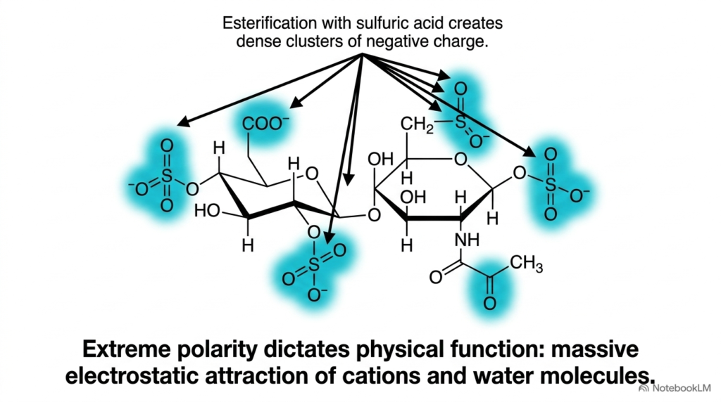
Glycosaminoglycans within the Extracellular Matrix exhibit extreme chemical polarity due to extensive enzymatic modifications during biosynthesis. During carbohydrate assembly, sulfuric acid residues are covalently esterified onto specific hydroxyl and amino groups along the sugar rings. This chemical modification creates dense clusters of negatively charged sulfate groups along the carbohydrate chain. Combined with negatively charged carboxylate groups on uronic acid residues, these sulfate clusters give the Extracellular Matrix one of the highest negative charge densities found in biological systems.
This extreme negative charge density directly dictates the physical behavior of the Extracellular Matrix in living tissues. Because charges repel, the negatively charged carbohydrate chains extend outward into stiff, space-filling conformations. At the same time, these clustered negative charges exert strong electrostatic attraction on positive sodium cations and polar water molecules. This intense electrostatic interaction locks water molecules into place, preventing fluid from being squeezed out under high pressure.
Water flows rapidly into the interstitial tissue space along this strong osmotic gradient, converting the Extracellular Matrix into a highly pressurized, hydrated gel that absorbs massive compressive forces during physical locomotion, joint articulation, and heavy mechanical load-bearing across human connective tissues during daily activity and strenuous exercise in healthy adults. Thus, the chemical charge density of glycosaminoglycans directly converts osmotic pressure into structural resistance across all connective tissues.
Slide 10: The Glycosaminoglycan Ledger in the Extracellular Matrix
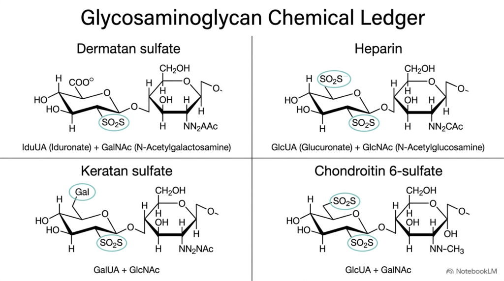
The chemical diversity of glycosaminoglycans within the Extracellular Matrix is demonstrated by four major sulfated polysaccharide structures. Dermatan sulfate consists of repeating iduronate and N-acetylgalactosamine units with specific sulfate modifications that support vascular tissue integrity. Heparin contains glucuronic acid and N-acetylglucosamine residues and features dense sulfation that confers potent anticoagulant activity. Keratan sulfate incorporates galactose and N-acetylglucosamine disaccharides, providing essential structural transparency in ocular corneal tissues governed by the Extracellular Matrix.
Chondroitin six-sulfate features glucuronic acid paired with N-acetylgalactosamine sulfated at the sixth carbon position and serves as a primary load-bearing component in articular cartilage. Each glycosaminoglycan has a distinct pattern of sugar-ring stereochemistry and sulfate-ester positions that determine specific ligand interactions and binding affinities. These subtle chemical variations dictate how each glycosaminoglycan interacts with specific proteins and signaling molecules.
This chemical ledger allows tissues to customize the local microenvironment of the Extracellular Matrix. By altering the relative composition of these sugar polymers, tissues create specialized extracellular environments that regulate growth factor distribution, ion diffusion, and mechanical flexibility across human physiology, tissue repair, cellular signaling, and metabolic function in clinical biochemistry. Students should recognize that subtle alterations in glycosaminoglycan structure can significantly impact tissue hydration, growth factor signaling, and disease states.
Slide 11: Hydration and Pathogen Resistance in the Extracellular Matrix
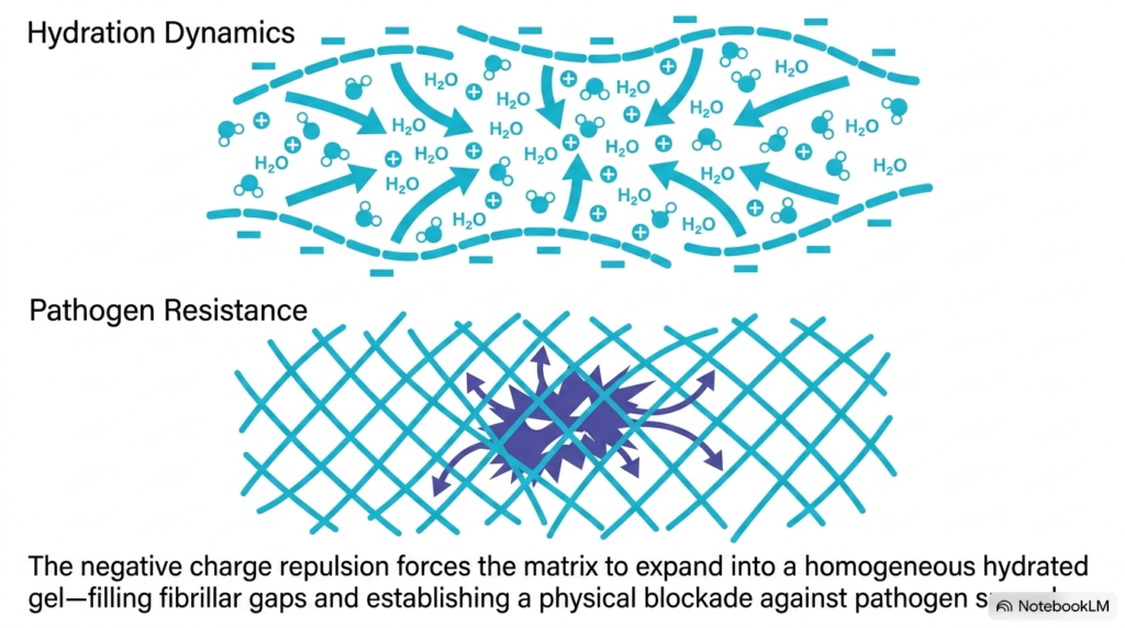
The unique chemical structure of glycosaminoglycans drives dynamic tissue hydration within the Extracellular Matrix. Because adjacent negative charges repel each other, carbohydrate chains expand outward into an open physical network. This expansion draws abundant water molecules and positive sodium cations into the interstitial space, converting the Extracellular Matrix into a homogeneous, fully hydrated gel that completely fills all gaps between structural collagen fibers.
This continuous hydrated gel provides an unexpected vital function by establishing robust resistance against invasive pathogenic microorganisms. Because the Extracellular Matrix forms a dense, pressurized carbohydrate network, it creates a formidable physical blockade that restricts the free migration of bacteria and viruses through biological tissues, preventing rapid bacterial dissemination into adjacent blood vessels. This physical barrier traps infectious agents within localized regions, preventing systemic dissemination throughout bodily tissues.
Invasive pathogens cannot easily penetrate this thick gel unless they secrete specialized enzymes like hyaluronidase to degrade the carbohydrate meshwork. Thus, the Extracellular Matrix functions as both a mechanical shock absorber and a primary physical barrier against systemic infection during pathogen encounters, microbial exposure, and localized tissue defense mechanisms. Understanding matrix hydration dynamics helps explain how pathogenic organisms use specific enzymes to break down extracellular barriers during infection.
Slide 12: The Unified Scaffold of the Extracellular Matrix
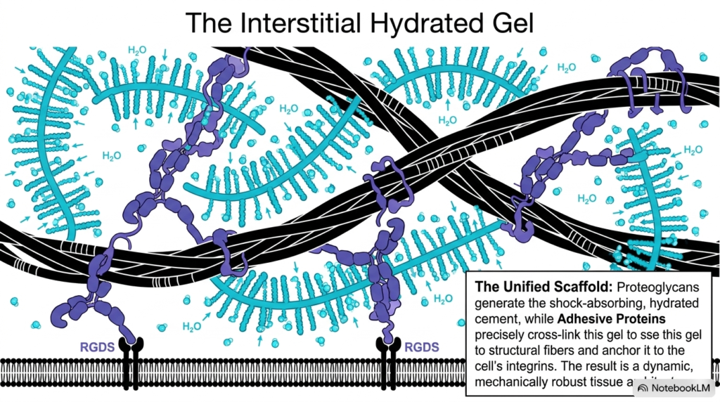
The Extracellular Matrix functions as a unified, dynamic scaffold that integrates structural fibers, hydrated carbohydrate gels, and cell membrane receptors. Proteoglycans generate a shock-absorbing, water-rich cement that fills interstitial spaces and resists compression. At the same time, adhesive proteins like fibronectin cross-link this hydrated gel to rigid collagen fibers. These adhesive proteins also anchor the entire matrix network to cell-surface integrin receptors through specific recognition sequences like RGDS, establishing a continuous structural link into the Extracellular Matrix.
This biochemical integration yields a mechanically robust tissue framework that can adapt to complex physiological stresses. Cells remain securely anchored to the matrix while receiving constant mechanical feedback that influences cell division, differentiation, gene expression, structural organization, and overall tissue survival across human organ systems. This continuous physical pathway allows mechanical forces to directly modulate nuclear gene expression and cellular behavior.
By uniting tensile strength, compressive resilience, and cell signaling, the Extracellular Matrix serves as the master architect of tissue form and cellular function. Understanding this integrated molecular scaffold provides a crucial foundation for medical research in tissue engineering, wound healing, regenerative medicine, and disease pathology across clinical disciplines and modern biomedical science. Mastering the biochemical principles of matrix organization allows biomedical researchers to design synthetic tissue scaffolds for regenerative medicine.
Please read our Content Disclaimer Statement.
Check out our social media channels: