94. Genetic Engineering in Medicine: Diagnostics and Therapeutics
Imagine holding the instruction manual for the human body and possessing the precise tools to fix any misprinted page. That is the incredible promise of modern molecular biology. This comprehensive slide deck breaks down the core concepts of diagnostic and therapeutic genetic techniques for aspiring healthcare professionals. The core purpose of these slides is to demystify complex biochemical structures and processes, bridging the gap between theoretical laboratory genetics and real-world clinical applications. Students will gain a robust foundation in how scientists read and rewrite the genetic code to cure disease.
Slide 1: Genetic Engineering in Medicine: Decoding the Blueprint of Life
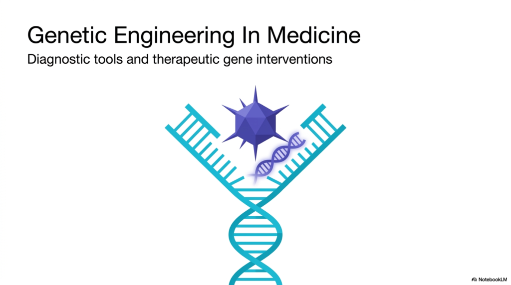
The modern healthcare landscape relies on the principles of molecular biology to solve complex clinical challenges. At the center of this scientific revolution is Genetic Engineering in Medicine, a dynamic field that blends basic biochemistry with life-saving clinical applications. For medical and college students, mastering these intricate concepts represents the future of targeted patient care. By understanding how scientists manipulate essential DNA and RNA structures, future medical professionals can appreciate the sophisticated molecular tools used in hospitals to diagnose mysterious illnesses, track viral outbreaks, and treat inherited genetic diseases.
The first slide introduces the dual nature of Genetic Engineering in Medicine, dividing the field into diagnostic tools and therapeutic interventions. Diagnostics involves reading the biological blueprint to identify pathogens or genetic defects with pinpoint accuracy. Therapeutics focuses on editing or supplementing that genetic blueprint to correct fundamental cellular malfunctions. The visual representation features the iconic double helix alongside a customized viral vector, symbolizing how natural biological components are repurposed for human health. Grasping this primary division is crucial for students transitioning from theoretical biochemistry into applied clinical sciences.
By exploring Genetic Engineering in Medicine, the scientific community shifts its focus from managing superficial disease symptoms to addressing root molecular causes. The integration of viruses as targeted delivery vehicles showcases the ingenuity of this discipline, turning former cellular enemies into microscopic delivery trucks for therapeutic genes. This introductory visual sets the stage for a deep dive into specific molecular techniques. As the lesson progresses, students will explore the biochemical mechanisms that make these clinical miracles possible, ranging from amplifying microscopic forensic DNA traces to silencing harmful metabolic pathways inside human cells.
Slide 2: Genetic Engineering in Medicine: Translating Biology into Clinical Practice
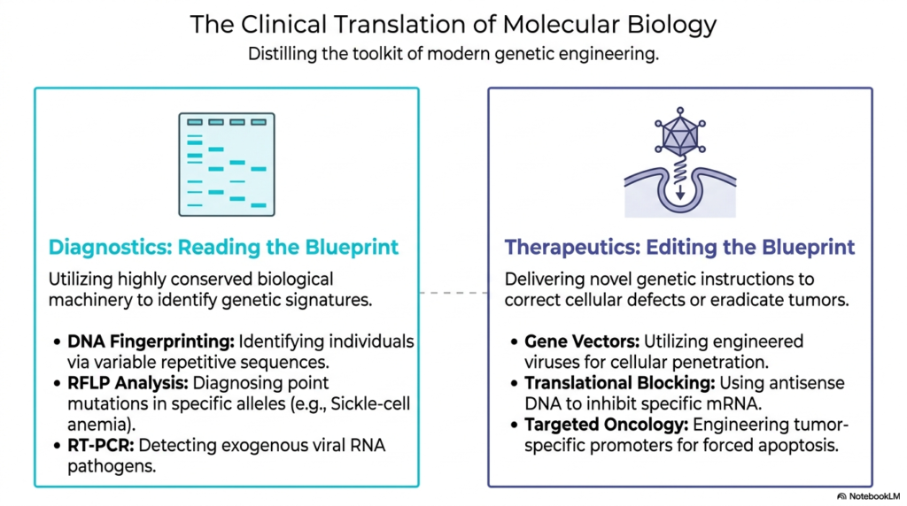
The transition from textbook biochemistry to clinical application requires a deep understanding of molecular tools. Slide two outlines the primary toolkit of Genetic Engineering in Medicine, dividing it into distinct diagnostic and therapeutic categories. Diagnostics focuses on reading the biological blueprint. Techniques like DNA fingerprinting, restriction fragment length polymorphism analysis, and reverse transcription-polymerase chain reaction allow clinicians to identify unique genetic signatures. These methods utilize conserved biological machinery to detect everything from forensic evidence to exogenous viral pathogens like SARS-CoV-2. Understanding these foundational diagnostic tools helps students appreciate how minute molecular differences translate into medical diagnoses.
Conversely, the therapeutic side of Genetic Engineering in Medicine focuses on editing the cellular blueprint. This involves delivering novel genetic instructions to correct fundamental cellular defects or to eradicate tumors. Instead of treating symptoms, scientists use engineered gene vectors, such as modified viruses, to penetrate target cells and deliver functional DNA payloads. Other advanced strategies include translational blocking, where antisense DNA inhibits specific messenger RNA, or targeted oncology, which forces cancer cells into programmed cell death. These approaches highlight the power of manipulating biochemical pathways directly to achieve therapeutic goals.
By mastering both the diagnostic and therapeutic arms of Genetic Engineering in Medicine, students prepare themselves for the future of personalized healthcare. Reading the blueprint provides the precise diagnosis, while editing the blueprint offers a targeted, molecular-level cure. The distinction between analyzing variable repetitive sequences and engineering tumor-specific promoters is fundamental to the discipline. This comprehensive overview sets the stage for a detailed examination of each specific technique, ensuring learners grasp the underlying biochemical principles that drive modern medical innovation and improve patient outcomes across the globe.
Slide 3: Genetic Engineering in Medicine: The Biochemistry of Individual Identification
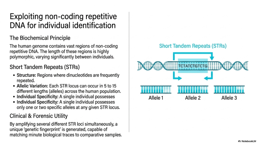
A foundational concept in Genetic Engineering in Medicine is the utilization of non-coding repetitive DNA for individual identification. The human genome consists largely of non-coding regions, which contain vast sequences of repetitive DNA. Specifically, short tandem repeats are regions where tiny dinucleotide sequences are repeated. Because the length of these repetitive regions is polymorphic, they vary between different individuals. Students analyzing this biochemical principle will discover that this natural variation provides a unique molecular signature, forming the basis for precise forensic and clinical identification techniques used worldwide.
The structural characteristics of short tandem repeats make them ideal targets for genetic profiling in genetic engineering in Medicine. Each specific locus can occur in five to fifteen different lengths, or alleles, across the broader human population. However, a single individual will possess only one or two specific alleles at any given locus, inherited from their parents. The slide illustrates how these varying lengths look at the molecular level, demonstrating alleles with different numbers of repeating units. This variation is the biochemical key to distinguishing one human being from another with near-absolute certainty.
The clinical and forensic utility of this phenomenon is a major milestone for Genetic Engineering in Medicine. By simultaneously amplifying several different loci, scientists generate a unique genetic fingerprint. This comprehensive molecular profile is capable of matching minute biological traces, such as a drop of blood or a hair follicle, to comparative reference samples. For future medical professionals, understanding the statistical power of multiplexing these repeats highlights how basic molecular variations are harnessed to solve complex real-world problems, ensuring accurate identification in both legal and medical contexts.
Slide 4: Genetic Engineering in Medicine: Amplifying and Analyzing Trace DNA
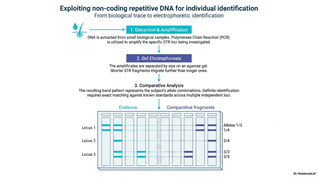
The practical workflow of DNA profiling demonstrates the hands-on laboratory techniques essential to Genetic Engineering in Medicine. The process begins with extracting DNA from minuscule biological samples. Because these trace samples often contain insufficient genetic material for direct analysis, biochemists utilize the polymerase chain reaction. This powerful technique amplifies the specific short tandem repeat loci being investigated. By focusing the amplification on these polymorphic regions, scientists ensure that enough targeted genetic material is synthesized to allow for clear detection. Students must understand that this targeted replication mimics natural cellular processes in a controlled environment.
Following amplification, the next crucial step in Genetic Engineering in Medicine involves gel electrophoresis. The amplified DNA fragments are loaded into a porous agarose gel matrix and subjected to an electrical current. Because DNA is negatively charged, the fragments migrate toward the positive electrode. The biochemical principle governing this movement is size separation; shorter fragments migrate faster and further through the gel than longer, bulkier ones. This differential migration separates the various alleles by length, creating a distinct visual pattern of bands. This spatial separation converts invisible molecules into interpretable diagnostic data.
The final stage of this process in Genetic Engineering in Medicine is the comparative analysis of the resulting electrophoretic band patterns. Definite identification requires exact matching between the evidence sample and known comparative standards across multiple independent loci. The slide provides a clear visual matrix comparing evidence fragments against known standards, showing exact alignment for a positive match. For students, mastering this workflow illustrates how theoretical molecular biology translates into reliable analytical procedures. Understanding this visual comparison ensures future scientists can interpret complex laboratory results with confidence in clinical settings.
Slide 5: Genetic Engineering in Medicine: Detecting Pathogenic Point Mutations
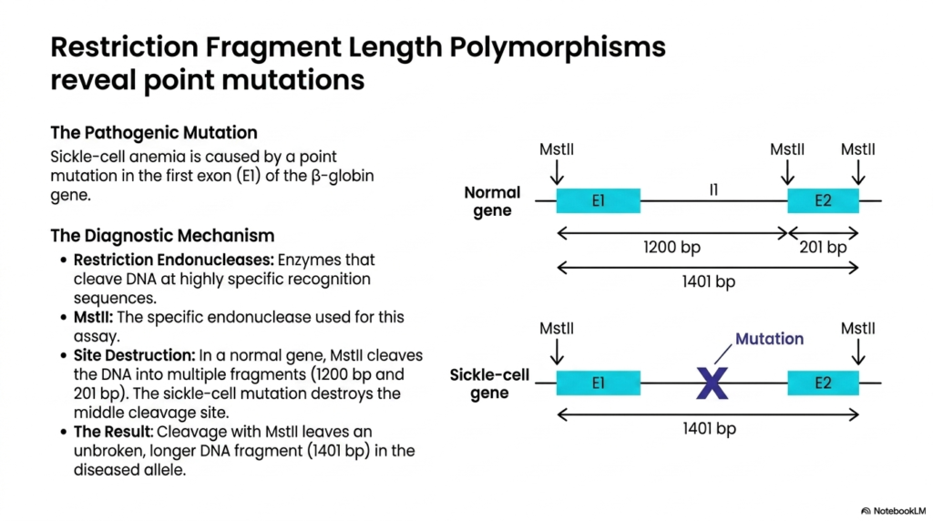
Identifying the exact molecular cause of inherited diseases is a primary objective of Genetic Engineering in Medicine. Slide five explores how restriction fragment length polymorphisms reveal pathogenic point mutations, using sickle-cell anemia as the classic biochemical model. Sickle-cell anemia is caused by a single point mutation in the first exon of the beta-globin gene. This tiny alteration in the nucleotide sequence has profound physiological consequences. For medical students, understanding how such a minuscule genetic error leads to systemic disease underscores the vital importance of precision molecular diagnostics in patient care.
To detect this specific mutation, Genetic Engineering in Medicine employs restriction endonucleases. These specialized enzymes act as molecular scissors, cleaving DNA at specific recognition sequences. The diagnostic assay for sickle-cell anemia utilizes the MstII endonuclease. In a normal, healthy gene, MstII successfully recognizes its specific sequence and cleaves the DNA into multiple shorter fragments, specifically a 1200-base-pair and a 201-base-pair fragment. This predictable cleavage pattern serves as the baseline diagnostic signature for the presence of the normal, functional beta-globin allele within the patient’s genome.
The fundamental diagnostic mechanism of Genetic Engineering in Medicine is illustrated when the mutation is present. The sickle-cell mutation destroys the precise middle cleavage site that MstII recognizes. Consequently, the enzyme cannot cut the DNA at that location. This site destruction results in an unbroken, longer DNA fragment of 1401 base pairs in the diseased allele. By analyzing these varying fragment lengths, clinicians can directly observe the presence of the genetic defect, showcasing how biochemical assays translate hidden nucleotide changes into visible, diagnostic molecular markers.
Slide 6: Genetic Engineering in Medicine: Genotyping Sickle-Cell Carriers
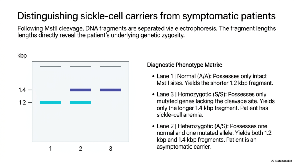
Following targeted enzymatic cleavage, Genetic Engineering in Medicine utilizes electrophoresis to distinguish sickle-cell carriers from symptomatic patients. The varying DNA fragment lengths reveal the patient’s underlying genetic zygosity. When the processed DNA is run on a gel, the resulting bands provide a clear diagnostic phenotype matrix. This visual readout is essential for clinicians to determine not just if a mutation is present, but how it is inherited. For medical students, interpreting these gel patterns is a critical skill for accurately diagnosing and counseling patients with hereditary conditions.
The diagnostic matrix provided by Genetic Engineering in Medicine outlines the three potential genetic states. A normal, healthy homozygous individual possesses only intact MstII cleavage sites, yielding solely the shorter 1.2 kilobase pair fragment on the gel. In contrast, a symptomatic patient who is homozygous for the sickle-cell trait possesses only mutated genes lacking the cleavage site. This state yields exclusively the longer 1.4 kilobase pair fragment. These distinct single-band patterns make identifying the extreme ends of the genetic spectrum straightforward for laboratory technicians and attending physicians.
The true power of Genetic Engineering in Medicine is demonstrated when identifying asymptomatic carriers. A heterozygous individual possesses one normal allele and one mutated allele. Consequently, the MstII enzyme cleaves the normal copy but leaves the mutated copy intact. The resulting electrophoretic gel displays both the 1.2 kilobase pair and the 1.4 kilobase pair fragments simultaneously. This dual-band pattern identifies the patient as an asymptomatic carrier. Understanding this biochemical readout allows future healthcare providers to offer critical genetic counseling and preventative care for prospective parents carrying the trait.
Slide 7: Genetic Engineering in Medicine: Amplifying Viral RNA Signatures
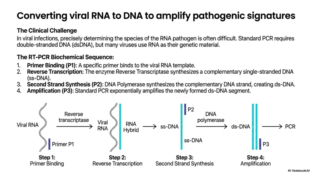
One of the greatest clinical challenges addressed by Genetic Engineering in Medicine is the rapid and precise identification of RNA viruses. Standard diagnostic amplification requires double-stranded DNA, but many dangerous viral pathogens utilize single-stranded RNA as their genetic material. To overcome this biochemical hurdle, scientists developed reverse transcription-polymerase chain reaction. This ingenious multi-step sequence allows clinical laboratories to convert unstable viral RNA into a stable DNA format, making it possible to amplify and detect elusive pathogenic signatures with extreme sensitivity. Understanding this fundamental difference between nucleic acid structures is vital for students studying molecular diagnostics.
The core biochemical sequence of this technique in Genetic Engineering in Medicine begins with specific primer binding to the target viral RNA template. Next, the specialized enzyme reverse transcriptase synthesizes a complementary single-stranded DNA molecule from the RNA template. This crucial step bridges the gap between RNA and DNA biology. Following this, DNA polymerase synthesizes the second complementary DNA strand, creating a stable, double-stranded DNA hybrid. Students must recognize that reverse transcriptase is an enzyme derived from retroviruses, showing how scientists have repurposed viral machinery for modern diagnostic purposes.
Once the double-stranded DNA is synthesized, Genetic Engineering in Medicine applies standard amplification protocols. The newly formed DNA segment undergoes exponential amplification, creating millions of copies of the specific viral signature. This mass amplification allows clinicians to detect even the smallest viral loads in patient samples. The ability to visualize the stepwise progression from single-stranded viral RNA to a massive pool of double-stranded DNA amplificates is central to understanding how theoretical biochemistry directly saves lives during global infectious disease crises by directing targeted antiviral therapies.
Slide 8: Genetic Engineering in Medicine: Confirming Pathogen Identity
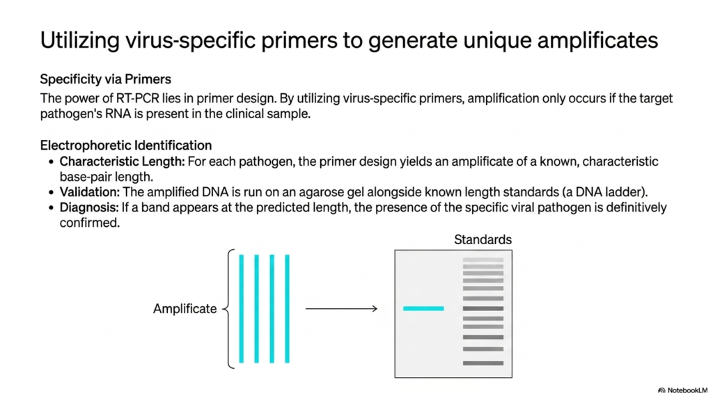
The extraordinary diagnostic specificity of Genetic Engineering in Medicine relies on intelligent molecular design. The true power of the amplification process lies in the meticulous design of viral primers. By using specialized, virus-specific primers, the biochemical reaction ensures that amplification occurs only if the target pathogen’s exact genetic sequence is present in the clinical sample. If the target RNA is absent, the primers have nothing to bind to, and the reaction remains silent. For medical students, understanding the thermodynamics of primer annealing and sequence complementarity is essential, as it dictates the success of the entire diagnostic assay.
To visualize this specific amplification, Genetic Engineering in Medicine once again employs gel electrophoresis. For each unique pathogen, the customized primer design yields an amplificate of a known, characteristic base-pair length. After the reaction is complete, the amplified DNA is run on an agarose gel alongside known length standards, referred to as a DNA ladder. This ladder provides a reliable molecular ruler. Understanding how to read these electrophoretic ladders is a fundamental skill for any biochemist, as it provides the critical spatial context needed to validate the success of the targeted amplification process.
The final diagnostic confirmation in Genetic Engineering in Medicine is remarkably straightforward yet biochemically profound. If a distinct band appears on the gel at the predicted molecular length, the presence of the specific viral pathogen is confirmed. This visual proof translates complex molecular hybridization events into a clear clinical answer. By utilizing these precise electrophoretic techniques, laboratories can confidently distinguish between closely related viral strains, ensuring that patients receive the most appropriate and effective therapeutic interventions based on their exact viral profile and molecular signature.
Slide 9: Genetic Engineering in Medicine: The Paradigm Shift to Gene Therapy
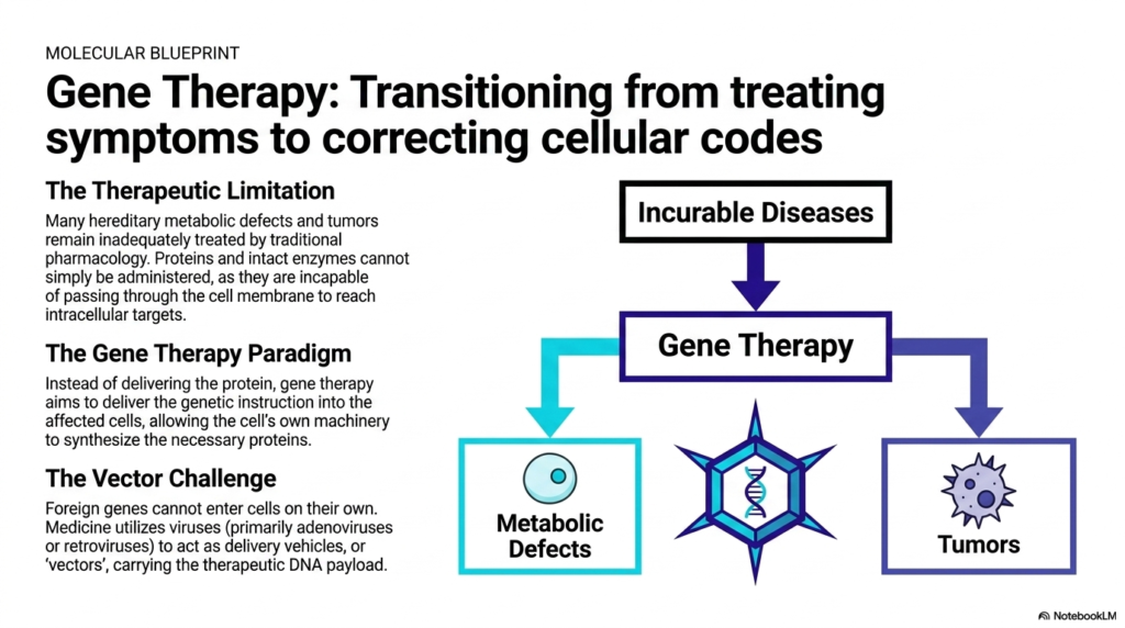
Moving beyond diagnostics, Genetic Engineering in Medicine introduces a paradigm shift in how we approach incurable diseases. Historically, many hereditary metabolic defects and aggressive tumors remained untreated by traditional pharmacology. The primary therapeutic limitation is that large proteins and intact corrective enzymes cannot be administered as conventional drugs because they cannot pass through the protective lipid bilayer of the cell membrane to reach their vital intracellular targets. Students must appreciate these biophysical properties of cell membranes to understand why traditional pharmacology fails, highlighting the need for innovative genetic delivery systems.
To overcome this barrier, Genetic Engineering in Medicine shifts the focus from treating surface symptoms to correcting cellular codes. Instead of attempting to deliver a bulky, pre-formed protein, modern gene therapy aims to deliver the fundamental genetic instructions directly into the affected cells. By providing the correct DNA sequence, this approach allows the patient’s own cellular machinery to synthesize the necessary missing proteins internally. Understanding the central dogma of molecular biology—how DNA is transcribed into RNA and translated into protein—is crucial here, as gene therapy leverages these innate cellular pathways.
The most significant hurdle in Genetic Engineering in Medicine is the delivery mechanism, known as the vector challenge. Foreign genes cannot autonomously enter human cells. To address this, modern medicine uses modified viruses, primarily adenoviruses or retroviruses, as microscopic delivery vehicles. By stripping these viruses of their pathogenic components and loading them with a therapeutic DNA payload, scientists create engineered vectors capable of efficiently penetrating cells to cure metabolic defects or target tumors. Analyzing the molecular anatomy of these viral vectors reveals how specific viral envelope proteins mediate vital cellular entry.
Slide 10: Genetic Engineering in Medicine: Correcting Metabolic Enzyme Deficiencies
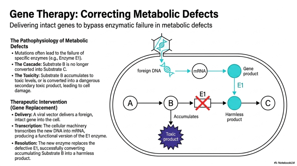
A prime clinical application of Genetic Engineering in Medicine is the correction of inherited metabolic defects through targeted gene replacement. The pathophysiology of these diseases usually involves mutations that lead to the complete failure of specific, vital enzymes. When an enzyme fails, the metabolic cascade halts; the upstream substrate is no longer converted into the downstream product. Consequently, this substrate accumulates to toxic levels within the cell, leading to severe cell damage. Understanding enzyme kinetics and metabolic flux is essential for students, as it explains why the absence of a single enzyme can cause devastating systemic illnesses.
To intervene, Genetic Engineering in Medicine employs a sophisticated mechanism of gene replacement. The therapy begins with a specialized viral vector that delivers a foreign, intact gene directly into the nucleus of the diseased cell. Once inside, the host’s own cellular machinery takes over. The new, functional DNA is transcribed into messenger RNA, which is then translated by ribosomes to produce a functional version of the previously missing enzyme. By studying this intracellular process, students visualize how therapeutic vectors hijack the natural transcriptional apparatus, turning the patient’s own cells into factories for their medicines.
The ultimate resolution achieved by Genetic Engineering in Medicine is the complete restoration of the disrupted biochemical pathway. The newly synthesized, functional enzyme replaces the defective one. It immediately begins processing the accumulated, dangerous substrate, converting it into a harmless product as originally intended by nature. Mastering the biochemistry behind this targeted enzyme replacement therapy empowers future clinicians to understand how advanced genetic interventions stabilize metabolic pathways, preventing irreversible tissue damage and dramatically improving the overall quality of life for patients suffering from these complex metabolic conditions.
Slide 11: Genetic Engineering in Medicine: Silencing Toxic Pathways
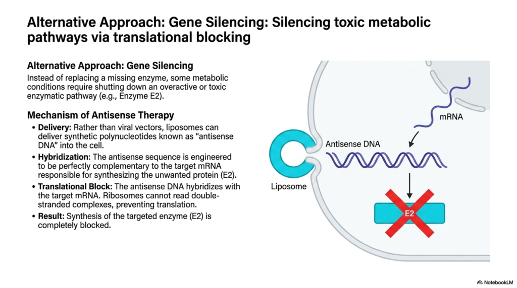
While some conditions require gene replacement, Genetic Engineering in Medicine also provides tools for gene silencing. This alternative approach is critical when metabolic conditions require shutting down an overactive or inherently toxic enzymatic pathway, rather than replacing a missing one. In these scenarios, the goal is to stop the synthesis of a specific, harmful protein. To achieve this, scientists utilize synthetic polynucleotides known as antisense DNA. Understanding the amphipathic nature of lipid-based liposomes is vital for students, as it explains how synthetic delivery vehicles integrate with the cell membrane to deposit highly polar nucleic acids directly into the cytoplasm.
The core biochemical mechanism of this technique in Genetic Engineering in Medicine relies on precise molecular hybridization. The synthetic antisense sequence is engineered to be perfectly complementary to the target messenger RNA responsible for synthesizing the unwanted protein. Once the liposome releases its payload, the antisense DNA rapidly seeks out and hybridizes with the target messenger RNA in the cytoplasm. Students must grasp the strict rules of Watson-Crick base pairing to understand the immense specificity of this therapy, ensuring that the antisense drug only neutralizes the specific disease-causing transcript without interfering with essential cellular functions.
The clinical result of this hybridization in Genetic Engineering in Medicine is a complete translational block. Cellular ribosomes, the molecular machines responsible for synthesizing proteins, are structurally incapable of reading double-stranded nucleic acid complexes. Because the target messenger RNA is trapped in this duplex, translation is physically prevented, and the synthesis of the toxic enzyme is blocked. This elegant application of molecular biology highlights the incredible versatility of nucleic acid therapeutics, proving that preventing the expression of a mutated gene can be just as clinically powerful as providing a functional replacement for a missing one.
Slide 12: Genetic Engineering in Medicine: Precision Targeting in Oncology
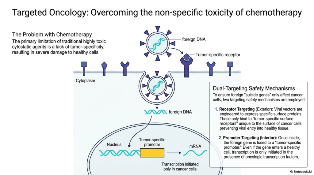
One of the most urgent applications of Genetic Engineering in Medicine is overcoming the severe limitations of traditional cancer treatments. The primary problem with conventional chemotherapy is its non-specific toxicity; systemic cytostatic agents cannot distinguish between cancerous and healthy tissue, resulting in devastating collateral cellular damage. To solve this, molecular biology offers targeted oncology. By employing engineered viral vectors, scientists deliver specific therapeutic genes, often referred to as suicide genes, exclusively to the malignant cell population. Students will realize that the transition from generalized systemic poisons to precise genetic weapons drastically reduces patient suffering.
To ensure maximum safety, Genetic Engineering in Medicine utilizes a sophisticated dual-targeting safety mechanism. The first layer of defense is exterior receptor targeting. The viral vectors are genetically engineered to express specific surface proteins that bind to unique tumor-specific surface receptors. Because these targeted receptors are absent on healthy tissue, the therapeutic virus is physically prevented from entering normal cells. Understanding the biochemistry of membrane receptor interactions is critical here, as it defines the precise tropism of the engineered vector, allowing scientists to exploit unique protein expression profiles to direct the therapy accurately.
The second layer of safety in Genetic Engineering in Medicine involves interior promoter targeting. Once the therapeutic payload enters the targeted cell, the foreign suicide gene remains inactive because it is fused to a specialized tumor-specific promoter. This promoter ensures that transcription is only initiated in the presence of specific oncologic transcription factors found only inside cancer cells. By studying this two-tiered safety system, future clinicians learn that even if a stray vector breaches a healthy cell, the absence of malignant internal transcription factors keeps the lethal gene permanently silenced to protect the patient.
Slide 13: Genetic Engineering in Medicine: Activating Cellular Destruction
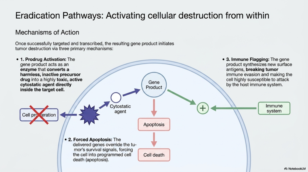
Once targeted delivery is successful, Genetic Engineering in Medicine employs specific biochemical pathways to eradicate the tumor from within. After the therapeutic gene is transcribed and translated inside the cancer cell, the resulting gene product initiates destruction via three primary mechanisms. The first is prodrug activation. The delivered gene acts as an enzyme that converts an administered, harmless precursor drug into a toxic, active cytostatic agent directly inside the cell. Understanding the biochemical kinetics of this localized enzymatic conversion demonstrates how scientists exploit the tumor microenvironment to create concentrated zones of cytotoxicity safely.
The second potent eradication pathway utilized in Genetic Engineering in Medicine is forced apoptosis. Cancer cells naturally thrive by aggressively overriding their own internal survival signals, allowing them to evade programmed cell death. The newly delivered therapeutic genes are designed to counteract these malignant survival signals. By biochemically overriding the tumor’s defensive pathways, the therapy forces the diseased cell to initiate its own programmed cell death cascade. Students must study the intrinsic and extrinsic apoptotic pathways to comprehend how these engineered genetic interventions dismantle complex molecular roadblocks, restoring the natural biological order of cellular turnover.
The third mechanism in Genetic Engineering in Medicine involves immune flagging to break the tumor’s evasion strategies. The newly introduced gene product synthesizes novel surface antigens that are displayed on the exterior of the cancer cell. These visible molecular flags break the tumor’s immune evasion capabilities, making the previously invisible cell susceptible to attack by the host’s innate immune system. By analyzing this immunological mechanism, future medical professionals learn how genetic therapies bridge the gap between molecular biology and systemic immunology, essentially vaccinating the patient against their own tumors by unmasking the hidden malignant cells.
Slide 14: Genetic Engineering in Medicine: Synthesizing the Molecular Toolkit
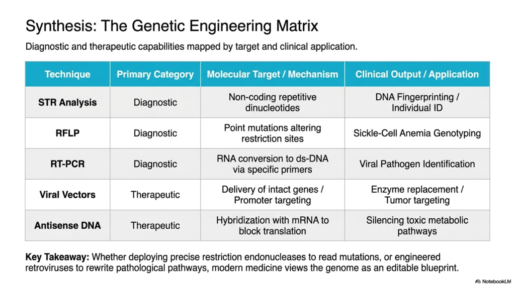
The final slide synthesizes the vast landscape of Genetic Engineering in Medicine into a clear, comprehensible matrix. This organizational tool maps both diagnostic and therapeutic capabilities by linking specific biochemical techniques to their molecular targets and ultimate clinical applications. From analyzing non-coding repetitive dinucleotides via STR analysis for individual identification, to identifying RNA viral pathogens using designed primers in RT-PCR, the matrix categorizes the reading of the biological blueprint. By studying this highly organized summary, students can rapidly cross-reference different methodologies, ensuring they understand which biochemical tool is most appropriate for a given clinical scenario.
Furthermore, the matrix highlights the therapeutic achievements of Genetic Engineering in Medicine. It contrasts the use of viral vectors to deliver intact genes and target tumors with the use of antisense DNA to hybridize with messenger RNA and silence toxic metabolic pathways. Understanding these categorizations allows future healthcare leaders to appreciate the sheer breadth of molecular interventions available. This demonstrates how diverse laboratory techniques all share the common underlying goal of manipulating nucleic acids to restore normal physiological function in patients, reinforcing the dual nature of the discipline.
The key takeaway from this entire exploration of Genetic Engineering in Medicine is profound. Whether deploying precise restriction endonucleases to read invisible genetic mutations, or utilizing engineered retroviruses to rewrite pathological cellular pathways, modern medicine now views the human genome as an editable blueprint. Ultimately, this comprehensive matrix proves that the integration of basic biochemistry and clinical medicine has created a new era of personalized therapies. Mastering these molecular targets empowers the next generation of scientists and physicians to actively rewrite the genetic destiny of their patients and revolutionize global healthcare.
Please read our Content Disclaimer Statement.
Check out our social media channels: